
Dipsas pavonina Schlegel, 1837
|
publication ID |
https://doi.org/10.5281/zenodo.189660 |
|
DOI |
https://doi.org/10.5281/zenodo.5664041 |
|
persistent identifier |
https://treatment.plazi.org/id/A54787C9-FFE3-350E-89B0-F8F4FCEFD39D |
|
treatment provided by |
Plazi |
|
scientific name |
Dipsas pavonina Schlegel, 1837 |
| status |
|
Dipsas pavonina Schlegel, 1837
Dipsas pavonina Schlegel, 1837: 280
Leptognathus pavoninus — Duméril, Bibron & Duméril, 1854: 474 Leptognathus catesbyi — Günther, 1858: 180
Leptognathus pavonina — Cope, 1868: 107
Cochliophagus pavoninus —Von Ihering, 1910: 330
Sibynomorphus pavoninus — Amaral, 1926: 8
Dipsas indica — Beebe, 1946: 24
Dipsas pavonina — Peters, 1960: 79; Peters, 1964: 47; Roze, 1966: 114; Peters & Orejas-Miranda 1970: 88; Dixon & Soini, 1977: 43; Cunha & Nascimento, 1978: 72; Duellman, 1978: 239; Gasc & Rodrigues, 1980: 574; Abuys, 1983: 118; Cunha et al., 1985: 48; Cunha & Nascimento, 1993: 46; Starace, 1998: 172; Harvey & Embert, 2008: 78.
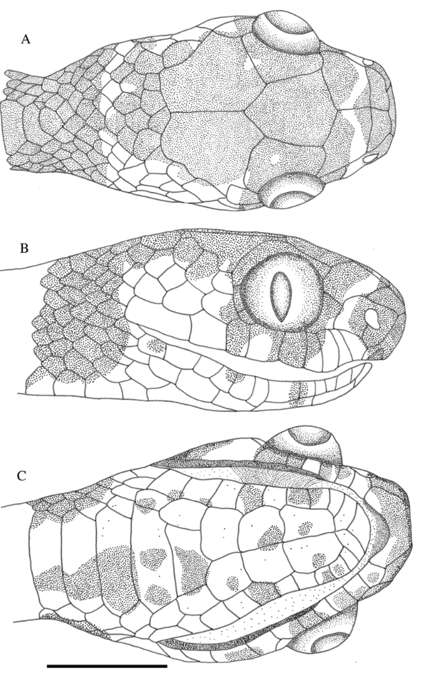
Diagnosis. Dispas pavonina differs from the other species of genus by the following combination of characters: coloration pattern with rectangular spots fused through the vertebral row; top of head black, with a white transverse stripe at the prefrontals, part of nasal and first, second and third supralabial scales; white collar; belly cream with irregular dark spots of different sizes; dorsal scales in 13 rows; 190–230 ventrals; 70– 130 subcaudals. It is distinct from D. catesbyi by the following characters: loreal entering the orbit; prefrontal in contact with the orbit; preocular absent or one; 3 or 2 postoculars; hemipenis non-bilobed, body strongly compressed; head distinct from neck, antero-posteriorly shortened head; Harder’s gland visible between levator anguli oris muscle and supralabial gland.
Description and variation ( Table 1 View TABLE 1 ). Smooth dorsal scales in 13 rows without reduction (n = 113), the vertebral row enlarged. Ventrals 190–230 (n = 69) in males, and 180–220 (n = 44) in females. Subcaudals divided: 80–130 (n = 67) in males and 70–130 (n = 42) in females. Cloacal plate single. Snout-vent length 180–570 mm (= 439.3; SD = 83.1; n = 70) in males, and 175–520 mm (= 396.9; SD = 89; n = 44) in females. Tail length 75–245 mm (= 183.4; SD = 38.4; n = 67) in males, and 55–209 mm (= 164.9; SD = 38.9; n = 42) in females. Head length 7.7–13.5 mm (= 11; SD = 1.3; n = 70) in males, and 7.7–13.5 mm (= 11; SD = 1.5; n = 44) in females. Frontal length 2.2–4.3 (= 3.6; SD = 0.4; n = 69) in males, and 2.6–4.1 (= 3.3; SD = 0.3; n = 44) in females. Eye diameter 1.8–4.0 mm (= 2.9; SD = 0.4; n = 69) in males, and 1.7– 3.4 mm (= 2.7; SD = 0.4; n = 44) in females. Head distinct from neck. Loreal scale contacting the orbit, the rostral is longer than high, nasal entire, preoculars one (n = 64), absent (n = 49) or two (n = 4); three postoculars (n = 59), two (n = 39), or four (n = 11). Anterior temporals two (n = 80), one (n = 8) or three (n = 18). Posterior temporals three (n = 77) or two (n = 29). Supralabials ten (n = 68), eleven (n = 34), or nine (n = 24). Supralabials in contact with orbit fourth to sixth (n = 54), fourth to seventh (n = 19), fifth to seventh (n = 12), fourth to fifth (n = 4), or fifth to sixth (n = 4). Infralabials twelve (n = 53), eleven (n = 26), thirteen (n = 15), ten (n = 6) or fourteen (n = 7). First to fifth supralabials (n = 60) entering the first pair of gular or first to sixth (n = 33).
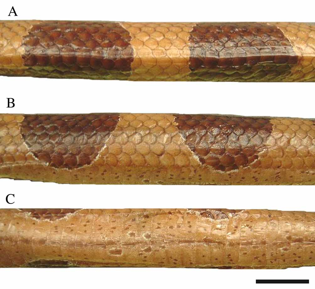
Color pattern in 70% alcohol ( Figs. 6 View FIGURE 6 and 7 View FIGURE 7 ). Top of head dark, mainly the rostral, internasals, part of the prefrontals, the parietals and adjacent scales are also dark, some small white dots can occur. White transverse stripe is present on the prefrontals, part of the nasals and on the first, second and third supralabials. This stripe interrupted in some individuals. The last supralabial scale touching the orbit sometimes with white rounded spots. White collar, sometimes interrupted or not very clear. Body with 15 to 35 rectangular spots, bordered with white and fused along the vertebral line. The spots narrow one the ventral region; usually the six first spots are fused ventrally. Tail with eight to 20 blotches. Venter cream with irregular dark spots of many different sizes.
FIGURA 8. Hemipenis of Dipsas pavonina (right organ, MPEG 8163). Sulcate (A) and asulcate (B) views. Scale bar = 5mm.
Hemipenis morphology (Fig. 8). Organ non-bilobed, cylindrical and slightly enlarged distally, about twice longer than wide at the level of the sulcus spermaticus division. The sulcus spermaticus is deep and divides at the base of the capitulum, with centrolineal branches terminating on the distal tips of the lobes. Capitular sulcus evident on both sides. Capitulum occupies half of the organ on the asulcate face and 70% on the sulcate side, being ornamented with calyces. On the proximal region of the capitulum the calyces are spines, and become papillate on the distal region. Large spines distributed on rows and being concentrated on the adjacent region of capitulum, at proximal portion of the body. Both sides of hemipenis have small spines among the large spines.
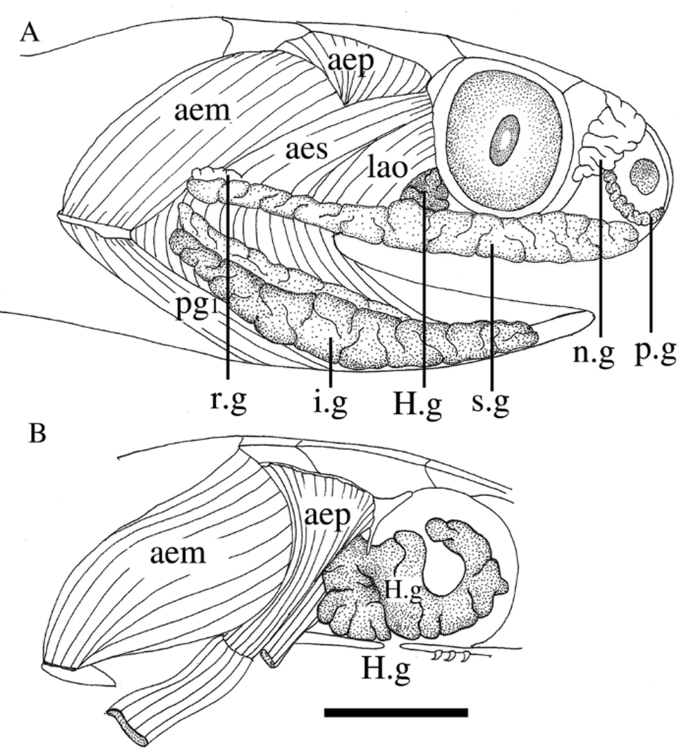
Harder´s gland ( Fig. 9 View FIGURE 9 ). Visible between M. levator anguli oris and the orbit; fibers of M. levator anguli oris and M. adductor mandibulae externus superficialis parallel its origin until reaching the supralabial gland, not creating a space as in D. catesbyi ; orbital lobe bigger than the postorbital one and U-shaped; originating on the superior region of the orbit and extending from the anterior region of the prefrontal bone to the postocular bone; postorbital lobe small and rounded, being limited by the postorbital bone, by the M. levator anguli oris, M. adductor mandibulae externus superficialis and M. adductor mandibulae externus profundus, and by the ectopterigoid bone.
Geographic distribution ( Fig. 9 View FIGURE 9 ). Dipsas pavonina occurs through the Amazonian forest of Bolivia, Peru, Ecuador, south of Venezuela, Guyana, Suriname, French Guyana ( Peters 1960) and Brazil (Par, Rondônia, Roraima, Maranhão and Mato Grosso states).
No known copyright restrictions apply. See Agosti, D., Egloff, W., 2009. Taxonomic information exchange and copyright: the Plazi approach. BMC Research Notes 2009, 2:53 for further explanation.
|
Kingdom |
|
|
Phylum |
|
|
Class |
|
|
Order |
|
|
Family |
|
|
SubFamily |
Dipsadinae |
|
Genus |
Dipsas pavonina Schlegel, 1837
| Lima, Ana Caroline De & Prudente, Ana Lúcia Da Costa 2009 |
Dipsas pavonina
| Harvey 2008: 78 |
| Starace 1998: 172 |
| Cunha 1993: 46 |
| Cunha 1985: 48 |
| Gasc 1980: 574 |
| Cunha 1978: 72 |
| Duellman 1978: 239 |
| Dixon 1977: 43 |
| Peters 1970: 88 |
| Roze 1966: 114 |
| Peters 1964: 47 |
| Peters 1960: 79 |
Dipsas indica
| Beebe 1946: 24 |
Sibynomorphus pavoninus
| Amaral 1926: 8 |
Cochliophagus pavoninus
| Ihering 1910: 330 |
Leptognathus pavonina
| Cope 1868: 107 |
Leptognathus pavoninus
| Gunther 1858: 180 |
| Dumeril 1854: 474 |
Dipsas pavonina
| Schlegel 1837: 280 |