
Dipsas catesbyi (Sentzen, 1796)
|
publication ID |
https://doi.org/10.5281/zenodo.189660 |
|
DOI |
https://doi.org/10.5281/zenodo.5664039 |
|
persistent identifier |
https://treatment.plazi.org/id/A54787C9-FFE7-3502-89B0-FF44FCA5D48E |
|
treatment provided by |
Plazi |
|
scientific name |
Dipsas catesbyi (Sentzen, 1796) |
| status |
|
Dipsas catesbyi (Sentzen, 1796)
Coluber catesbeji Sentzen, 1796: 66 (typographic error), type-locality: “ America ”.
Dipsas catesbyi — Boie, 1827: 560; Schlegel, 1837: 279
Stremmatognathus catesbyi — Duméril, Bibron & Duméril, 1854: 522.
Leptognathus catesbyi — Günther, 1858: 180; Boulenger, 1896: 449; Berg, 1898: 29
Colchliophagus catesbyi – Berg, 1901: 291
Sibynomorphus catesbeji — Barbour & Noble, 1920: 620
Sibynomorphus catesbyi — Amaral, 1926: 8
Sibynomorphus catesbyei — Amaral, 1929: 196
Dipsas catesbyi — Beebe, 1946: 24
Dipsas catesbyi — Peters, 1956; Peters, 1960: 56; Peters & Orejas-Miranda, 1970: 86; Dixon & Soini, 1977: 43; Duellman, 1978: 237; Cunha & Nascimento, 1978: 68; Gasc & Rodrigues, 1980: 573; Abuys, 1983: 117; Cunha et al., 1985: 46; Pérez-Santos & Moreno, 1988: 141; Cunha & Nascimento, 1993: 44; Starace, 1998: 165; Lehr, 2001: 131; Lehr & Lara, 2002: 35; Alves et al., 2005: 573; Argôlo, 2004: 33; Harvey & Embert, 2008: 72.
Diagnosis. Dipsas catesbyi differs from the others species of the genus by the following combination of characters: coloration pattern with rounded spots on the body and tail that narrow the vertebral region; top of head black; belly white; dorsal scales in 13 rows; two prefrontals; 160–220 ventrals; and 60–120 subcaudals. Distinguished from D. pavonina by: loreal present, not entering the orbit; 2 preoculars; 1 or 2 postoculars; hemipenis lightly bilobed with cylindrical body, lightly compressed; little distinction head of the remaining portion of the body and the antero-posterior shortening of the head absent; postorbital lobe of Harder's gland visible between levator anguli oris and adductor mandibulae externus superficialis muscles and extending until the posterior extremity of the infralabial gland.
Description and variation ( Table 1 View TABLE 1 ). Smooth dorsal scales in 13 rows without reduction (n = 386), the vertebral row moderately to broadly enlarged. Ventrals 160–220 (n = 140) in males; and 160–200 (n = 120) in female. Subcaudals divided: 70–120 (n = 113) in males; and 60–100 (n = 126) in females. Cloacal plate single. Snout-vent length 177–598 mm (= 414.9; SD = 82.8; n = 212) in males, and 210–560 mm (= 399.5; SD = 80.3; n = 211) in females. Tail length 72–221 mm (= 158.9; SD = 33.9; n = 205) in males, and 74–221 mm (= 143; SD = 27.6; n = 194) in females. Head length 7.7–15.1 mm (= 1.3; SD = 1.3; n = 212) in males, and 7.7–16.5mm (= 11.3; SD = 1.4; n = 210) in females. Rostral longer than high. Nasal single. Frontal length 2.7–5.0 mm (= 3.7; SD = 0.4; n = 210) in males, and 1.4–4.7 (= 3.5; SD = 0.4; n = 209) in females. Eye diameter 1.8–4.3 mm (= 2.7; SD = 0.3; n = 209) in males, and 1.9–3.4 mm (= 2.5; SD = 0.2; n = 210) in females. Loreal not contacting the orbit. Two preoculars (n = 386, but also can be three (n = 14) or one (n = 12). Two postoculars (n = 338), but also can be one (n = 57) or three (n = 24). One anterior temporal (n = 342) or two (n = 63). Two posterior temporal (n = 383) or one (n = 13). Nine supralabials (n = 235) or eight (n = 117) or ten (n = 74). Fourth and fifth supralabials (n = 135), or fourth and sixth (n = 95) or fifth and sixth (n = 150), or fourth to seven (n = 3) in contact with the orbit. Eleven infralabials (n = 171) or ten (n = 143), and rarely nine (n = 31) or eight (n = 3). First to fifth infralabials (n = 290) or first to fourth (n = 102) in contact to the first pair of gulars.
Color pattern in 70% alcohol ( Figs. 1 View FIGURE 1 and 2 View FIGURE 2 ). Top of head dark, especially the rostral, internasals, second half of prefrontals, the posterior region of the parietals, the pre and postoculars, as well the supralabials that touch the orbit. White transverse stripe on the prefrontals, loreal, second and third supralabials. Whitish color is evident. Gular region is white with black spots. The body pattern is constituted by 10–40 rounded and paired spots, sometimes alternated and with irregular form; most of them are bordered with white. Many of these spots are fused along the vertebral row, some touch the ventrals. First spots on the body (close to the head) fused through venter. Venter white with black paired spots intercalated with lateral spots of the body. Each one of these ventral spots occupies part of three ventral scales. Some small dark spots can occur along the body, between the rounded spots. On young, the body spots can be all fused through the vertebral row, giving a superficial impression of a banded pattern, these spots not bordered with white. Dark spots on the adult venter sometimes absent on young.
Character Dipsas catesbyi Dipsas pavonina
Hemipenis morphology ( Fig. 3 View FIGURE 3 ). Organ strongly capitate and slightly bilobed, about twice as long as wide at the level of the sulcus spermaticus division. Slightly divergent lobes, about 10% of the total length.
Sulcus spermaticus division at the base of the capitulum, centrolineal branches terminating on distal tips of the lobes. On the sulcate side, there is a deep capitular sulcus delimitating the capitulum, which occupies half of the organ on the non-sulcate face, and approximately 70% on the sulcate face, being ornamented with spine calyces on the proximal region. Calyces become bigger and papillate on the distal portion. Capitular sulcus very evident in both sides. Body covered by several spines of many forms and size. On the sulcate face, spines irregularly distributed, the ones on the proximal portion being slightly smaller. On asulcate surface, body spines larger and prominent, distributed in diagonal rows; body spines on the distal portion are concentrated in transversal rows adjacent to capitulum.
Harder´s gland ( Fig 4 View FIGURE 4 ): visible between the M. levator anguli oris and M. adductor mandibulae externus superficialis; the fibers of these muscles diverge after origin, creating a space to accommodate the Harder’s gland; inverted L-shape; orbital lobe thin and concave, originating on the inferior region of the orbit and extending to the region of prefrontal bone until postocular bone; postorbital lobe bigger and rounded, being limited posteriorly by the postorbital bone and by the M. adductor mandibulae externus medialis, above by the parietal and by the M. adductor mandibulae externus profundus, below by the ectopterigoid bone and by the M. superficialis pterigoideus, where it contacts the posterior region of the anterior component of the infralabial gland.
Geographic distribution ( Fig. 5 View FIGURE 5 ). Dipsas catesbyi occurs in the Andean slopes of Bolivia, Peru, Ecuador, Colombia, Venezuela, Guyana, Suriname, French Guiana ( Peters 1960) and Brazil (Acre, Amapá, Amazonas, Pará, Rondônia, Roraima, Maranhão, Mato Grosso, and Tocantins states). Besides the wide distribution on the Amazonian forest, Dipsas catesbyi is represented in different portions of Atlantic rainforest, Bahia state (Feira de Santana, Ilhéus, Porto Seguro and Itabuna) ( Argôlo 2004).
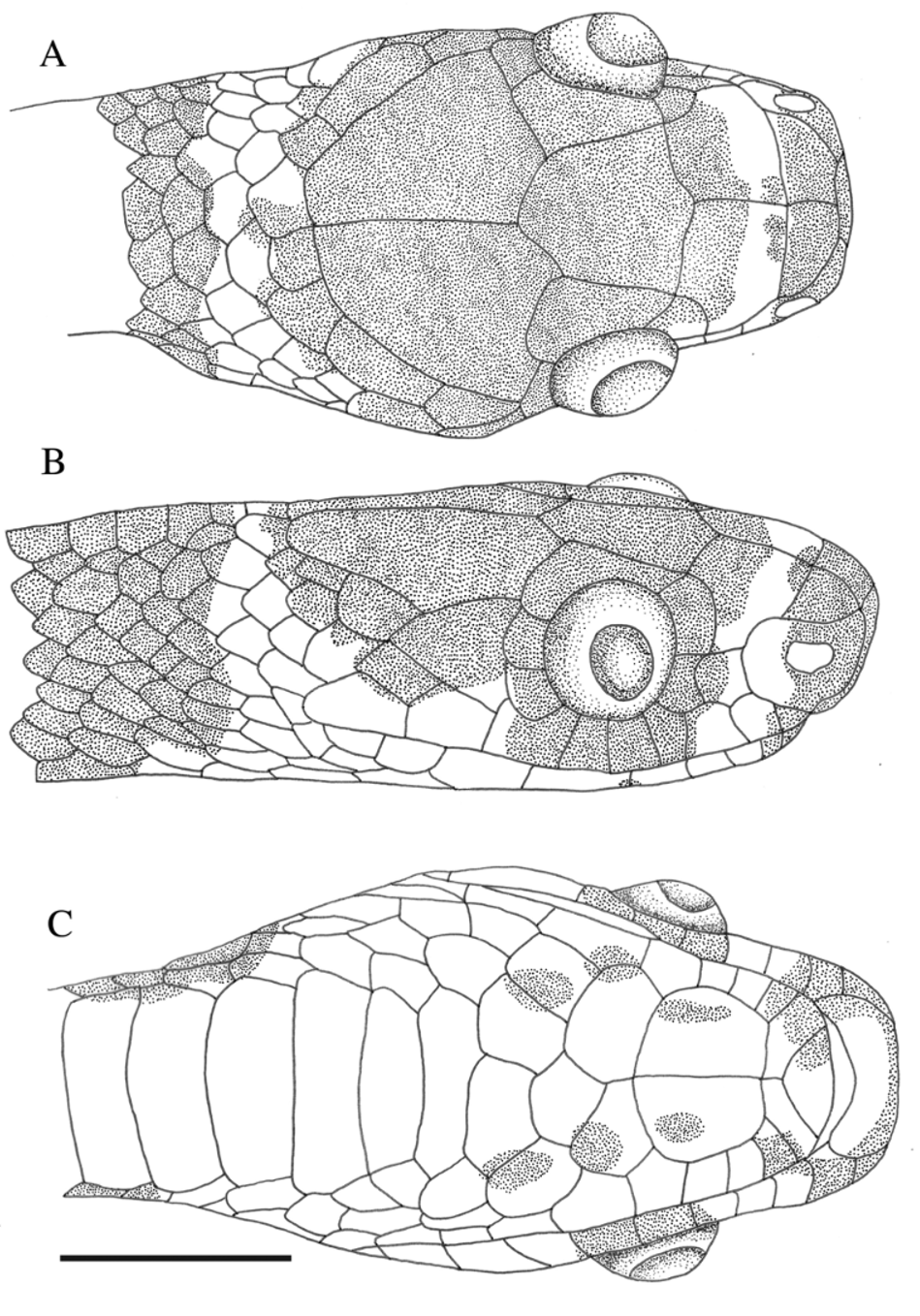
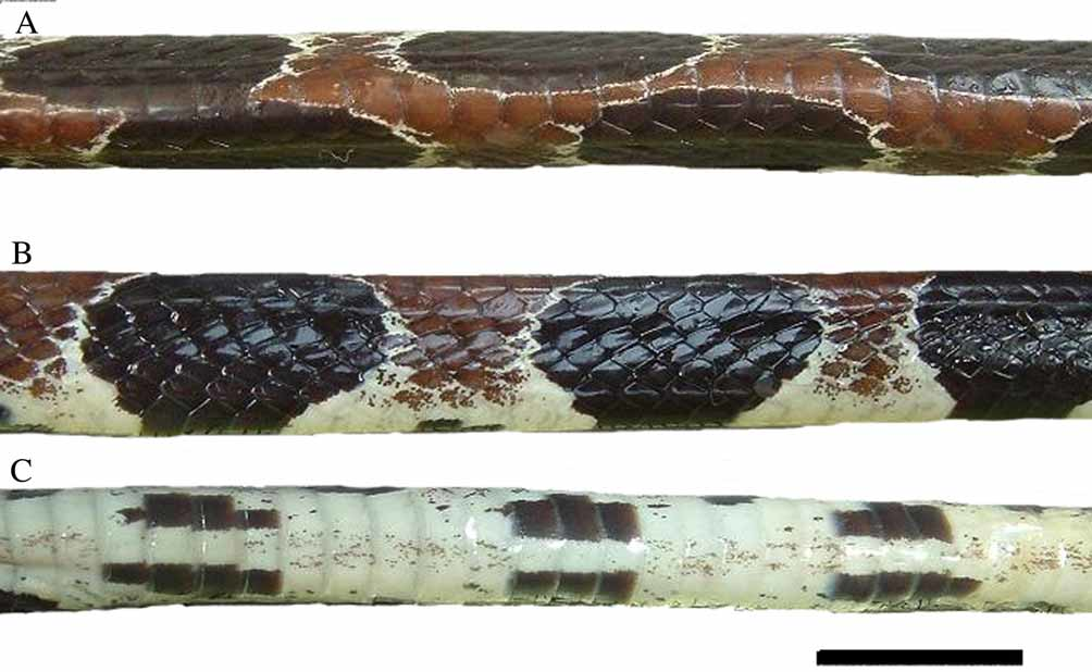
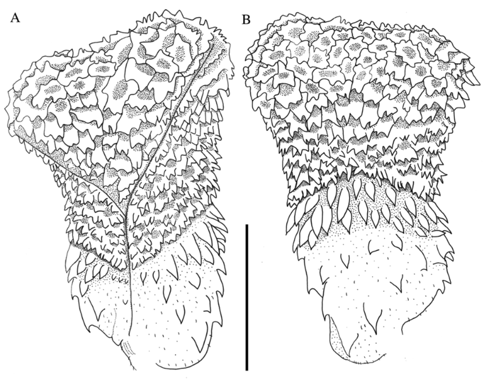
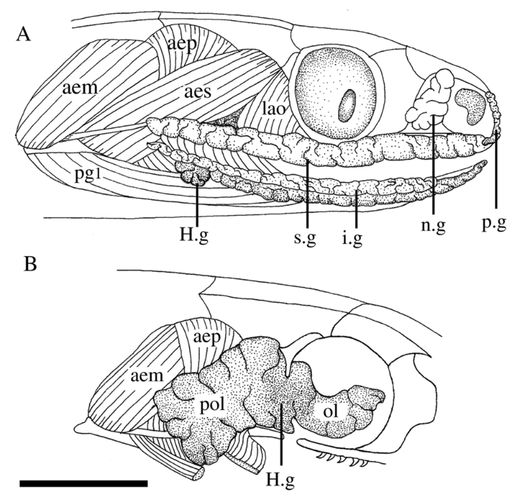
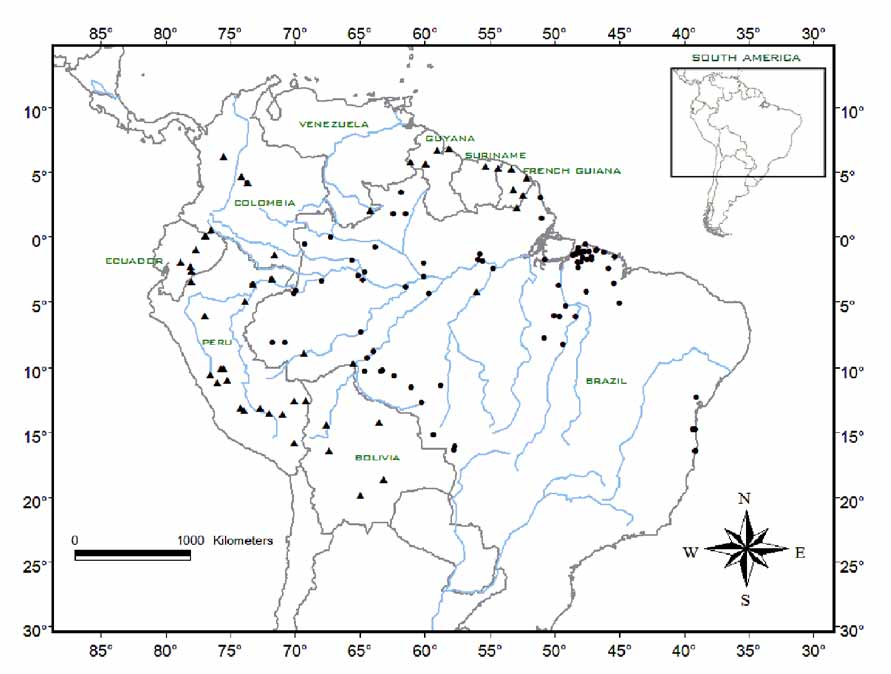
TABLE 1. Variation of some external characters of Dipsas catesbyi and D. pavonina. F: females; M: males; VE: ventrals; SC: subcaudals; SL: supralabials; SLO: supralabials contacting the orbit; IL: infralabials; ILG: infralabials contacting the first pair of gulars; TA: temporal anterior; TP: temporal posterior; PO: preocular; PTO: postocular; TB: tail blotches; BB: body blotches; HL: head length; FL: frontal length.
| Peters (1959; 1960) | Present study | Peters (1960) | Present study | |
|---|---|---|---|---|
| VE SC | 164–202 M/167–189 F 86–116 M/77–102 F | 160–220 M/160–200 F 70–120 M/60–100 F | 203–210 M/190–200 F 119–131 M/101–122 F | 190–230 M/180–220 F 80–130 M/70–130 F |
| SL | 8 or 9 | 8, 9 or 10 | 9–11 | 9–11 |
| SLO IL | 4–5/5–6 8–11 | 4–5/4–6 or 5–6 8–11 | 4 – 5 11 or 12 | 4–5/4–6/4–7/5–6/5–7 10 to 14 |
| ILG | 1–4/1–5 | 1–4/1–5 | 1–5 or 1–6 | 1–5 or 1–6 |
| TA TP | 1 or 2 2 | 1 or 2 1 or 2 | 1 or 2 2 or 3 | 1,2 or 3 2 or 3 |
| PO | 2 | 1 or 2 | 1 or 2 | 0, 1 or 2 |
| PTO TB | 1 or 2 7 to 20 | 1, 2 or 3 6 to18 | 2 or 3 – | 2, 3 or 4 8 to 18 |
| BB | 14 to 40 | 10 to 40 | 14 to26 | 15 to 35 |
| HL (mm) FL (mm) | - - | 7.7–15.1 M/7.7–16.5 F 2.7–5.0 M/1.4–4.7 F | - - | 7.7–13.5 M/ 7.7–13.5 F 2.2–4.3 M/2.6–4.1 F |
No known copyright restrictions apply. See Agosti, D., Egloff, W., 2009. Taxonomic information exchange and copyright: the Plazi approach. BMC Research Notes 2009, 2:53 for further explanation.
|
Kingdom |
|
|
Phylum |
|
|
Class |
|
|
Order |
|
|
Family |
|
|
SubFamily |
Dipsadinae |
|
Genus |
Dipsas catesbyi (Sentzen, 1796)
| Lima, Ana Caroline De & Prudente, Ana Lúcia Da Costa 2009 |
Dipsas catesbyi
| Harvey 2008: 72 |
| Alves 2005: 573 |
| Argolo 2004: 33 |
| Lehr 2002: 35 |
| Lehr 2001: 131 |
| Starace 1998: 165 |
| Cunha 1993: 44 |
| Perez-Santos 1988: 141 |
| Cunha 1985: 46 |
| Gasc 1980: 573 |
| Duellman 1978: 237 |
| Cunha 1978: 68 |
| Dixon 1977: 43 |
| Peters 1970: 86 |
| Peters 1960: 56 |
Dipsas catesbyi
| Beebe 1946: 24 |
Sibynomorphus catesbyei
| Amaral 1929: 196 |
Sibynomorphus catesbyi
| Amaral 1926: 8 |
Sibynomorphus catesbeji
| Barbour 1920: 620 |
Colchliophagus catesbyi
| Berg 1901: 291 |
Leptognathus catesbyi
| Berg 1898: 29 |
| Boulenger 1896: 449 |
| Gunther 1858: 180 |
Stremmatognathus catesbyi
| Dumeril 1854: 522 |
Dipsas catesbyi
| Schlegel 1837: 279 |
| Boie 1827: 560 |