
Loricaseius, Plumari, Massimo & Mašán, Peter, 2014
|
publication ID |
https://doi.org/10.11646/zootaxa.3802.1.1 |
|
publication LSID |
lsid:zoobank.org:pub:61C5EA02-4BEC-4AF4-9956-3E1EB9C5BE01 |
|
DOI |
https://doi.org/10.5281/zenodo.6139371 |
|
persistent identifier |
https://treatment.plazi.org/id/A713364E-3516-FF89-F28F-FA21FC997D99 |
|
treatment provided by |
Plazi |
|
scientific name |
Loricaseius |
| status |
gen. nov. |
Loricaseius gen. nov.
( Figs 1–31 View FIGURE 1 View FIGURE 2 View FIGURES 3 – 6 View FIGURES 7 – 10 View FIGURE 11 View FIGURES 12 – 14 View FIGURES 15 – 17 View FIGURE 18 View FIGURE 19 View FIGURES 20 – 25 View FIGURES 26 – 27 View FIGURES 28 – 32 )
Type species: Loricaseius lepontinus sp. nov., by monotypy.
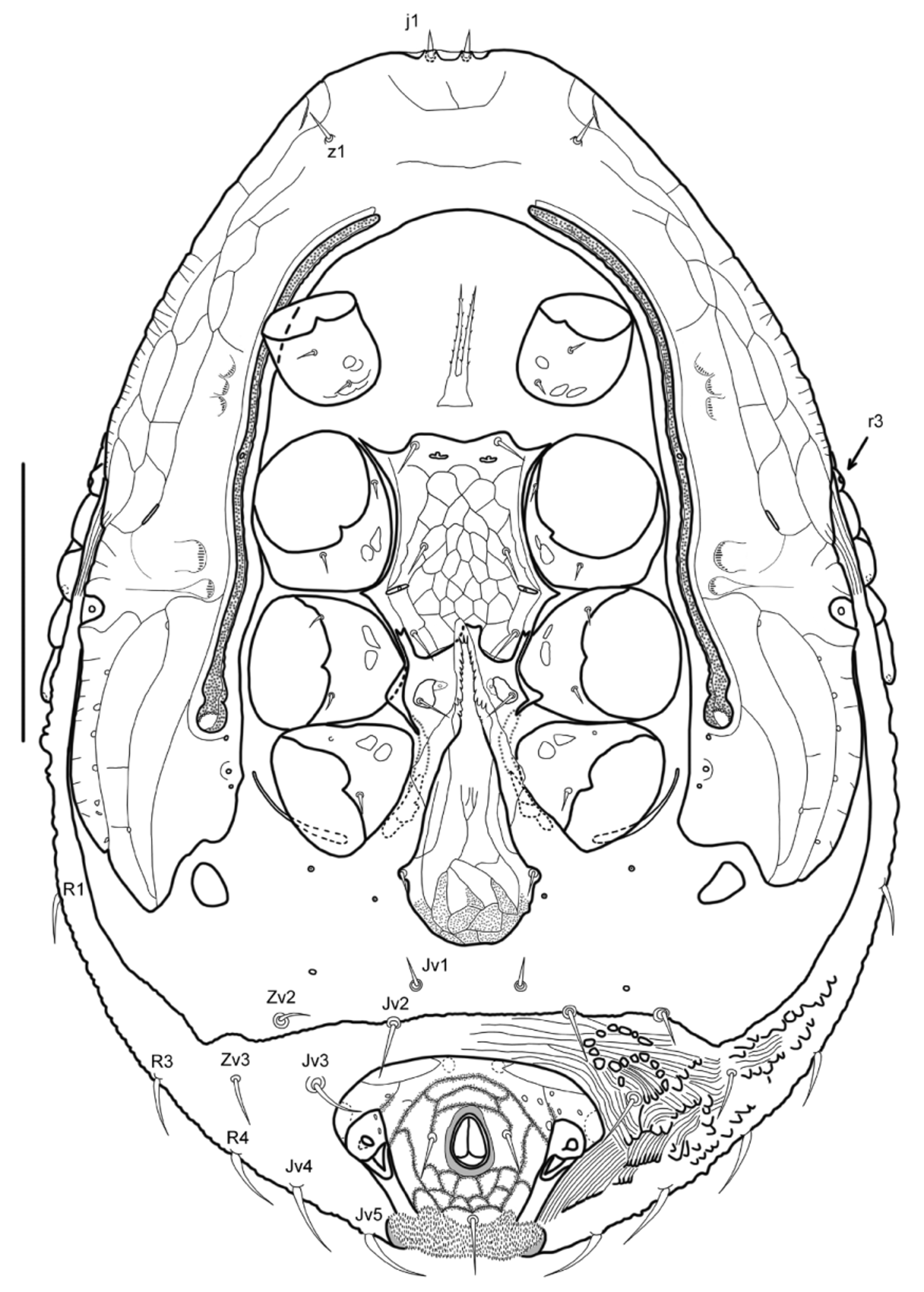
Diagnosis (adults). Idiosomal vertex wide and truncate. Dorsal shield with punctate-reticulate sculpture in female, fused to anterior parts of peritrematal shields to form a ventral arch-shaped shield structure; dorsal setae j2 widely spaced, j1 and z1 ventrally placed. Intercoxal region narrowed and together with gnathosomal cavity displaced more posteriorly. Endopodal platelets II-III largely incorporated in sternal shield. Metasternal platelets present. Epigynal shield subtriangular, produced into a narrow anterior projection. Peritrematal shields extensively expanded laterally, with post-stigmatic section expanded and elongated. Anal shield surrounded in mature females by a large area of strongly sclerotized skin that resembles a ventri-anal shield, having well developed anterolateral extensions projecting between dorsal and peritrematal shields and posterolaterally fused with dorsal shield. Gnathosoma deeply retractile, not reaching beyond the anterior margin of idiosoma. Palptarsus without macroeupathidia, palpgenu with six setae. Cheliceral movable digit bidentate in female. Tarsus I dorsally concave.
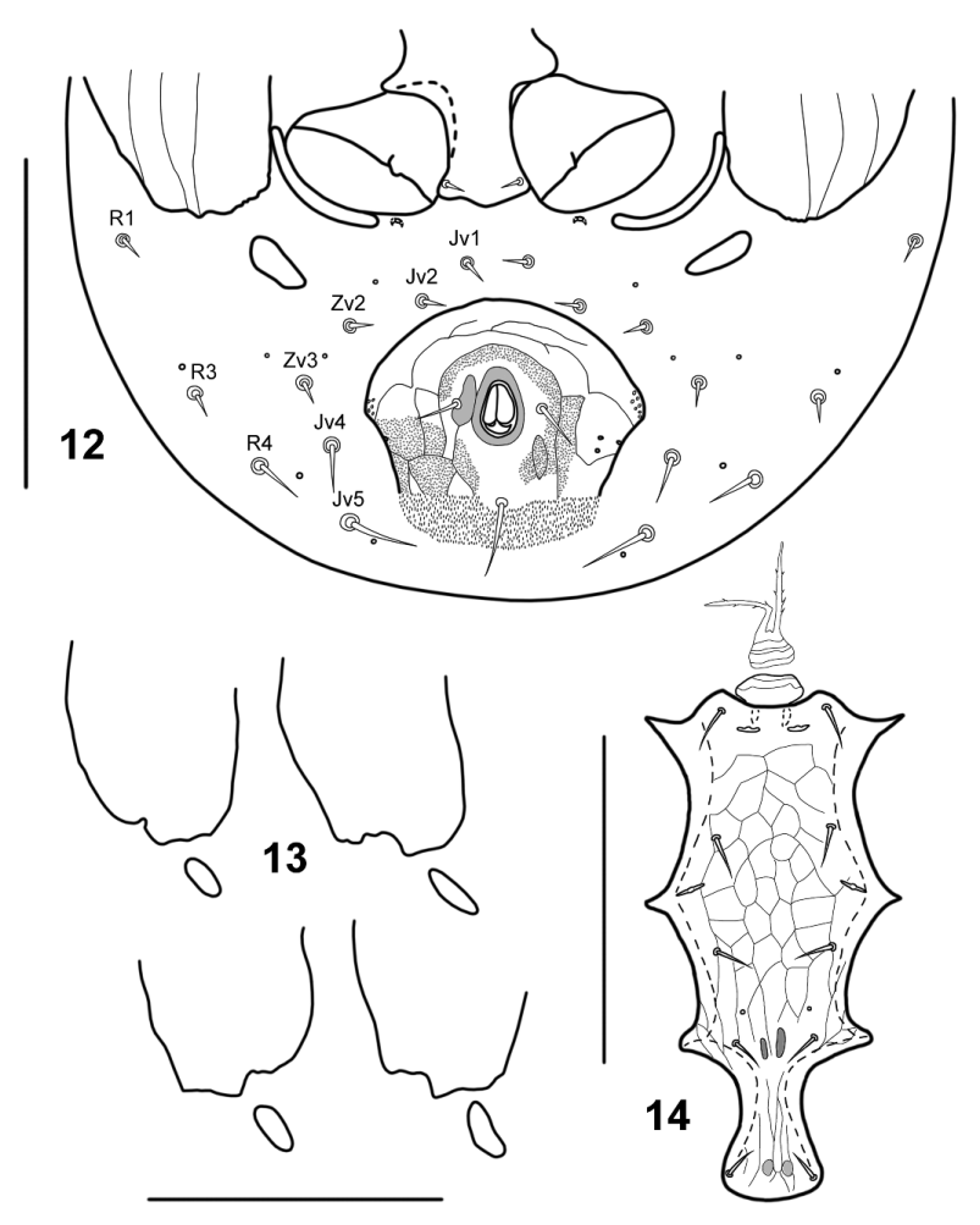
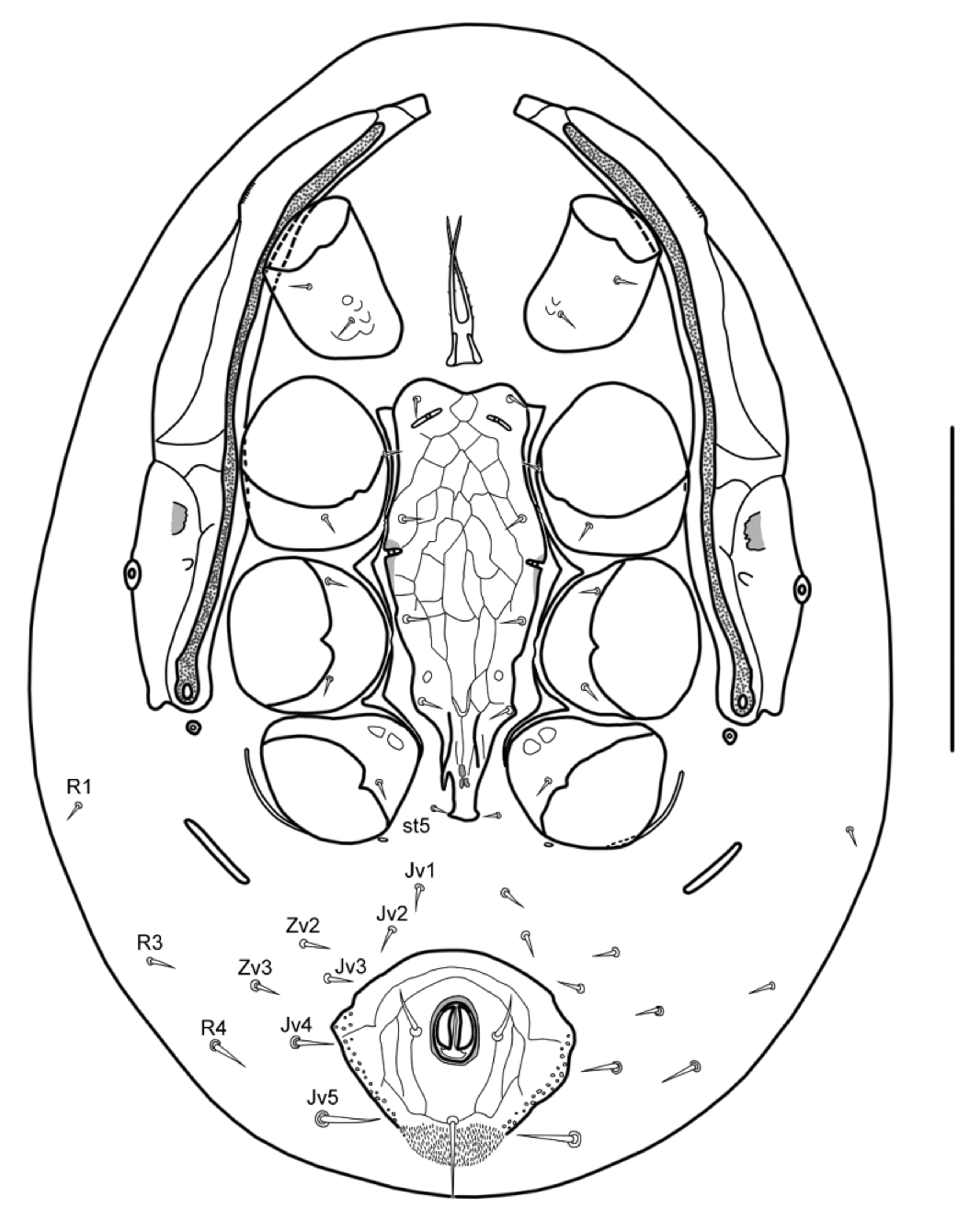
Description (adults and deutonymph). Dorsal idiosoma ( Figs 1 View FIGURE 1 , 11 View FIGURE 11 , 18 View FIGURE 18 ). Idiosoma suboval, dorsoventrally flattened, with vertex wide and truncate. Dorsal shield entire, completely covering dorsal surface, slightly overlapping onto ventral idiosoma, strongly sclerotized in female, crenulated along posterolateral margins; anterior extension of dorsal shield overlapping onto ventral surface beyond vertex, fused in adults to anterior parts of peritrematal shields to form an arched ventral structure. Dorsal area of the shield bearing 28 pairs of setae; vertical setae j1 thickened and acuminate, z1 thin and needle-like, both pairs positioned on ventral extension of dorsal shield, conspicuously shorter than setae on dorsum (except setae J 5 in males); other dorsal setae smooth, needlelike, subequal in length and shape; setae j2 widely spaced; dorsal surface with distinct punctate-reticulate sculptural pattern in female, smooth and very weakly ornamented with irregular polygonal pattern in male and deutonymph. Ventral idiosoma ( Figs 2 View FIGURE 2 , 3 View FIGURES 3 – 6 , 12, 14 View FIGURES 12 – 14 , 19 View FIGURE 19 , 20 View FIGURES 20 – 25 , 28–31 View FIGURES 28 – 32 ). Tritosternum with subrectangular base and two free laciniae, serrated. Presternal platelets absent. Intercoxal region narrowed, inner margins of coxae II and lateral margins of sternal shield closely abutting each other. Female sternal shield well sclerotized, narrow, longer than wide, with three pairs of setae and two pairs of lyrifissures; first pair of lyrifissures parallel to anterior margin of shield, second pair oriented at 45°. Male sterno-genital shield well sclerotized, narrow, with five pairs of setae and three pairs of lyrifissures; first pair slit-like and oriented at 90° to body axis, second pair slit-like and oriented at 45°, third pair smaller and rounded; post-sternogenital sclerites absent. Deutonymphal sternal shield well sclerotized, narrow, with four pairs of setae, two pairs of slit-like lyrifissures, first pair oriented at 45°, second pair oblique, and a pair of rounded lyrifissures between sternal setae st3 and st4; sternal setae st5 situated on soft integument close to posterior end of sternal shield. In female, endopodal platelets I-II and II-III largely fused to sternal shield to form pointed anterolateral and lateromedial corners of the shield, endopodal platelets II-III connected to largely free endopodal platelets III-IV by posterior constricted and partially free ends; in male, endopodal platelets fused to sterno-genital shield; in deutonymph, endopodal platelets I-II fused to sternal shield, but discernible, endopodal platelets II-III and III-IV free, well-separated from lateral margins of sternal shield. Metasternal platelets present, each bearing metasternal seta st4 and lyrifissure. Epigynal shield narrow and subtriangular, with rounded posterior margin, lateral margins anteriorly convergent and hyaline anterior margin produced into narrow, tapering projection, anteriorly reaching beyond posterior margin of sternal shield; a pair of genital setae on epigynal shield; genital lyrifissures situated on soft integument, beside genital setae. Post-genital sclerites absent. Peritrematal shields well developed along whole peritreme; in adults, fused anteriorly with dorsal shield and with each other ventrally, extensively expanded laterally, with outer margin almost overlapping onto lateral margin of dorsal shield; post-stigmatic section expanded and posterior margin wide; in female, with end reaching far beyond posterior margin of coxa IV; in male, post-stigmatic section shorter than in female, with end reaching close to posterior margin of coxa IV; post-stigmatic gland pores distinct, not hypertrophied, situated close to stigmata. In deutonymph, peritrematal shields not fused anteriorly with dorsal shield and with each other ventrally, clearly narrower than in adults, tapered anteriorly and posteriorly, with outer margins widely separated from lateral margins of dorsal shield; post-stigmatic sections not well developed, very short, with posterior ends reaching posterior margins of coxae III; post-stigmatic gland pores outside shields, each on a small platelet placed on soft integument close to stigma. Anterior ends of peritremes projecting beyond coxae I. Metapodal platelets behind coxae IV; in adults, from oval to subtriangular, closely associated with posterior margin of peritrematal shields; in deutonymph, elongated and straight, widely separated from posterior end of peritrematal shields. Exopodal platelets between coxae I–IV absent; parapodal platelets narrow and curved, obscured by margins of coxae IV, slightly larger in male. Anal shield relatively well developed, more strongly sclerotized in mature females, with very well developed cribrum, three circum-anal setae, of which post-anal seta longer than adanal setae, a pair of adanal gland pores, hypertrophied in mature females, and anteromedial anus; female anal shield subtriangular, with widely rounded anterior margin and concave posterolateral margins; male anal shield subcircular, markedly wider than long, transversely more ovoid than in female (posterior margin more prolonged), with widely rounded anterior margin, pronounced anterolateral corners and concave posterolateral margins; deutonymphal anal shield subtriangular, with widely rounded anterior margin and obtusely pointed or rounded anterior corners. Ventral setae smooth and needle-like, subequal in length. Sexual dimorphism of ventral chaetotaxy well developed: female adult and female deutonymph on soft integument of ventral opisthosoma with more setae than male adult and male deutonymph (setae Jv3 missing in male adult and male deutonymph). In mature females, a large area of strongly sclerotized skin, posterolaterally fused with dorsal shield and with complex tuberculated and striated pattern of ornamentation, surrounds anal shield and extends between dorsal and peritrematal shields with two narrow anterolateral projections.
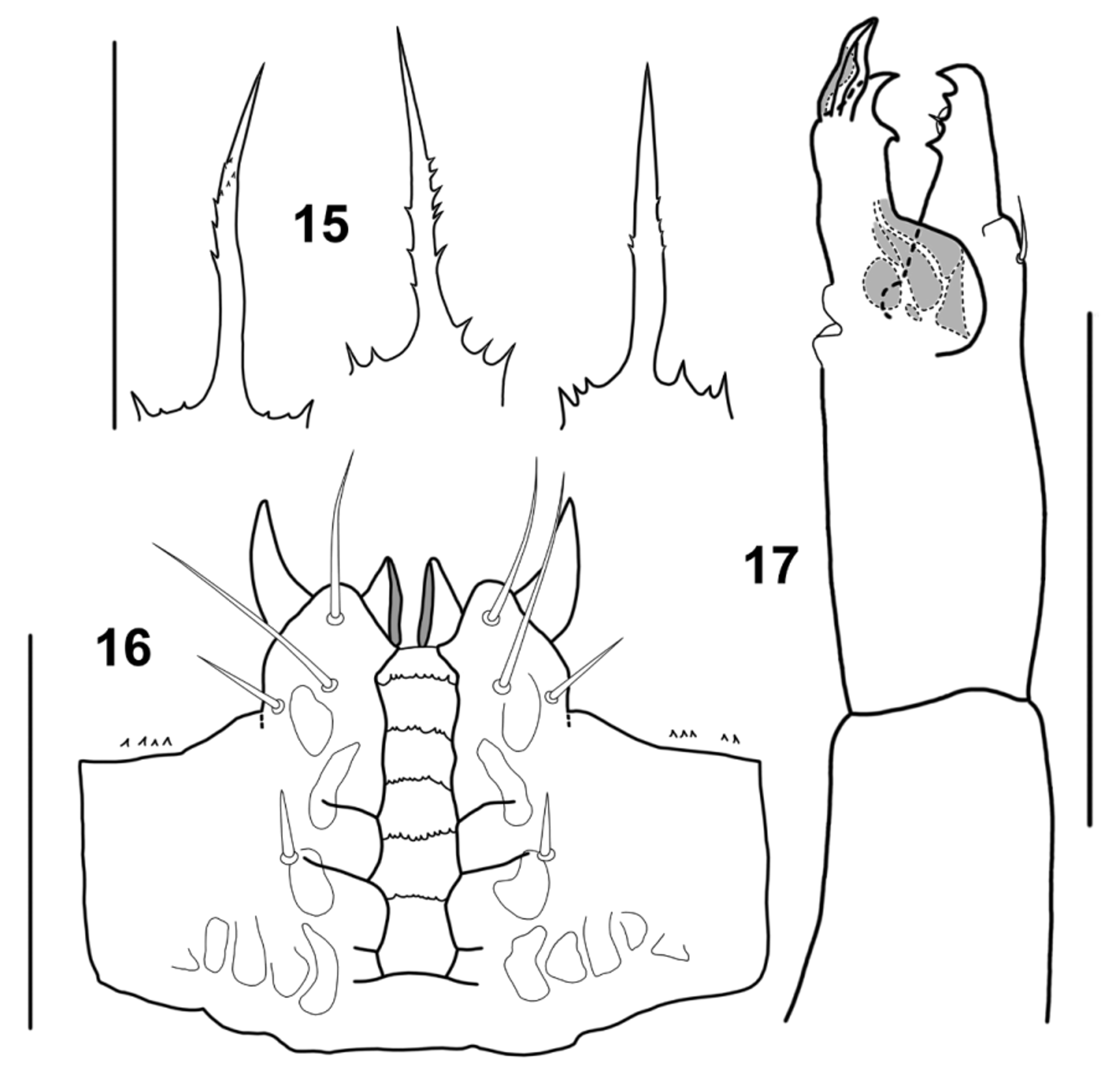
Gnathosoma ( Figs 7–10 View FIGURES 7 – 10 , 15–17 View FIGURES 15 – 17 , 21, 23–25 View FIGURES 20 – 25 ). Gnathosoma deeply retractile, never reaching beyond anterior margin of idiosoma. Hypostome with three pairs of smooth and needle-like setae; h2 longest, h1 moderately shorter than h2, h3 shortest; palpcoxal setae short and stout; corniculi horn-like; deutosternal groove with five transverse rows of denticles; hypopharyngeal walls strongly sclerotized; internal malae not well discernible. Epistome with elongated central projection and very short, pointed lateral prongs, and usually with some small denticles between central projection and lateral prongs. Palptarsus without paired macroeupathidia, all sensory setae with pointed tip, tarsal apotele two-tined; palpgenu with six setae. Chelicera with shortened proximal segment; dorsal cheliceral seta stout and arthrodial membrane represented by a low rounded flap; cheliceral digits robust, movable digit bidentate in female and deutonymph, unidentate in male; fixed digit tridentate in adults, bidentate in deutonymph, with pilus dentilis fine and short, without hyaline appendage; spermatodactyl simple, digitiform and projecting beyond distal end of movable digit.
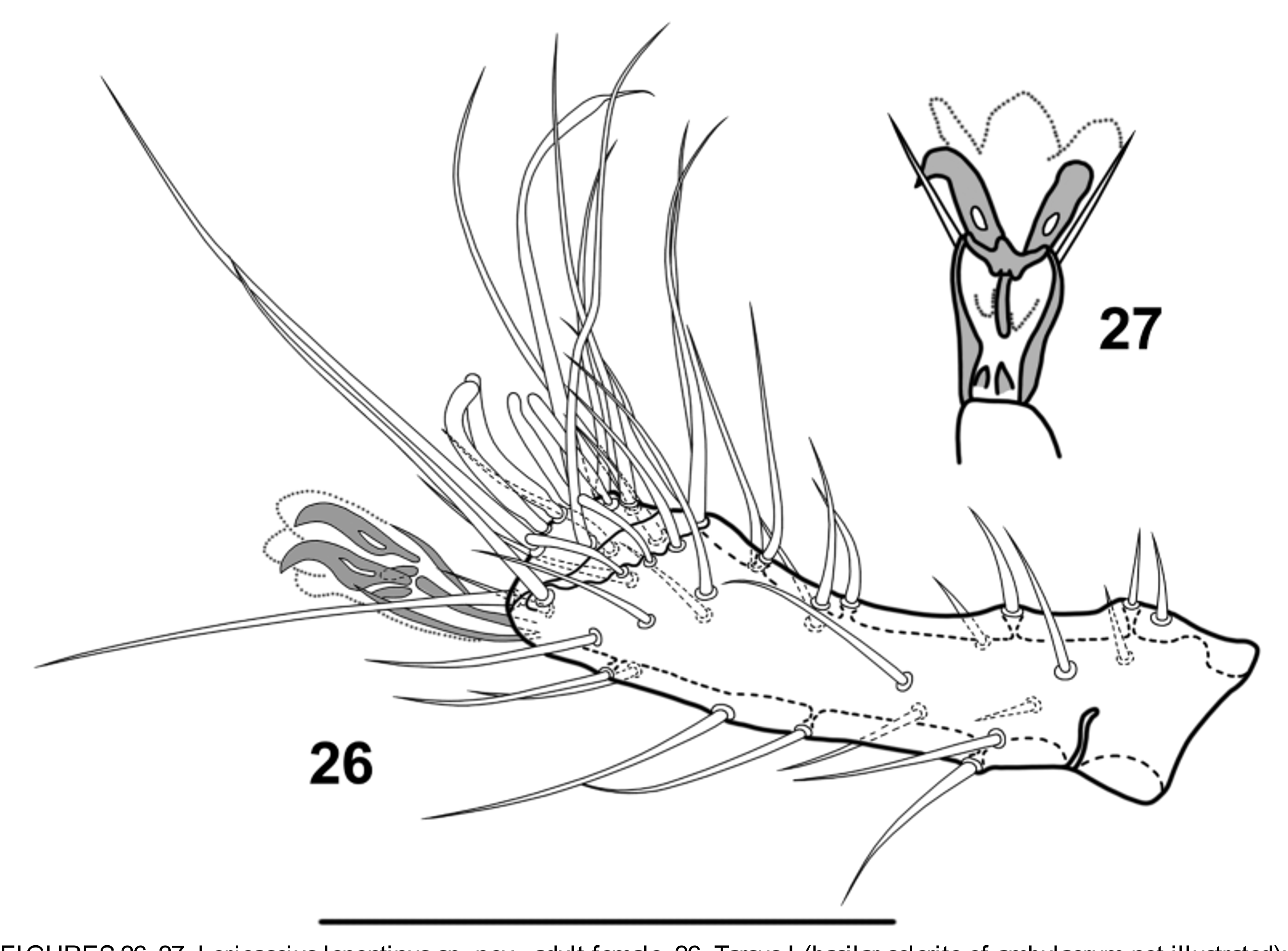
Legs ( Figs 26–28 View FIGURES 26 – 27 View FIGURES 28 – 32 ). Legs I–IV relatively robust, subequal in length, short (markedly shorter than width of idiosoma), with pretarsus having a well developed ambulacral stalk, a pair of claws and three rounded pulvillar lobes, not conspicuous, slightly projecting beyond claws; pretarsus of legs II–IV with paired paradactyli, longer than claws; tarsus I dorsally concave; coxal setae unmodified, setiform; coxae II not expanded and subequal in size to other leg coxae. Legs of male with no spur-like setae. Setation of legs I–IV: coxae 2, 2, 2, 1; trochanters 6, 5, 5, 5; femora 13 (2-5/4-2), 11 (2-5/3-1), 6 (1-3/1-1), 6 (1-3/1-1); genua 11 (1-5/3-2), 11 (2-5/2-2), 8 (1-4/2-1), 7 (1-4/1- 1); tibiae 11 (1-5/3-2), 10 (2-4/2-2), 7 (1-3/2-1), 7 (1-3/2-1).
Etymology. The name Loricaseius is composed of the Latin name lorica (armor), referring to the armored appearance of the female, that is due to large extension of the strongly sclerotized areas of the dorsal and ventral idiosoma, and from Seius , a proper noun used in the names of many mite taxa.
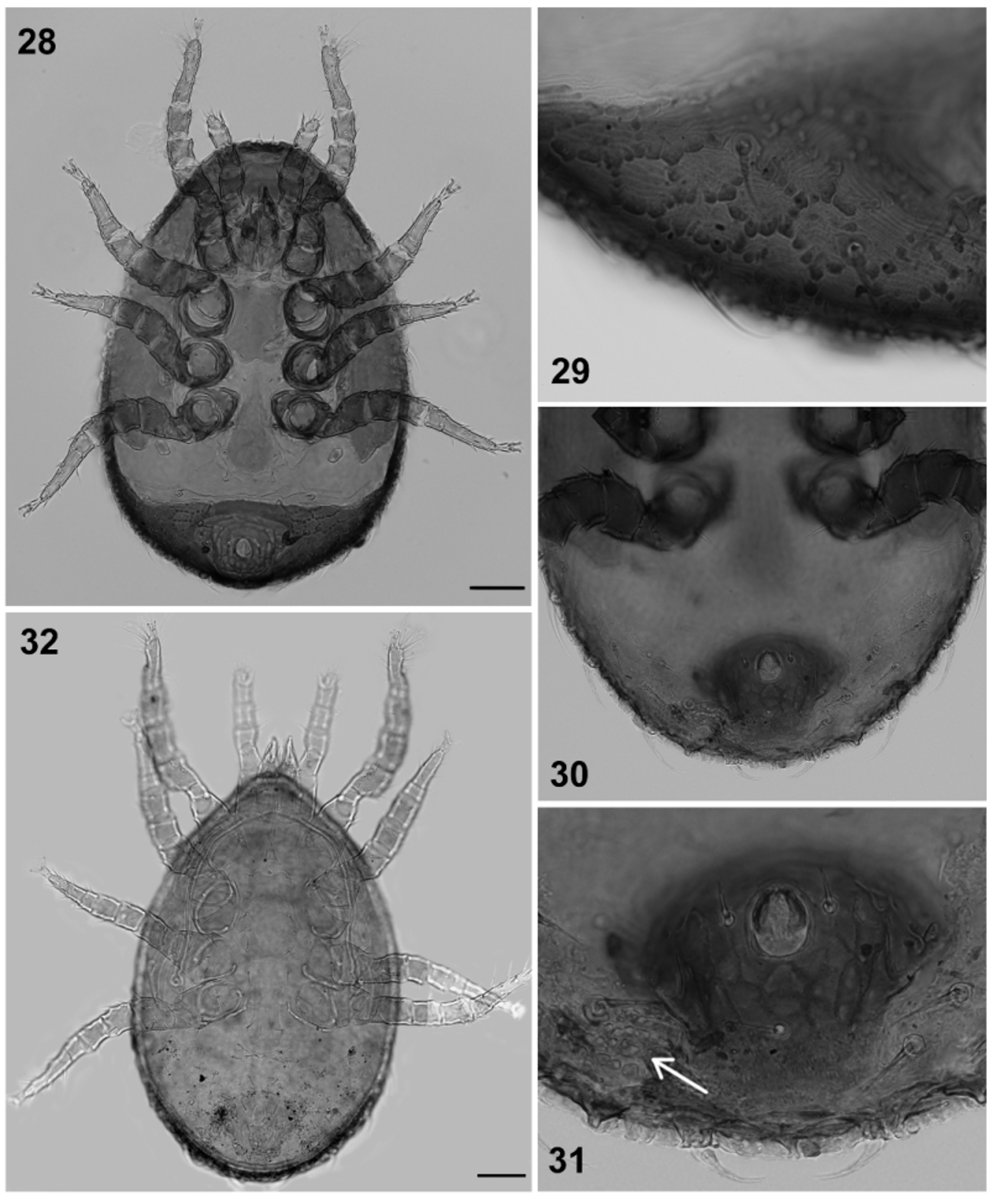
Notes. In adult females with strongly sclerotized skin surrounding the anal shield, the latter is always well demarcated from surrounding integument ( Fig. 2 View FIGURE 2 ). The adult females, in which the anal shield is surrounded by soft integument ( Figs 3 View FIGURES 3 – 6 , 30 View FIGURES 28 – 32 ), closely match more strongly sclerotized females ( Fig. 28 View FIGURES 28 – 32 ) for all other characters. No such differences were observed in male and deutonymphal specimens. However, in females with unsclerotized opisthogastric integument, the general coloration varies from ochre to brown, and in darker females, some small fragmented areas of incipient strong sclerotization can be observed along posterior margin of ventral opisthosoma, to the sides of anal shield ( Figs 30–31 View FIGURES 28 – 32 ). On the basis of these observations, we have concluded that specimens with unsclerotized integument of ventral opisthosoma, have varying degrees of sclerotization and they are immature females, while specimens with anal shield surrounded by strongly sclerotized skin are mature females belonging to same species. Therefore, the development of a large area of strongly sclerotized integument surrounding anal shield may be considered to be a case of age-related secondary sclerotization, although experiments of breeding in laboratory are necessary in order to verify our hypothesis.


No known copyright restrictions apply. See Agosti, D., Egloff, W., 2009. Taxonomic information exchange and copyright: the Plazi approach. BMC Research Notes 2009, 2:53 for further explanation.
|
Kingdom |
|
|
Phylum |
|
|
Class |
|
|
Order |
|
|
Family |