
Paracanthopoma daemon, Pinna & Dagosta, 2022
|
publication ID |
https://doi.org/ 10.11606/1807-0205/2022.62.072 |
|
publication LSID |
lsid:zoobank.org:pub:A32FD3AF-C87F-4C75-9100-D695C3578283 |
|
DOI |
https://doi.org/10.5281/zenodo.10839406 |
|
persistent identifier |
https://treatment.plazi.org/id/A81A87C0-FFF4-FC72-FF03-17E924B1AA94 |
|
treatment provided by |
Felipe |
|
scientific name |
Paracanthopoma daemon |
| status |
sp. nov. |
Paracanthopoma daemon , new species ( Fig. 18 View Figure 18 )
Holotype: MZUSP 103047 View Materials , 20.4 mm SL, Brazil, Mato Grosso, Gaúcha do Norte, rio Coronel Vanick ( rio Xingu drainage), ca. 20 km from Vila do Culuene (13°31′34″S, 52°43′52″W), col., F.C. T. Lima, C. R. Moreira, A.C. Ribeiro & C.M.C. Leite, 08 Oct 2007. GoogleMaps
Paratypes: MZUSP 95597, 5 ex (2 c&s), 14.7-18.6 mm SL, collected with holotype.
Diagnosis: Distinguished from all congeners except Pc. carrapata , Pc. parva , and Pc. truculenta by the presence of 9 or 10 teeth on the median premaxilla (vs. 3 to 5 or 11 and more); by the presence of a single median s6 pore, visible on the middle of skull posterior to transverse line through posterior margin of eyes (vs. paired s6 pores, posterior to posterior margin of eye, distant from midline of skull), and by the supraoccipital anteriorly produced into large pointed spike (vs. either anteriorly concave or straight across skull roof). Distinguished from Pc. carrapata , Pc. parva , and Pc. truculenta by the caudal fin truncate with round corners or only slightly concave (vs. deeply emarginate, bilobed); by the expanded caudal peduncle having an even depth along its length (vs. peduncle less deep anteriorly and expanding close to caudal fin); by the more numerous ventral caudal-fin procurrent rays (20 or 21; vs. 14-18); by the origin of the anal fin anterior to the vertical through the middle of dorsal-fin base (vs. origin of anal fin posterior to that point); and by the fewer vertebrae (35 or 36; vs. 37-40).
Description: Morphometric data for the holotype and paratypes are provided in Table 6 View Table 6 . Body moderately elongate (HL 16.9-20.4% SL). Cross-section of body as deep as broad or slightly deeper than broad at pectoral-fin insertion and increasingly compressed posterior to that point, tapering to caudal fin. Dorsal profile of body gently convex from head to origin of dorsal fin ( Fig. 18 View Figure 18 ). Dorsal and ventral profiles of caudal peduncle straight or gently convex. Caudal peduncle spatulate, expanded by procurrent rays along nearly its entire length ( Fig. 18 View Figure 18 ). Ventral profile of body straight or gently convex until pelvic-fin origin ( Fig. 18 View Figure 18 ), with some specimens with distended abdomens due to full ovaries. Myotomes and longitudinal skeletogenous septum clearly visible along whole body. Axillary gland comparatively small, elongate in shape, not protruding markedly on surface of body and extending maximally to end of adpressed pectoral fin. Gland tapering to fine posterior tip, its large round pore located approximately at vertical through midlength of pectoral fin. Condition of gland posterior to pore evidently related to amount of secretion stored. In some specimens, postpore part of gland appearing as nearly absent, clearly due to empty condition of its lumen.
Dorsal profile of head continuous with that of dorsum, sometimes broken by slight muscle constriction or change in angle ( Fig. 18 View Figure 18 ). Head longer than broad (head width 61.0-64.0% HL), snout broad, semicircular in dorsal view. Muscles covering only lateral portion of dorsal aspect of head, with skull roof mostly exposed. Head deep for Paracanthopoma (head depth 42.6-53.0% HL), with dorsal profile straight and sloped dorsally until eye in lateral view, then angled to horizontal and straight to trunk. Eye large (14.8-17.0% HL), without free orbital rim, located dorsolaterally on head and directed dorsolaterally, covered by thin and transparent integument ( Fig. 18 View Figure 18 ). Middle of eye slightly anterior to middle of HL, interorbital width approximately equal to or slightly shorter than longitudinal diameter of eye. Eyelens occupying central portion of lateral surface of eye and constricted by iris marginally, with large round pupil in specimens examined. Anterior nostril small, located in narrow teardrop-shaped slit on surface of skin and surrounded by short tubule of integument produced posteriorly into small pointed process, with double elastin cores. Anterior internarial width slightly larger than interorbital. Posterior naris larger than anterior one, partly occluded by anterior flap of integument. Posterior naris positioned anteromesially and adjacent to eye, their middle at transverse line through anterior margin of eye. Posterior internarial width narrower than interorbital.
Opercular odontodophore medium-sized, laterally located on head, on dorsal half of head depth in lateral view. Opercular odontodes 5, closely positioned as two very large ones juxtaposed posteriorly and three smaller anterior ones. Main axis of opercular odontodes oriented horizontally in lateral view, with their distal portion curved dorsomedially. One or two caps of replacement odontodes posteriorly to mature ones. Interopercular odontodophore either similar-sized, or slightly larger than opercular one, located ventrolaterally on head, immediately ventral to horizontal through origin of pectoral fin, with 4 or 5 odontodes closely positioned in single row of four near posterior edge of interopercle, plus single smaller one anteriorly (when 5). Odontodes of posterior row strongly angled medially. Interopercular odontodophore much closer to opercular one than to eye. One or two replacement tooth caps located posteromesially to mature ones. Opercular and interopercular periodontodal folds thin and transparent. Epiodontodeal velum not visible in specimens available.
Mouth inferior (ventral). Each premaxilla with single scalpelloid teeth attached to its distal tip ( Figs. 4E View Figure 4 , 19 View Figure 19 ), but actually two adjacent tooth sockets, one of which vacant, corresponding to half-formed replacement tooth adjacent to mature one. Vacant socket position varying among specimens and between sides of same specimen, being either lateral or mesial one. One or two additional initial-stage replacement caps suspended in soft tissue dorsally to mature one and its incomplete neighbor. Mature scalpelloid tooth with distal portion disproportionately reduced and very strongly curved over rest of teeth, with pungent tip nearly adpressed to margin of basal plate. Scalpelloid tooth deeply hidden in labial tissue, but its distal surface easily emerging when premaxilla forcibly abducted. Conical teeth absent in premaxilla. Upper lip thick, ventral surface not plicate. Median premaxilla very large, with 9 or 10 teeth disposed in one anterior row (convex anteriorly) of four or five,one posterior row (convex posteriorly) of four plus single middle tooth ( Figs. 4E View Figure 4 , 19 View Figure 19 ). All teeth perpendicular to ventral surface of median premaxilla basally, but strongly curved posteriorly at distal pungent portion, those on anterior row taller than on posterior one. All median premaxillary teeth strongly laterally compressed basally. Median premaxillary dentition occupying most of exposed upper jaw and most of interior of mouth. Many replacement tooth caps posterodorsally to mature dentition, creating confusing aspect to posterior limit of median premaxillary dentition. Median premaxillary velum irregular. Hypodontal pad of median premaxilla thickly cushioning teeth. Lower jaw wide, occupied mostly by large dentary lobes largely continuous with each other and continuous with mental region posteriorly. Dentary diastema well differentiated, represented by small concave or angulate area at midline. Rami of mandible very close together at midline. Jaw cleft deep and strongly directed posteriorly, approaching parallel to longitudinal axis and forming broad space separating lower jaw laterally from inner margin of upper jaw. Dentary teeth 4, closely packed at mesial end of dentary and disposed in two aligned pairs, one dorsal and one ventral, with only latter visible in ventral view ( Figs. 4E View Figure 4 , 19 View Figure 19 ). Axis of dentary teeth anteroventrally-directed at base, but strongly curved dorsally distally. Branchiostegal velum forming large, continuous, round and posteriorly concave, free fold across whole of mental region ( Fig. 18 View Figure 18 ). Dorsal portion of branchial membrane reaching anterior margin of pectoral-fin base. Branchial openings small, located anteriorly to pectoral-fin base, spanning approximately for area between ventral margin of opercular odontodophore and dorsal margin of interopercular odontodophore. Maxillary barbel very short and proximally broad,its base flap-like,only distal portion filamentous. Posterior point of is base anterior to vertical through anterior margin of eye, its tip extending posteriorly approximately to vertical through posterior margin of eyes, or slightly anterior to that, in lateral view. Mesial (or ventral) part of maxillary-barbel base continuous with membranous outgrowth extending posteriorly from corner of mouth. Rictal barbel vestigial, located mesially to base of maxillary one, its base immersed in membranous expansion at corner of mouth. Rictal barbel sometimes difficult to identify among irregularities of surrounding integument folds, but its homology with trichomycterid rictal barbel evident by well-developed internal core in cleared and stained specimens. In some specimens, no clear external component of rictal barbel. Nasal barbel vestigially represented by posterior elongated portion of fold around anterior naris described above.
Lateral line short and straight, curved dorsally near posterior end in some populations its terminal pore slightly anterior to vertical through midlength of pectoral-fin, near dorsal margin of axillary pore. Short secondary branch splitting off ventrally from anterior portion of canal, with corresponding pore opening approximately at anterior third of main canal. Single lateral-line tubule extending for more than half of sector of canal posterior to bifurcation.
Pectoral fin short (67.3-85.0% HL),with i +5 rays.Distal margin of pectoral fin gently convex, nearly straight, its base immediately ventral to midline of body in lateral view. Pelvic fins small, well-separated from each other at base,with i + 4 rays.Pelvic splint present.Origin of pelvics located well anterior to vertical through origin of dorsal-fin, entirely covering anus and extending posteriorly to origin of anal fin or beyond. Posterior margin of pelvic fin round. Dorsal fin small, triangular with broadly round apex, gently convex distal margin and i + 6 (holotype) or ii + 6 fin rays, plus 4 or 5 procurrent ones. Anal fin small, slightly more elongate in shape than dorsal one, with gently convex distal margin and i + 5 (holotype) or ii + 5 fin rays, plus 4 procurrent ones. Origin of anal fin slightly posterior to vertical through origin of dorsal-fin. Caudal fin truncate or slightly concave. Principal caudal-fin rays 5 + 7, 6 + 6 (holotype) or 6 + 7. Procurrent caudal-fin rays 19 or 20 dorsally and 20 or 21 ventrally.
Vertebrae 35 (n = 3) or 36 (n = 3, holotype). First dorsal-fin pterygiophore subsequent to neural spine of vertebra 17 (n = 1) or 18 (n = 1). First anal-fin pterygiophore subsequent to haemal spine of vertebra 19 (n = 1) or 20 (n = 1). Dorsal-fin pterygiophores 7 (n = 2). Anal-fin pterygiophores 6 (n = 2). Branchiostegal rays 3.
Pigmentation in preservative: Body almost entirely white. Neurocranium dark with brain pigment seen by transparency. Faint dark field along lateral and middle regions of snout, anterodorsally to maxillary barbel base. Dark spots on bases of opercular and interopercular odontodophores. Sparse dark spots along dorsum, more concentrated near dorsal fin. Dark spots on hypural plate and adjacent region of caudal-fin base. Irregular series of dark spots along dorsal part of abdomen, particularly visible in specimens with abdominal distention.
Etymology: Daemon is a latinized form of the Greek daimon, referring to the supernatural entities hierarchically between gods and mortals, including inferior divinities and ghosts of some dead men. The word was incorporated into the Judean-Christian tradition by its usage in Greek translations of sacred texts.
Geographical distribution: Paracanthopoma daemon is known to occur in a single locality in the rio Coronel Vanick, tributary to the upper rio Xingu in central Brazil ( Fig. 20 View Figure 20 ).
Notes on ecology: Paracanthopoma daemon seems to be a small inhabitant of psammic environments. One of the collectors of the type series (C. Moreira) informs that the specimens were captured during daytime at a river sector with mild current, in a small fine-sand bank close to the margin of the river,surrounded by fields of mud on one side and coarse gravel on the other. It occurred syntopically with Mastiglanis asopos (Heptapteridae) . Other vandelliines caught in the same site, but on muddy substrate, included two undescribed species of Vandellia .
Remarks: The holotype and the largest paratype of this species (20.4 and 18.6 mm SL, respectively) are mature females with large eggs, indicating that Pc. daemon matures at a considerably smaller size than its closest relatives Pc. carrapata , Pc. parva and Pc. truculenta . The distinctive characteristics of Pc. daemon are unambiguous at all comparable sizes.
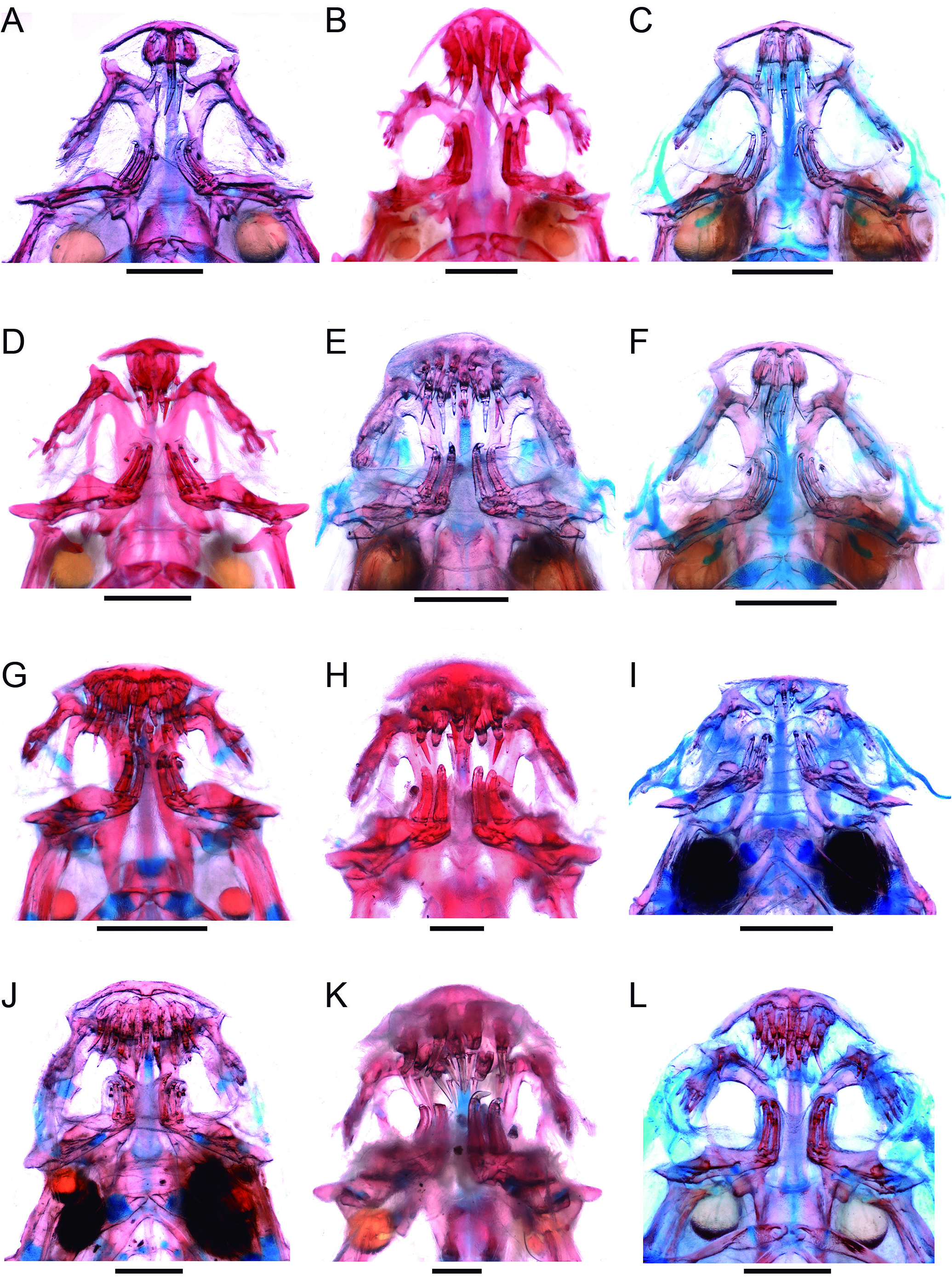
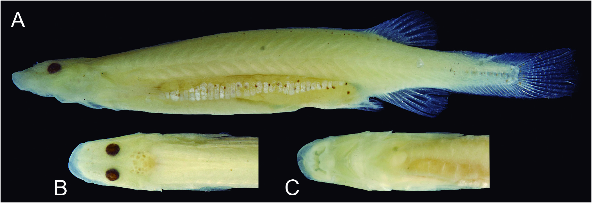
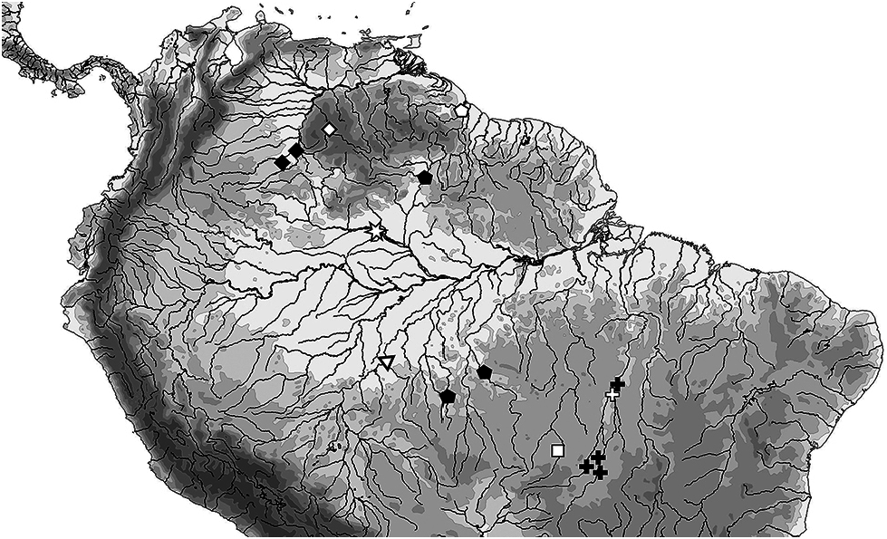
Table 6. Morphometric data of Paracanthopoma daemon. Ranges,mean and SD includeholotype.Head subunitswere obtained withanocular micrometer and therefore as projections.Abbreviations:min = minimum value;max = maximum value;n = number of specimens;SD = standard deviation.
| n | holotype | min | max | mean | SD | |
|---|---|---|---|---|---|---|
| Standard length (mm) | 4 | 20.4 | 14.7 | 20.4 | 17.8 | |
| Percentages of SL | ||||||
| Total length | 4 | 1.1 | 1.1 | 1.1 | 1.1 | 0.0 |
| Body depth | 4 | 15.4 | 12.3 | 15.5 | 14.3 | 1.5 |
| Caudal peduncle length | 4 | 20.5 | 18.6 | 21.1 | 19.9 | 1.2 |
| Caudal peduncle depth | 4 | 8.3 | 7.9 | 9.7 | 8.6 | 0.8 |
| Predorsal length | 4 | 69.9 | 69.9 | 71.8 | 70.4 | 0.9 |
| Preanal length | 4 | 72.4 | 71.8 | 74.6 | 72.8 | 1.2 |
| Prepelvic length | 4 | 66.0 | 64.8 | 66.7 | 66.0 | 0.8 |
| Dorsal-fin base length | 4 | 8.3 | 7.7 | 9.7 | 8.4 | 0.9 |
| Anal-fin base length | 4 | 6.4 | 6.4 | 8.0 | 7.5 | 0.7 |
| Pectoral-fin length | 4 | 11.5 | 11.5 | 14.2 | 13.3 | 1.2 |
| Head length | 4 | 18.6 | 16.9 | 20.4 | 18.8 | 1.5 |
| Percentages of HL | ||||||
| Head width | 4 | 61.0 | 61.0 | 64.0 | 62.3 | 1.5 |
| Head depth | 4 | 42.6 | 42.6 | 53.0 | 45.5 | 5.0 |
| Pectoral-fin length | 4 | 72.1 | 67.3 | 85.0 | 74.5 | 7.5 |
| Interorbital | 4 | 13.1 | 11.7 | 14.0 | 12.8 | 1.0 |
| Eye diameter | 4 | 14.8 | 14.8 | 17.0 | 15.8 | 1.0 |
| Snout length | 4 | 40.2 | 39.4 | 45.0 | 41.1 | 2.6 |
| Mouth width | 4 | 28.7 | 20.4 | 32.0 | 25.9 | 5.4 |
| Anterior internarial width | 4 | 17.2 | 16.0 | 20.0 | 17.9 | 1.7 |
| Posterior internarial width | 4 | 9.0 | 6.4 | 9.0 | 8.1 | 1.2 |
| T |
Tavera, Department of Geology and Geophysics |
| R |
Departamento de Geologia, Universidad de Chile |
| MZUSP |
Museu de Zoologia da Universidade de Sao Paulo |
No known copyright restrictions apply. See Agosti, D., Egloff, W., 2009. Taxonomic information exchange and copyright: the Plazi approach. BMC Research Notes 2009, 2:53 for further explanation.
|
Kingdom |
|
|
Phylum |
|
|
Class |
|
|
Order |
|
|
Family |
|
|
Genus |