
Archaeorthis groenlandica Poulsen, 1937
|
publication ID |
https://doi.org/ 10.11646/zootaxa.3076.1.1 |
|
persistent identifier |
https://treatment.plazi.org/id/A87D878B-FFAB-FFA4-0BA8-F908FD6EFC74 |
|
treatment provided by |
Felipe |
|
scientific name |
Archaeorthis groenlandica Poulsen, 1937 |
| status |
|
Archaeorthis groenlandica Poulsen, 1937
Pl. 24, Figs. 14–15; Pl. 25, Figs. 1 View FIGURE 1 –6; Table 32
1937 Archaeorthis groenlandica n. sp. ―Poulsen, pp. 42–43, Pl. 4, Figs. 1–4 View FIGURE 1 View FIGURE 2 View FIGURE 3 .
1958 Archaeorthis groenlandica Poulsen ―Hallam, pp. 73–74.
1960 Archaeorthis cf. groenlandica Poulsen ―Gobbett & Wilson, p. 451.
Holotype. Ventral valve ( MGUH 3666 View Materials ); Cape Weber Formation ( Lower Ordovician ), 1 km south of Devon Canyon, East Greenland.
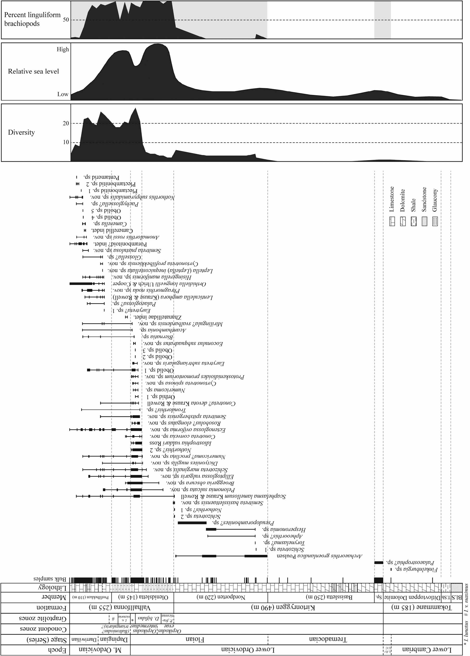
Material. Holo- and paratypes from Greenland ( MGUH 3666–3669) and the other specimens on which Poulsen (1937) based his study; 1195 specimens from Spitsbergen, including specimens A46819–21, A46823–24, A46835 View Materials –36, A50853–63, A50865 View Materials –78, A50885 View Materials , A50898 View Materials , A50904 View Materials , A50908 View Materials –9, A50916–31, and unnumbered specimens in the samples above and specimens from samples F4302, F4305, F4315, F4454, F4504, F4591, F4620, F4633, JH-34, JH-62–65, JH-67–72, JH-170 and JH-187. Many specimens are partly silicified. The figured specimens are MGUH 29704 View Materials , TSGF16755 , TSGF16756 , TSGF16881 , TSGF17075 , TSGF17086 and TSGF17087 .
Diagnosis. Archaeorthis with weak dorsal sulcus; 5–9 costellae per mm at 1-mm valve length; nearly planar ventral interarea; low callosity in front of ventral muscle field, up to 35% as wide as muscle field; nearly vertical brachiophore plates.
Description. Shell subcircular or transversely oval, moderately thick-walled and ventribiconvex. Maximum valve width located at 29–58% of valve length, moving forward with increasing size. Interarea 73–74% as wide as valve. Cardinal extremities obtuse but occasionally nearly rectangular. Largest specimen 7.7 mm long. Anterior margin rectimarginate or weakly unisulcate. Multicostellate, occasionally ramicostellate or fascicostellate, with 5– 9 (rarely 4) costellae per mm at 1-mm valve length and about 28 costellae per 2.5 mm at the 2.5-mm growth stage. Costellae subequal and slightly rounded to subangular. Filae absent but fine capillae present. Growth lines strong distally on larger specimens. Costellae impressed within distal 25–50% of valve length.
Dorsal valve moderately convex to nearly planar, with shallow sulcus developing at umbo. Sulcus often disappearing distally on larger specimens. L/W ratio 0.61–0.85. Maximum width located at 40–44% of valve length and maximum depth at about 35% of valve length. Interarea short, anacline and planar or slightly concave. Notothyrium wide and open. Brachiophores high, with base of brachiophore plates extending anterior to brachiophore tops. Brachiophore plates subparallel or gently diverging in both ventral and anterior directions, forming a deep Ushaped notothyrial cavity with sharply defined sides. Fulcral plates short, supporting brachiophores. Notothyrial platform low, generally slightly raised above low median ridge and reaching 14–24% of valve length. Cardinal process absent or rudimentary but with impressions of diductor muscle scars. Median ridge narrow and low, normally restricted to posterior part of valve. Adductor scars weak or obscure. Mantle canal system generally not impressed but otherwise appearing pinnate or digitate.
Ventral valve slightly to moderately (rarely strongly) convex, deepest just behind mid-length of valve. L/W ratio 0.66–1.09, increasing with size. Maximum width located at 40–50% of valve length. Ventral interarea short to moderately high, planar to slightly concave, moderately apsacline to nearly catacline, 82–96% as wide as valve. Delthyrium open, about 40°. Triangular teeth thin, with rather weakly developed crural fossettes. Dental plates thin, recessive, slightly diverging. Muscle field slightly raised on thickened platform, with moderately impressed muscle scars. Adductor scars elongate, extend anterior to teardrop-shaped diductor scars. Median callosity in front of muscle field low, proximally up to about 35% as wide as muscle field and generally fading completely at about 65% of valve length. Vascula media normally subparallel proximally to muscle field but soon diverging at about 50˚.
Remarks. Hallam (1958) assigned specimens from the southern part of Ny Friesland, Spitsbergen to the Lower Ordovician Archaeorthis groenlandica Poulsen, 1937 from Greenland. Two years later, Gobbett & Wilson (1960) studied the same material together with newly collected material and listed the species as A. cf. groenlandica , suggesting the existence of some differences. A re-examination of the type specimens and topotypes on which Poulsen (1937) based his description, including specimens showing some of the valve interior, which was not illustrated in the original description, revealed no differences from the Spitsbergen material. An illustration of the dorsal interior of one of the specimens from Greenland is included here for comparison (Pl. 25, Fig. 5). The material from Greenland does not include any specimens showing the delthyrial cavity. Seven of the nine specimens on which Hallam (1958) based his study, together with the specimens listed by Gobbett & Wilson (1960) and some previously overlooked specimens in the same collections, were also re-examined, revealing no important differences from those collected in northeastern Ny Friesland.
Archaeorthis presently includes more than 25 described species from the Lower and Middle Ordovician of Asia, Australia, Europe, Greenland and North and South America. A. greenlandica Poulsen resembles the four species A. biconvexa Cooper, 1956 , A. electra (Billings, 1862) , A. opima Nikitin & Popov, 1984 and A. scotia Curry & Williams, 1984 . A. biconvexa and A. electra have a wider median ridge on the ventral valve floor and apparently coarser costellae. A. opima has a higher L/W ratio and a cardinal process, while A. scotia has strongly divergent brachiophore plates and impressed adductor scars.
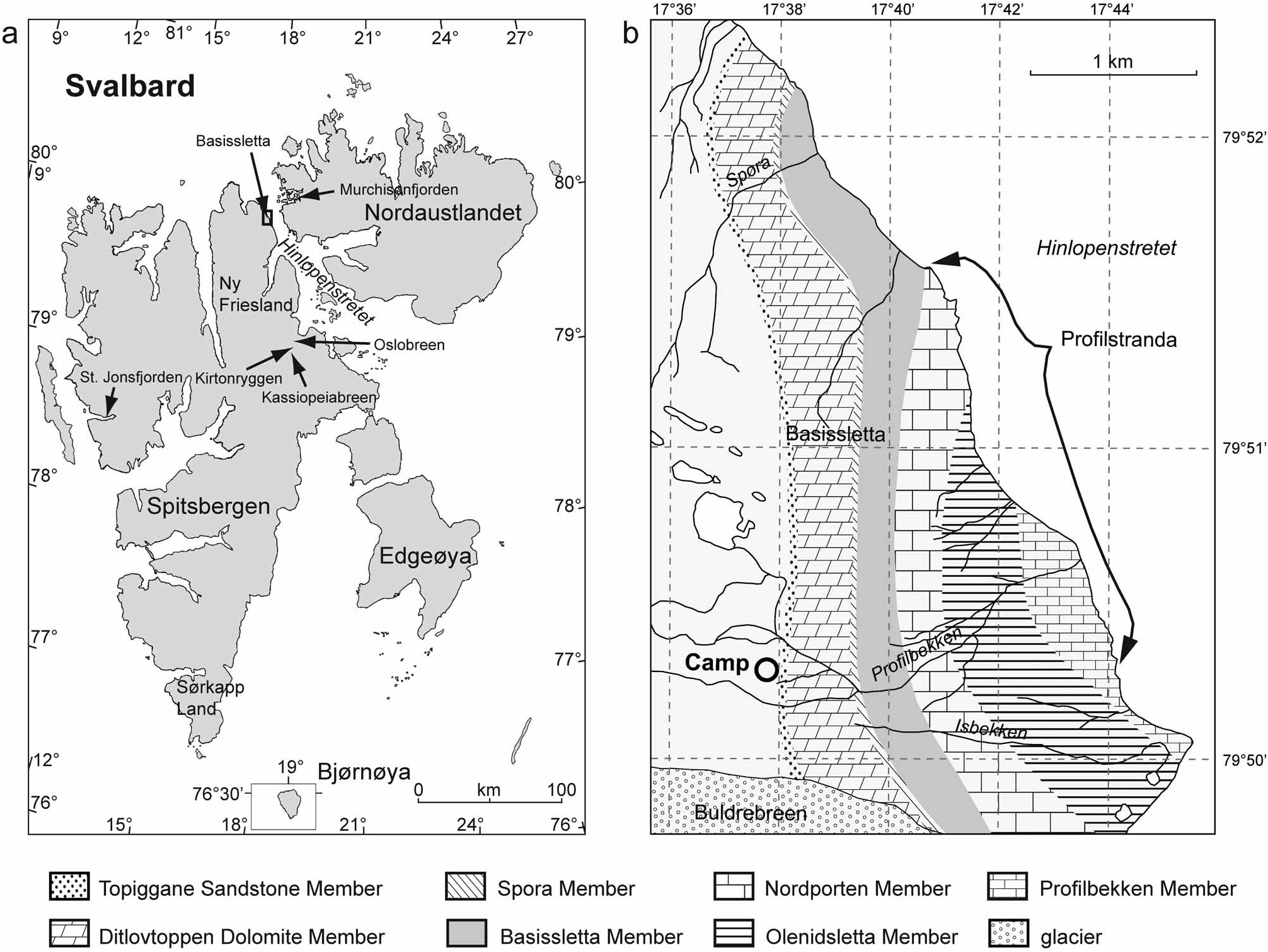
Occurrence. Middle Oslobreen Limestone, Kirtonryggen Formation, at Kassiopeiaisen, Kirtonryggen and Oslobreen in central Ny Friesland. In horizons throughout Nordporten Member, Kirtonryggen Formation, Basissletta in northeastern Ny Friesland, Spitsbergen. Cape Weber Formation (Lower Ordovician); Cape Weber, Mountain Gunvor, upper part of Devon Canyon and summit 1 km south of Devon Canyon, East Greenland.

| MGUH |
Museum Geologicum Universitatis Hafniensis |
No known copyright restrictions apply. See Agosti, D., Egloff, W., 2009. Taxonomic information exchange and copyright: the Plazi approach. BMC Research Notes 2009, 2:53 for further explanation.