
M. pterygoideus dorsalis
|
publication ID |
https://doi.org/10.1093/zoolinnean/zlaa163 |
|
persistent identifier |
https://treatment.plazi.org/id/AB191354-7A28-D47E-FCED-478D808CFA46 |
|
treatment provided by |
Plazi |
|
scientific name |
M. pterygoideus dorsalis |
| status |
|
M. pterygoideus dorsalis (mPTd)
Origin: In lepidosaurs, turtles, birds and crocodiles this muscle originates on the dorsal surface of the pterygoids and palatines, also extending onto the ectopterygoids and interorbital septum in lepidosaurs, turtles and crocodiles, and also onto the postorbital, jugal and maxilla in turtles.
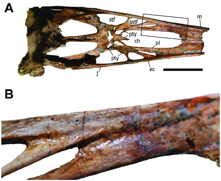
The dorsal surfaces of the palatal elements are totally covered by matrix in the holotypes of Anhanguera blittersdorffi , Anhanguera santanae , Tapejara wellnhoferi , Thalassodromeus sethi , Tropeognathus mesembrinus and Tupandactylus navigans . In the tapejarid Caupedactylus ybaka , in which the dorsal surfaces of the palatal bones are exposed, well-defined muscle scars can be seen in the dorsal surfaces of the ectopterygoids, pterygoids and also the palatines slightly anterior to the anterior margin of the choanae ( Fig. 6 View Figure 6 ), supporting the origin of mPTd in this region in pterosaurs. It should be noted that the dorsal surface of the pterygoid, in pterodactyloids, is partially overlain by the pterygoid process of the ectopterygoid ectopterygoid of C. ybaka , muscle scars can be seen only on the main body ( Fig. 6 View Figure 6 ), which includes the broadest region of the bone. In this way, the origin area of mPTd in pterodactyloids seems to have been restricted to a region that is anterior to the lateral process of the pterygoid (in the forms that exhibit such structure) and that, therefore, such muscle would pass through the secondary subtemporal fenestra and not the main subtemporal fenestra. In pterodactyloids that lack a lateral process of the pterygoid dividing the subtemporal fenestra, passage of mPTd would thus likely be restricted to the anterior region of such fenestra.
(e.g. Ösi et al., 2010). The origin area for mPTd in the pterodactyloid pterygoid is thus restricted to the anterior region of the main body of the bone. In the Insertion: In lepidosaurs and crocodylians this muscle passes through the subtemporal fenestra and inserts on the medial surface of the articular and angular bones ( Holliday & Witmer, 2007). However, in dinosaurs it was likely limited to the medial surface of the articular ( Holliday, 2009), including birds, in which the insertion is often located on the medial mandibular process of the articular or slightly anterior to it ( Holliday & Witmer, 2007). In turtles, the insertion varies between the prearticular, articular, surangular and coronoid process ( Werneburg, 2011).
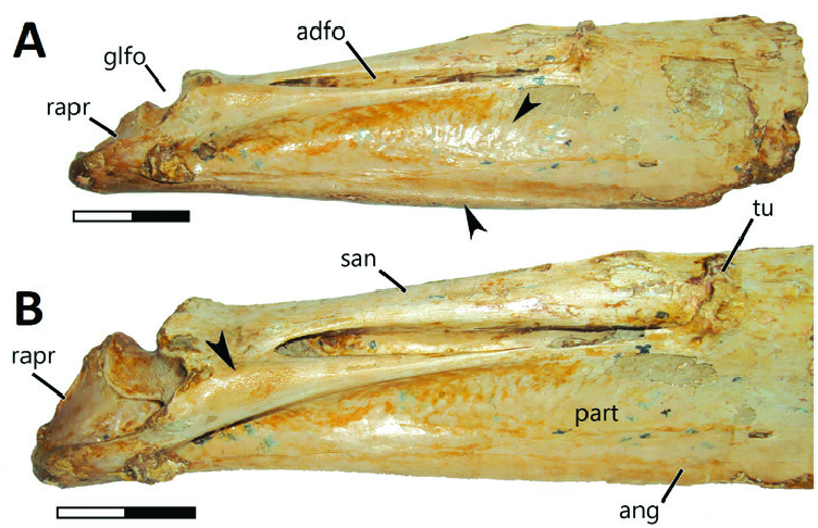
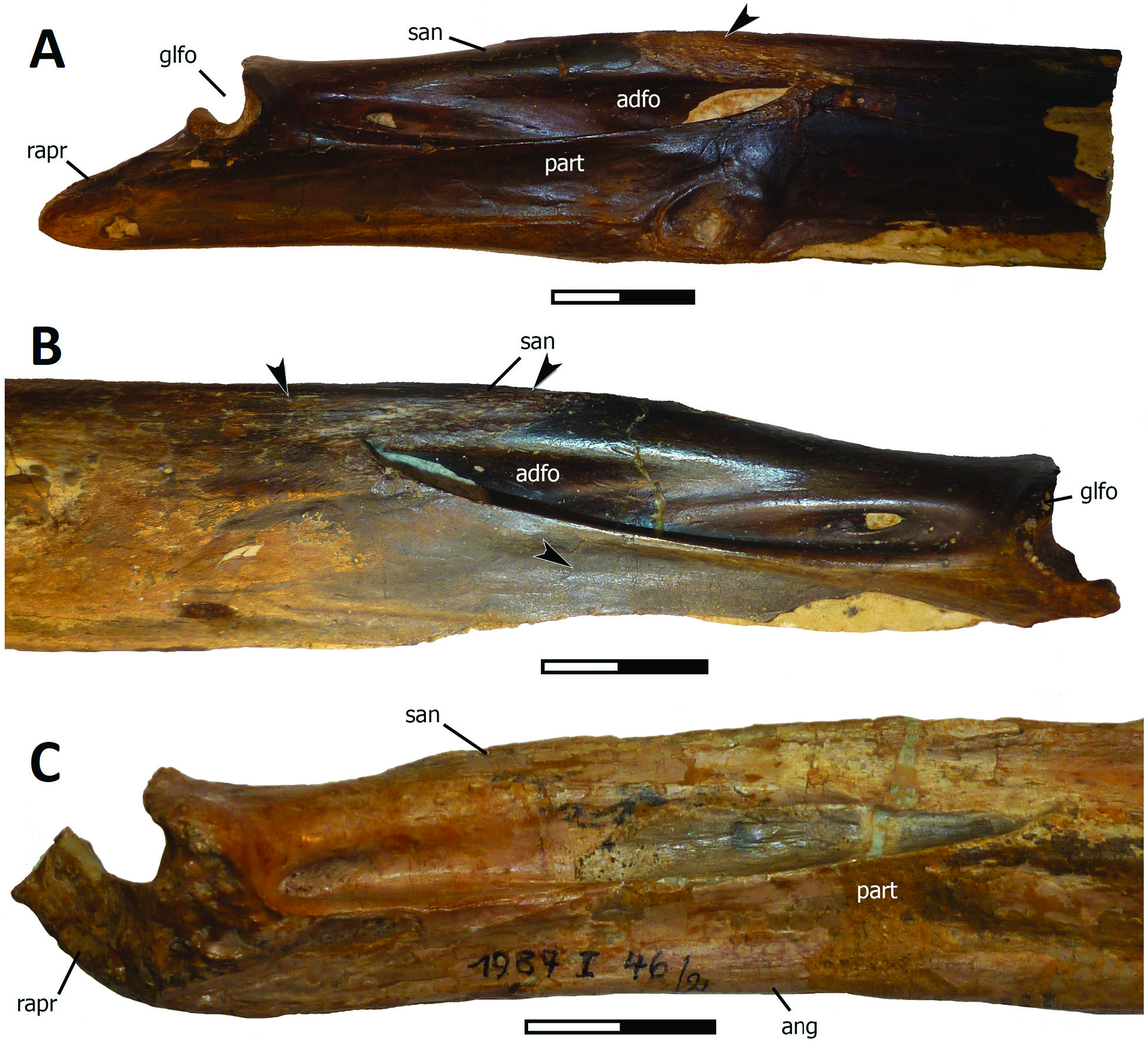
In An. araripensis (BSP 1982 89), Cearadactylus atrox , Th. sethi and Tr. mesembrinus (BSP BSP 1987 46) a depression (better developed in Th. sethi ) with well-defined muscle scars is present on the medial side of the angular and prearticular ( Figs 10 View Figure 10 , 11 View Figure 11 ), suggesting that the angular participated in the insertion surface for mPTd, as in the Lepidosauria and the Crocodylia, but also the prearticular, as in the Testudines, as well as the articular as in all living reptiles. Muscle scars can be seen in this depression in Th. sethi and An. araripensis (BSP 1982 89).
No known copyright restrictions apply. See Agosti, D., Egloff, W., 2009. Taxonomic information exchange and copyright: the Plazi approach. BMC Research Notes 2009, 2:53 for further explanation.