
Kryptoperidinium foliaceum (F. Stein) Lindemann
|
publication ID |
https://doi.org/ 10.1515/bot-2017-0041 |
|
DOI |
https://doi.org/10.5281/zenodo.11472870 |
|
persistent identifier |
https://treatment.plazi.org/id/AB4F87BD-AA58-FF8C-6018-37C3FC5EFC95 |
|
treatment provided by |
Felipe |
|
scientific name |
Kryptoperidinium foliaceum (F. Stein) Lindemann |
| status |
|
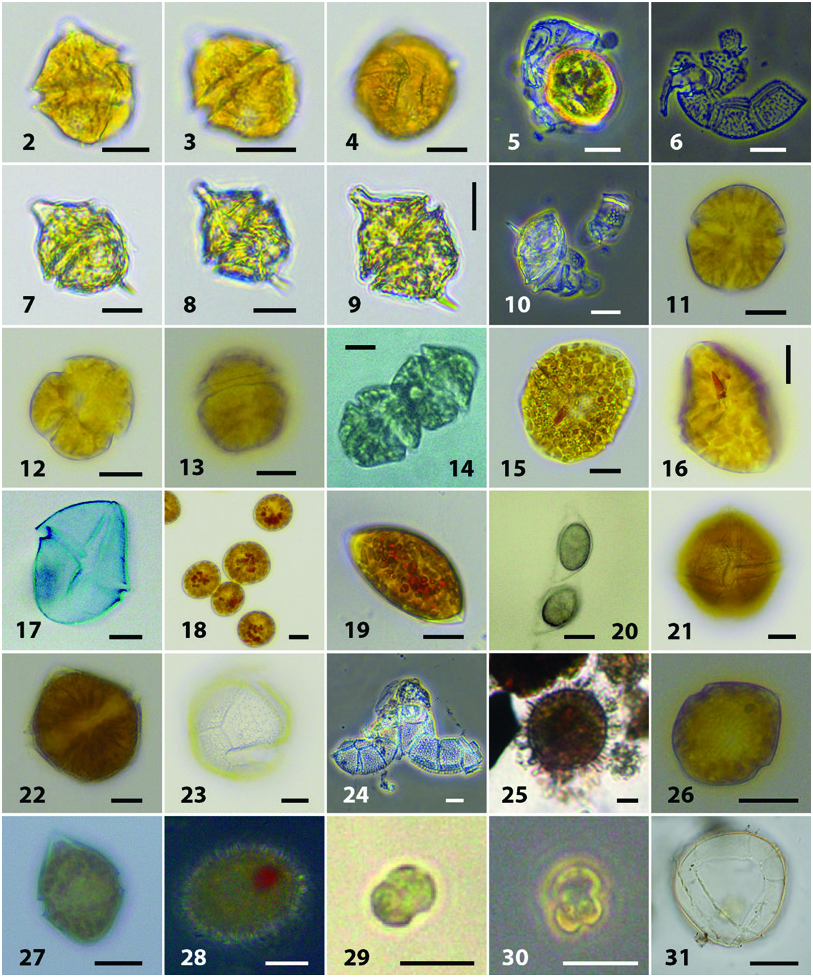
Kryptoperidinium foliaceum (F. Stein) Lindemann
( Figures 15–20 View Figures 2–31 )
Cells were pale brown in colour with a red eye spot and a central nucleus ( Figures 15 and 16 View Figures 2–31 ). Cells were 30–50 µm long and 28–45 µm wide. Cells were strongly dorso-ventrally flattened and broadly circular in dorsal view ( Figures 15 and 16 View Figures 2–31 ). The cingulum was median and not offset ( Figure 15 View Figures 2–31 ). Cells had very thin thecae on which it was very difficult to discern any thecal tabulation ( Figure 17 View Figures 2–31 ) although it has been reported by Figueroa et al. (2009). Cysts were formed within our cultures – these were ovoid to spherical in dorsal view ( Figure 18 View Figures 2–31 ) and narrowly elliptical in apical view ( Figures 19 and 20 View Figures 2–31 ). Two strains from the Caspian were successfully sequenced which match those for Kryptoperidinium foliaceum ( Figure 32 View Figure 32 ).

No known copyright restrictions apply. See Agosti, D., Egloff, W., 2009. Taxonomic information exchange and copyright: the Plazi approach. BMC Research Notes 2009, 2:53 for further explanation.
|
Kingdom |
|
|
Phylum |
|
|
Class |
|
|
Order |
|
|
Family |
|
|
Genus |