
Epipeltephilus kanti, González-Ruiz, Laureano R., Scillato-Yané, Gustavo J., Krmpotic, Cecilia M. & Carlini, Alfredo A., 2012
|
publication ID |
https://doi.org/10.5281/zenodo.210310 |
|
DOI |
https://doi.org/10.5281/zenodo.5662825 |
|
persistent identifier |
https://treatment.plazi.org/id/AC14242F-F617-BD7E-FF7B-559BFB36F84A |
|
treatment provided by |
Plazi |
|
scientific name |
Epipeltephilus kanti |
| status |
sp. nov. |
Epipeltephilus kanti new species
( Figures 2–3 View FIGURE 2 View FIGURE 3 A)
Etymology. “ kanti ” in honor of the Prussian philosopher Emmanuel Kant ( 1724–1804), brilliant creator of the criticism and precursor of the modern scientific philosophy.
Holotype. MLP 92-XI-19-7, three fixed osteoderms of the dorsal shield ( Figure 2 View FIGURE 2 A–C).
Geographic and stratigraphic occurrence of the holotype. Arroyo Chasicó ( 38°37´06.10´´S, 62°59´14.53´´W), Buenos Aires Province, Vivero Member, Arroyo Chasicó Formation, Chasicoan SALMA. Material collected during a field trip organized by the Facultad de Ciencias Naturales y Museo de La Plata ( Argentina) and Duke University ( USA).
Referred material. MLP 28-X-11 -66, three complete, fixed osteoderms and an additional fragment of the dorsal shield ( Figure 2 View FIGURE 2 D–G); MLP 60-VI-18-3, one molariform and osteoderms of an indeterminate region of the dorsal shield; MMP 339-M, one movable osteoderm of the dorsal shield ( Figure 2 View FIGURE 2 H). All specimens were collected in the Vivero Member, Arroyo Chasicó Formation ( Figure 1 View FIGURE 1 ).
Differential diagnosis. Osteoderms larger than those of Peltephilus nanus and Peltephilus pumilus , and similar in size to those of Epipeltephilus recurvus , Peltecoelus praelucens , and the other species of Peltephilus . Longitudinal crests (two lateral and one central) are higher and more developed than in Epipeltephilus recurvus , Peltephilus giganteus , Peltephilus pumilus , Peltephilus strepens and Peltephilus protervus . These crest are absent in Peltecoelus praelucens , Peltephilus depressus , Peltephilus granosus and Peltephilus nanus . Exposed surface of the osteoderms roughed like Epipeltephilus recurvus much more than the species of Peltephilus and Peltecoelus praelucens .
Comparative description. Molariform. Scillato-Yané (1982) described the molariform MLP 60-VI-18-3 1 as very small and lower because the main wear pit is located externally, as in Peltephilus according to Scott (1903), and it would correspond to the third or fourth molariform of the left dental series. It is broken in its base and the preserved part measures 7.6 mm of maximum antero-posterior diameter, and 3.5 mm of maximum cross-sectional diameter.
According to Scillato-Yané (1982) the molariform section is subtriangular, less compressed than in Anantiosodon , whereas in Epipeltephilus ( E. recurvus ) teeth are subeliptic. In addition, Vizcaíno and Fariña (1997: 81) remarked that the molariforms of Peltephilus are “ chiefly triangular in section and slenderly built ”. Lower molariforms of Peltecoelus and Parapeltecoelus are not known.
Osteoderms of the dorsal shield. Shape and size. The known osteoderms of Epipeltephilus kanti are rectangular or quadrangular ( Figure 2 View FIGURE 2 A–H). They cover a large range of sizes ( Table 1 View TABLE 1 ), indicating that osteoderms from different regions of the dorsal shield are represented; also, that the size variation of the osteoderms was probably remarkable at intraspecific level, depending on the regions of the dorsal shield, as it happens in Eutatus ( Burmeister, 1883; Scillato-Yané, 1982).
In relation to other species of Peltephilidae , the osteoderms of Epipeltephilus kanti are larger than those of Peltephilus nanus , and generally larger than those of Peltephilus pumilus , but within the range of variation of the remaining species of Peltephilus , as well as Epipeltephilus recurvus and Peltecoelus praelucens ( Table 1 View TABLE 1 ).
Foramina. Osteoderms of Epipeltephilus kanti generally have one or two big oval foramina, which can be associated to glandular cisterns ( Scillato-Yané, 1979; Croft et al., 2007). In the case of Epipeltephilus kanti , the largest foramina correspond to the largest osteoderms ( Figure 2 View FIGURE 2 A–H, 3A). In some osteoderms, there is a smaller foramen between and over the previous ones; this character is mentioned by Croft et al. (2007) for the osteoderms of a specimen of cf. Peltephilus sp. One movable osteoderm of Epipeltephilus kanti has a third big foramina underneath the two bigger ( Figure 2 View FIGURE 2 H).
Much variation has been documented for the number of osteoderm foramina in different species of Peltephili- 1.: * We searched this specimen several times in the MLP collections with Dr. Marcelo Reguero (Collection Manager) and with Dr. Scillato-Yané (who originally described it in 1982) but we could not find it. Unfortunately, the molariform is lost in the Museum and the illustrations given by Scillato-Yané (1982: plate 1, figura 4) do not have enough resolution for comparisons.
dae: Peltephilus strepens has two or four ( Ameghino 1887) ( Figure 3 View FIGURE 3 F–G); Peltephilus pumilus , P. nanus , P. giganteus , P. protervus and P. granosus have two ( Figure 3 View FIGURE 3 C, D, E, H, J); Peltephilus depressus has two or three; Ameghino (1897) mentioned four foramina for that species, but the type and assigned material have two or three ( Figure 3 View FIGURE 3 I); Peltecoelus praelucens has three foramina ( Ameghino, 1902) ( Figure 3 View FIGURE 3 K), and Epipeltephilus recurvus has generally four foramina ( Croft et al., 2009) ( Figure 3 View FIGURE 3 B), although there are also osteoderms with one, two or three foramina.
Crests. The osteoderms of Epipeltephilus kanti have one middle longitudinal crest and two lateral longitudinal crests. All of them are elevated, wide and have rough margins, with the subsequent development of deep and ample valleys among them. The middle longitudinal crest may or may not reach the posterior border while the two laterals always do. The posterior border is elevated and has rough margins.
Some osteoderms of Epipeltephilus recurvus have a middle longitudinal crest, but unlike Epipeltephilus kanti , it is a narrow and not high. Peltephilus giganteus has three longitudinal crests, but they are rounded, without roughness and without ample valleys between them ( Figure 3 View FIGURE 3 E). Peltephilus protervus has a smooth and well developed crest, which extends only one half the surface of the osteoderm, being anteriorly wide and disappearing posteriorly ( Ameghino, 1897) ( Figure 3 View FIGURE 3 H). A longitudinal crest is developed in Peltephilus strepens ( Figure 3 View FIGURE 3 F–G) ( Ameghino, 1887), but it is low instead of marked as in P. giganteus or Epipeltephilus kanti . Some osteoderms assigned to Peltephilus pumilus have a high and narrow longitudinal middle crest ( Ameghino, 1887) ( Figure 3 View FIGURE 3 C), whereas in other specimen assigned to this species this crest is absent. The osteoderms of Peltephilus nanus , Peltephilus depressus , Peltephilus granosus and Peltecoelus praelucens lack the longitudinal crests ( Ameghino, 1897; Ameghino, 1902) ( Figure 3 View FIGURE 3 D, I, J, K).
Surface roughness. The exposed surface of the osteoderms of Epipeltephilus kanti is rougher than any other Peltephilidae . The osteoderms of Epipeltephilus recurvus are not as rough as in E. kanti . Peltephilus pumilus , P. nanus , P. giganteus , and P. strepens has osteoderms with similarly rough surfaces ( Figure 3 View FIGURE 3 C–G), but rougher than in Peltephilus protervus , P. depressus , and Peltecoelus praelucens ( Figure 3 View FIGURE 3 H, I, K), and less than E. recurvus and E. kanti ( Figure 3 View FIGURE 3 A–B). Although P. granosus has an anterior rough region, Peltephilus protervus and P. granosus has smooth and punctuate surfaces ( Ameghino, 1897, 1902). Finally, although Ameghino (1902) remarks that the osteoderms of Peltecoelus praelucens are completely smooth, we observed that they are also punctuate.
Geochronology and biostratigraphy. The first fossils from Arroyo Chasicó ( Figure 1 View FIGURE 1 ) were collected by S. Roth and his assistant B. Eugui around 1915 ( Kraglievich, 1934; Pascual, 1961; Bondesio et al., 1980). Cabrera (1928) and Kraglievich (1934) were the first to mention this locality, followed by works of Reig (1957), Pascual (1961, 1965), and Pascual et al. (1965).
Kraglievich (1934) recognized the Chasicoan (“ Chasicoense ”) geologic horizon and assigned a Miocene age to its fauna, while indicating that: “… esta fauna es casi equivalente a la más antigua de Entre Ríos y su edad puede considerarse Miocena ” ( Kraglievich, 1934: 89). Pascual (1961, 1965) and Pascual et al. (1965) defined the Arroyo Chasicó Formation indicating the existence of outcrops in the headwaters of Arroyo Chasicó and in the cliffs of the Chasicó lagoon ( type area). They also recognized a Chasicoan SALMA on the basis of the presence of “relictual” mammals from the Santacrucian SALMA and the primitive character of the Pan-Araucanian predominant taxa.
Bondesio et al. (1980) summarized the data on the geology and fossil mammals of the area and divided Arroyo Chasicó Formation into two Members: the lower Vivero Member and the upper Las Barrancas Member. According to these authors, these members are related to two different biozones: 1) Biozone of Chasicotherium rothi Ameghino , which is a local representation of the “Viverense” (lower Chasicoan); and 2) Biozone of Chasicotatus ameghinoi Scillato-Yané , which represents the “Barranquense” (upper Chasicoan). Both units were deposited during the earliest part of the late Miocene ( Tonni et al., 1998; Cione et al., 2000). Zárate et al. (2007) conducted a geologic study of the Chasicoan deposits recognizing different lithofacies and paleosols. This lithofacial adjustment does not fit with the lithostratigraphic division of the Arroyo Chasicó Formation in two members.
In accordance with the more recent biostratigraphic scheme proposed by Verzi et al. (2008), Chasicotatus ameghinoi was recorded in the Biozones of Chasichimys bonaerense Pascual , Chasichimys scagliai (Pascual) , Xenodontomys simpsoni Kraglievich and Xenodontomys elongatus (Verzi, Montalvo & Tiranti) . For that reason, Verzi et al. (2008) suggested to change the Biozone of Chasicotatus ameghinoi , since it would be not an exclusive taxon for that biozone. Finally, Schultz et al. (2004) presented an 40Ar/39Ar age of 9.23 ± 0.09 Ma for Arroyo Chasicó Formation, placing the Chasicoan fauna between the Mayoan (ca.10.0–11.8 Ma) and the Huayquerian (6.8–9.0 Ma) faunas, in the time interval of ca. 9.0–10.0? Ma based on the scheme by Flynn and Swisher (1995).
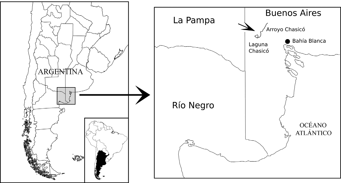

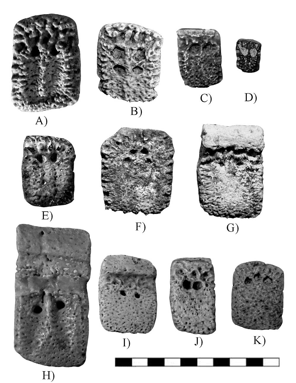
TABLE 1. Measurements of osteoderms and their associated foramina (mm).
| Taxon | Osteoderm type | Osteoderm surface Width Length (min-max) (min-max) | Foramina Width (min-max) | Length (min-max) |
|---|---|---|---|---|
| Epipeltephilus kanti | Fixed Movable | 12.4–27.1 11.5–29.5 16.1 24.6 | 1.4–3.1 1.4–1.9 | 1.6–3.5 0.8–1.0 |
| Epipeltephilus recurvus ? | Fixed | 12.1–19.0 11.2–22.6 | 2.0–2.8 | 2.2.–2.5 |
| Peltephilus strepens | Movable Fixed | 17.6–21.5 22.3–23.9 13.8–21.1 16.4–25.4 | 1.0–1.6 1.2–1.5 | 2.1–2.6 1.7–1.5 |
| MLP |
Museo de La Plata |
No known copyright restrictions apply. See Agosti, D., Egloff, W., 2009. Taxonomic information exchange and copyright: the Plazi approach. BMC Research Notes 2009, 2:53 for further explanation.
|
Kingdom |
|
|
Phylum |
|
|
Class |
|
|
Order |
|
|
Family |
|
|
Genus |