
Cletocamptus albuquerquensis ( Herrick, 1894 )
|
publication ID |
https://doi.org/ 10.11646/zootaxa.4272.3.1 |
|
publication LSID |
lsid:zoobank.org:pub:0ECB3A74-2E13-4713-95FF-D5739035EE09 |
|
DOI |
https://doi.org/10.5281/zenodo.6002068 |
|
persistent identifier |
https://treatment.plazi.org/id/AF5D87B3-4A0E-FFD7-AF8C-1E47C68BF8CF |
|
treatment provided by |
Plazi |
|
scientific name |
Cletocamptus albuquerquensis ( Herrick, 1894 ) |
| status |
|
Cletocamptus albuquerquensis ( Herrick, 1894)
( Figs. 4–14 View FIGURE 4 View FIGURE 5 View FIGURE 6 View FIGURE 7 View FIGURE 8 View FIGURE 9 View FIGURE 10 View FIGURE 11 View FIGURE 12 View FIGURE 13 View FIGURE 14 )
Synonymy. Marshia albuquerquensis: Herrick 1894: 41 –43, Figs. 1–11 View FIGURE 1 View FIGURE 2 View FIGURE 3 View FIGURE 4 View FIGURE 5 View FIGURE 6 View FIGURE 7 View FIGURE 8 View FIGURE 9 View FIGURE 10 View FIGURE 11 , 1895: 136–137, Plates XXXI–XXXII, Figs. 1–5 View FIGURE 1 View FIGURE 2 View FIGURE 3 View FIGURE 4 View FIGURE 5 ; Dodds 1915: 296, Fig. 80, 1919: 73, Fig. 10 View FIGURE 10 , 1920: 95, Table I; Marsh 1918: 780; Weckel 1914: 201; Willey 1923: 7, 1925: 206; Young 1924: 93.
Wolterstorffia albuquerquensis: Brehm 1913 .
Cletocamptus albuquerquensis: Lang 1936 ; Lang 1948 (part.); Comita 1951; Brehm 1954, 1965; Ringuelet 1958; Wilson & Yeatman 1959; Pallares 1962; Fleeger 1980.
Material examined. Type material: One female neotype preserved in alcohol, USNM No. 1422182; col. Wayne Wurtsbaugh (Utah State University), April 30, 2009; id. Ray Gerber.
Additional material: The vials housed in the US National Museum (Smithsonian Institution) that were analyzed are labeled as follows. Supplementary information about the content of each vial and about the collection site is provided.
Cletocamptus albuquerquensis , USNM No . 310388; Canada; Saskatchewan ; Dead Moose Lake; collector U. T. Hammer, 14 July 1990; identification by J. W. Reid. Notes: This vial contained 13 females and 5 males. Dead Moose Lake surface area is about 10.5 km2 and has an average salinity of about 30 gL -1 ( Last & Slezak 1986).
Cletocamptus albuquerquensis , USNM No . 310389, Accession No . 392560; Canada; Saskatchewan; Big Quill Lake; collector U. T. Hammer, May 29, 1990; identification by J. W. Reid, 2001. Notes: This vial contained 20 females and 10 males. Big Quill Lake is about 307 km 2 ( Schmutz 2001) and salinity ranges from 40–85 gL -1 ( Hammer & Hurlbert 1992).
Cletocamptus albuquerquensis , USNM No . 119498, Accession No . 266592; Wyoming ; Albany County; Twelvemile Lake; collector W. W. Rice, 1966; identification by T. E. Bowman. Notes: This vial contained 9 males and 1 copepodite. Twelvemile Lake is described as a small (0.08 km2) hypersaline pond, surrounded by salt flats, that fills with water in the spring and dries out by July (Frank Rahel (University of Wyoming) personal communication).
Cletocamptus albuquerquensis , USNM No . 1297024; Utah, Great Salt Lake, Farmington Bay ; collector Wayne Wurtsbaugh (Utah State University), June 2, 2008; salinity, 38 gL -1; identification by Ray Gerber.
Cletocamptus albuquerquensis , USNM No . 1297025; Utah, Great Salt Lake, Farmington Bay ; collector Wayne Wurtsbaugh (Utah State University), June 26, 2008; salinity, 36 gL -1; identification by Ray Gerber.
Cletocamptus albuquerquensis , USNM No . 1297026; Utah, Great Salt Lake, Farmington Bay ; collector Wayne Wurtsbaugh (Utah State University), April 15, 2009; salinity, 20 gL -1; identification by Ray Gerber.
Cletocamptus albuquerquensis , USNM No . 1297027; Utah, Great Salt Lake, Farmington Bay ; collector Wayne Wurtsbaugh (Utah State University), April 30, 2009; salinity, 15 gL -1; identification by Ray Gerber.
Type locality. Utah, Great Salt Lake, Farmington Bay (41°3.982’N, 112°13.804’W); salinity, 15 gL -1. GoogleMaps
Distribution. Argentina: Luro lagoon (La Pampa Province) ( Pallares 1962), Lucio V. Mansilla and Totoralejos (Córdoba Province) ( Brehm 1954, 1965). Canada: Dead Moose Lake and Big Quill Lake (Saskatchewan) (present study). US: Colorado ( Dodds 1915, 1919, 1920), North Dakota (Devils Lake) ( Willey 1923; Young 1924), Rio Grande Valley (New Mexico) ( Herrick 1895), west of Mission (Texas) ( Comita 1951), Salt Lake City (Utah) ( Comita 1951); Farmington Bay (Great Salt Lake, Utah) (present study), Twelvemile Lake (Albany County, Wyoming) (present study).
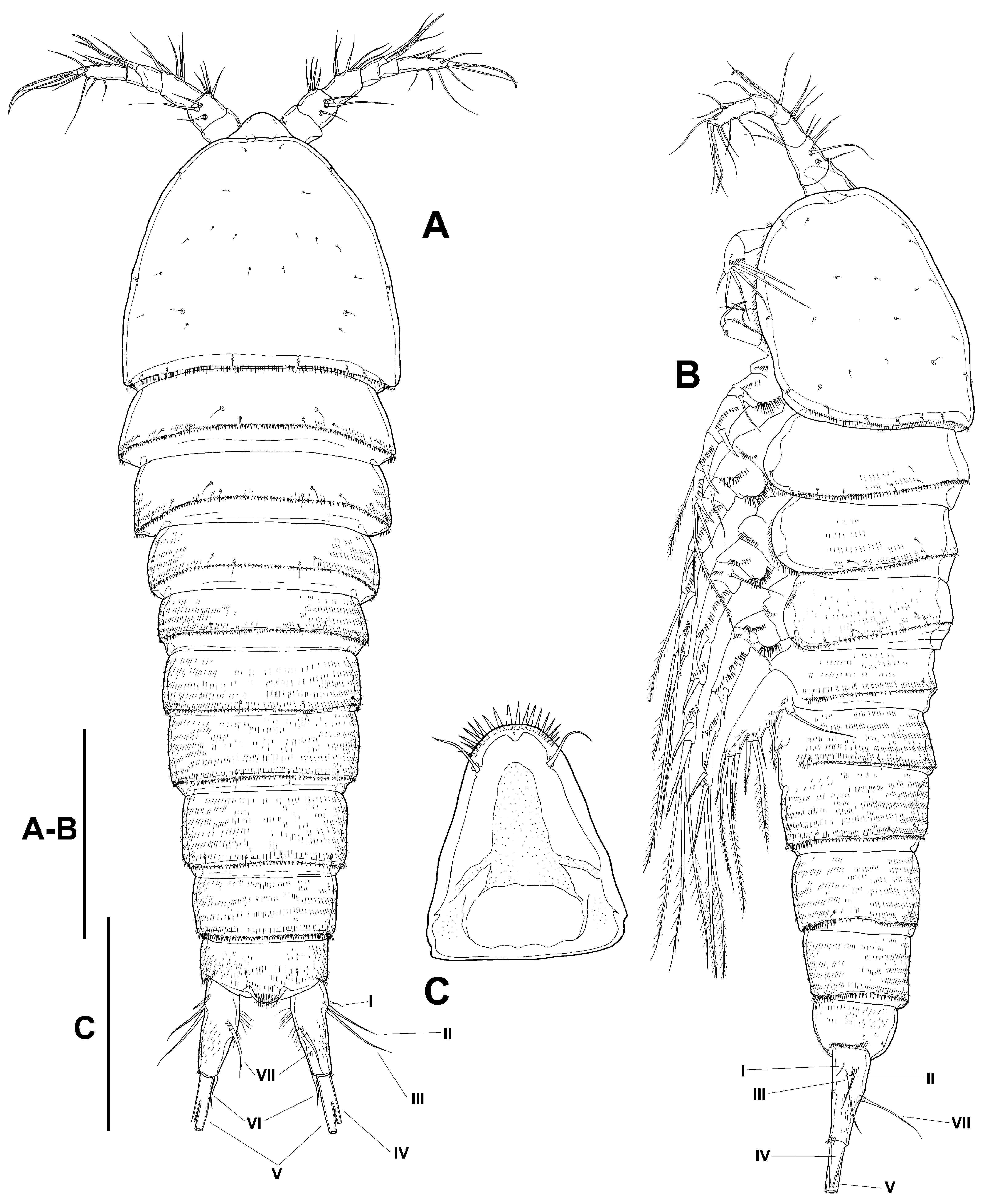
Redescription (based on specimens from Farmington Bay). Female. Habitus ( Fig. 4 View FIGURE 4 A, B) tapering posteriorly; total body length measured from tip of rostrum to posterior margin of caudal rami ranging from 694 µm to 905 µm (mean= 836 µm; n= 7). Rostrum set off, triangular, with pair of setules subapically and with row of spinules distally ( Fig. 4 View FIGURE 4 C). Cephalic shield with small spinules along margin dorsolaterally ( Fig. 4 View FIGURE 4 A, B). Dorsal and lateral surface of free thoracic somites (P2–P4 bearing-somites) with transverse rows of minute spinules, with short spinules along posterior margin ( Fig. 4 View FIGURE 4 A, B). Dorsal and lateral surface of first urosomite (P5 bearing-somite) with transverse rows of minute spinules, with row of small spinules along posterior margin ( Fig. 4 View FIGURE 4 A, B). Second and third urosomites distinct dorsally and laterally ( Fig. 4 View FIGURE 4 A, B), completely fused ventrally forming genital doublesomite ( Figs. 4 View FIGURE 4 B, 5A); dorsal and lateral surface of first and second half of genital double-somite with transverse rows of minute spinules, and row of larger spinules along posterior margin of second and third urosomites; ventrally with longer spinules ( Fig. 5 View FIGURE 5 A); copulatory pore in the middle of genital somite. Ornamentation of fourth and fifth urosomites as in second genital somite dorsally ( Fig. 4 View FIGURE 4 A, B); ventral surface with transverse rows of minute spinules, with larger spinules along posterior margin ( Fig. 5 View FIGURE 5 A). Dorsal surface of anal somite ( Fig. 4 View FIGURE 4 A) with transverse rows of minute spinules and with dorsolateral strong spinules close to joint with caudal rami; rounded anal operculum furnished with spinules ( Fig. 4 View FIGURE 4 A). Caudal rami ( Figs. 4 View FIGURE 4 A, B, 5A) about 2.4 times as long as wide; dorsal surface with sparse small spinules, smooth ventrally except for row of spinules close to insertion of caudal setae distally; with seven elements in all ( Fig. 4 View FIGURE 4 A, B, 5A); seta I small, situated laterally on proximal part of ramus, close to setae II and III, the latter setae longer; setae IV and V fused basally, the former 17%, the latter 54% of total body length; seta VI situated on distal inner corner; seta VII situated dorsally midway length of ramus on inner edge ( Fig. 4 View FIGURE 4 A, B).
Antennule ( Fig. 6 View FIGURE 6 A): six-segmented; surface of segments smooth except for two rows of spinules on first segment. Armature formula, 1-(1), 2-(9), 3-(6), 4-(1+[1+ae]), 5-(1), 6-(9+[1+ae]).
Antenna ( Fig. 6 View FIGURE 6 B, B’): with small coxa ornamented with spinules as shown. Allobasis with two abexopodal setae. Free endopodal segment with inner strong spinules proximally and subdistally; with two lateral inner spines and a slender seta, and five distal elements, two of them geniculate. Exopod small, one-segmented; with few spinules and with 3 somewhat stiff and thick small setae (indicated in Fig. 6 View FIGURE 6 B’).
Mandible ( Fig. 7 View FIGURE 7 A): robust, with spinules proximally; chewing edge with teeth as figured, with one pyriform element and one lateral pinnate seta. Palp one-segmented, with two long setae unequal in length.
Maxillule ( Fig. 7 View FIGURE 7 B, C): robust; arthrite of praecoxa with two rows of spinules and one surface seta, eight spines and two slender setae. Coxa ornamented with some spinules, with two slender setae. Basis with spinules as figured, with two apical setae. Exopod and endopod incorporated to basis, represented by two setae each.
Maxilla ( Fig. 7 View FIGURE 7 D): syncoxa with spinules along inner and outer margin, and close to joint with allobasis; with two endites, each bearing three setae. Allobasis drawn into strong claw with one accompanying seta. Endopod represented by three setae.
Maxilliped ( Fig. 7 View FIGURE 7 E): subchelate. Syncoxa with row of spinules, with one seta on inner distal corner. Basis without armature, with longitudinal row of inner spinules, and with few outer spinules proximally and subdistally. Endopod drawn into long and slender claw with one accompanying small seta.
P1 ( Fig. 8 View FIGURE 8 A): praecoxa with three groups of spinules close to joint with coxa. The latter with anterior transverse rows of spinules, and with stronger spinules close to outer distal corner. Basis with inner and outer spine; with median rows of spinules and with stronger spinules at base of exopod near outer spine, between exopod and endopod, and at base of inner spine. Exopod three-segmented; EXP1 without, EXP2 with inner seta; EXP3 with four elements. Endopod two-segmented, reaching proximal third of EXP3; first segment with inner seta relatively short, barely as long as both segments combined, with brush tip; second segment with one inner and two apical elements.
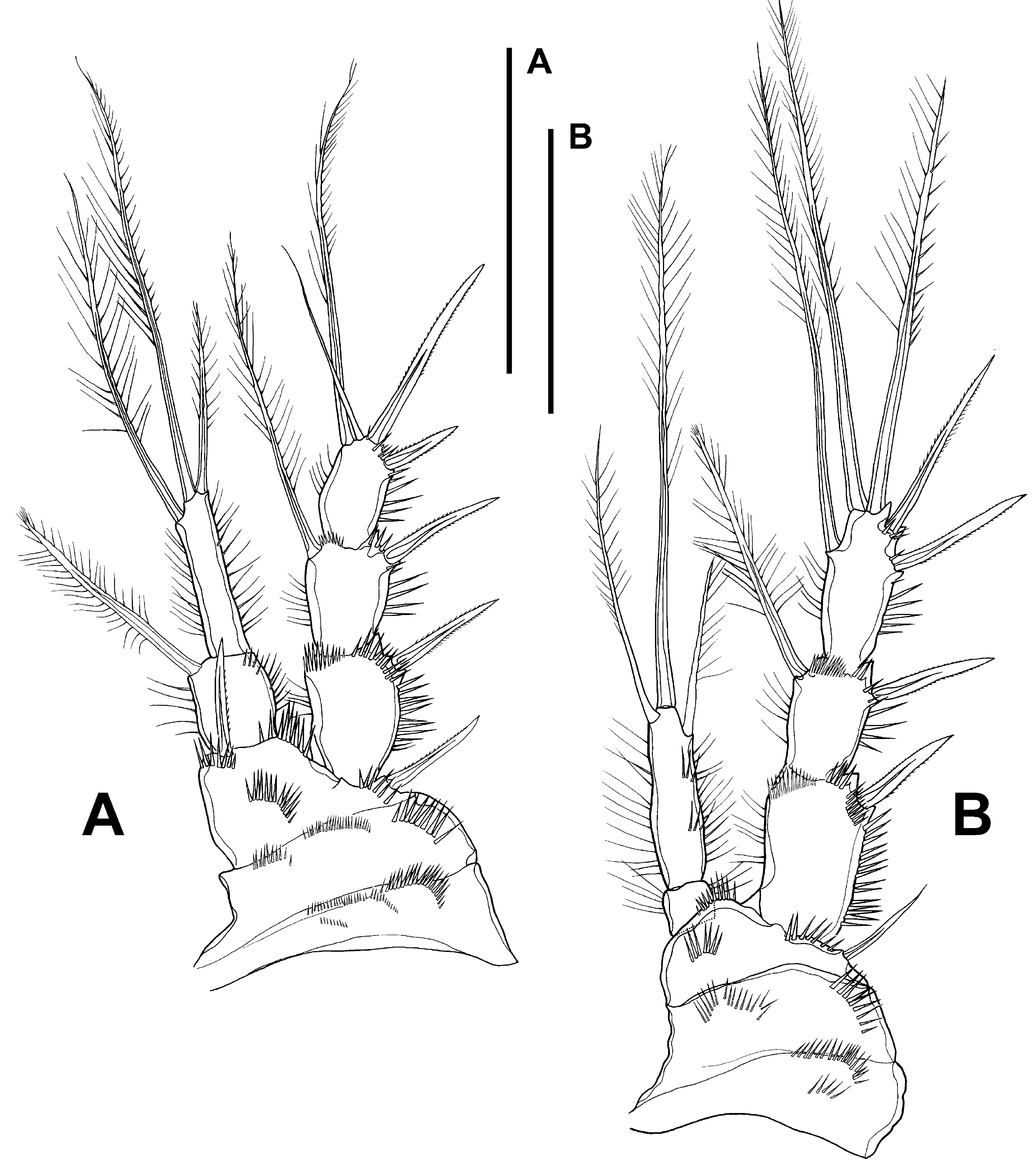
P2 ( Fig. 8 View FIGURE 8 B): praecoxa ornamented as figured; coxa with median row of small spinules and with strong spinules close to outer distal corner. Basis with medial spinules anteriorly, between rami and at base of exopod; outer element spiniform, thin, shorter than those of P3 and P4. Exopod three-segmented and ornamented as shown; EXP1 without inner seta; inner seta of EXP2 relatively short, nearly 0.6 times as long as outer apical seta of EXP3, with brush tip; EXP3 with five elements, of which inner seta 1.1 times as long as outer apical seta, without brush tip. Endopod two segmented, reaching about the middle of EXP2; ENP 1 small, slightly wider than long, with outer and inner spinules, without armature; ENP 2 about 3.7 times as long as ENP 1, with inner and outer long spinules, with two outer transverse rows of spinules, with one inner, one apical and one outer element, the latter shortest, apical seta longest.
P3 ( Fig. 9 View FIGURE 9 A): praecoxa with one row of spinules close to joint with coxa. The latter as in P2. Basis as in P2 except for setiform outer element. Exopod as in P2; inner seta on EXP2 relatively short, nearly 0.6 times as long as outer apical seta of EXP3, with brush tip; inner seta of EXP3 about 1.1 times as long as outer apical seta, without brush tip. Endopod two-segmented, reaching slightly beyond EXP1; first segment nearly as long as wide, with long inner spinules; second segment with long spinules as shown, with two inner elements, two apical setae and one outer element, the two inner setae shortest, apical setae longest.
P4 ( Fig. 9 View FIGURE 9 B): praecoxa, coxa and basis as in P3. Exopod as in P3, except for armature formula (without inner seta in P4EXP3); inner seta on EXP2 about 0.6 times as long as outer apical seta of EXP3, with brush tip. Endopod two-segmented, barely reaching middle of EXP1; ENP 1 small, slightly wider than long; ENP 2 about five times as long as ENP 1, with slender spinules, with two long apical setae, of which inner shorter, reaching tip of EXP3.
P5 ( Fig. 5 View FIGURE 5 C): exopod and baseoendopod fused, barely separated by small notch. Baseoendopod with outer seta of basis; endopodal lobe longer than exopod, with outer small spinules, and long spinules along inner margin, with one outer, two apical and three inner setae, relative length of setae as shown. Exopod with outer spinules, with five setae in all.
P6 ( Fig. 5 View FIGURE 5 A, B): represented by median plate in anterior half of first genital somite, each vestigial leg represented by two small spiniform setae.
Male. Total body length measured from tip of rostrum to posterior margin of caudal rami, ranging from 786 µm to 1033 µm (mean= 899 µm; n= 7). Habitus as in female except for somewhat more clear distinction between prosome and urosome, and for separate second and third urosomites ( Fig. 10 View FIGURE 10 A, B); anal somite ( Figs. 10 View FIGURE 10 A, B, 11B, C) as in female; caudal rami ( Figs. 10 View FIGURE 10 A, B, 11B, C) about 2.8 times as long as wide; caudal seta IV and V 20 % and 61% of total body length, respectively. Ventral ornamentation of third, fourth and fifth urosomites ( Fig. 11 View FIGURE 11 B) denser than in female.
Rostrum ( Fig. 11 View FIGURE 11 A): sexually dimorphic, elongate.
Antennule ( Fig. 12 View FIGURE 12 A–C): six-segmented; subchirocer; smooth, except for the presence of spinules on first segment ( Fig. 12 View FIGURE 12 A1); last segment with two acute teeth ( Fig. 12 View FIGURE 12 A, A5, B, C). Armature formula difficult to define; most probably as follows: 1-(1), 2-(9), 3-(9), 4-(8+[1+ae]), 5-(2), 6-(6+[1+ae]). The armature on the last segment arises from a plate-like swelling, it seems not to be a true segment. The nature of this structure as well as an indepth analysis of the segmentation of the antennule deserves further investigation.
Antenna, mandible, maxillule, maxilla and maxilliped (not shown) as in female.
P1 ( Fig. 13 View FIGURE 13 A) as in female except for dimorphic projection on inner distal corner of basis and for relatively thinner inner spine of basis.
P2 ( Fig. 13 View FIGURE 13 B) proportionally shorter than in female and with relatively stouter outer spines of exopod; armature of ENP 2 relatively shorter than in female.
P3 ( Fig. 14 View FIGURE 14 A, B) as in female except for relatively stouter outer spines, and for longer EXP1. Endopod clearly two-segmented; first segment slightly longer than wide, with long inner spinules, unarmed; second segment with inner distal apophysis very short and bent outwards at an angle of almost 90°, and with two apical setae relatively shorter than in female (innermost shorter); with paired asprothekes (see Discussion for definition) at base of apophysis posteriorly ( Fig. 14 View FIGURE 14 B).
P4 ( Fig. 14 View FIGURE 14 C) as in female, except for stouter outer spines of exopod, relatively longer EXP1, and relatively shorter setae of ENP 2. Two-segmented endopod somewhat shorter than half the length of EXP1; first segment very small, about as long as wide; second segment elongate, about 3 times as long as wide and about 2.5 times as long as ENP 1, with two elements, of which inner shorter, reduced, not reaching tip of EXP1.
Both P5 fused ( Fig. 12 View FIGURE 12 D); exopod and baseoendopod fused; division between rami indicated by slight notch. Exopod with spinules at base of setae of basis; with four elements. Baseoendopod with outer seta of basis; endopodal lobe with outer and inner spinules as shown, with three elements in all.
P6 ( Fig. 11 View FIGURE 11 B) represented by plate; without armature.
Armature formula in Table 1.
Leg P1 P2 P3 P4 P5 Female EXP I-0; I-1;I,I1,1 I-0; I-1;II,2,1 I-0; I-1;II,2,1 I-0; I-1;II,2,0 5 ENP 0-1;0,I1,1 0-0;I,1,1 0-0;I,2,2 0-0;0,2,0 6 Male EXP I-0; I-1;I, I1; 1 I- 0; I-1;II,2,1 I-0; I-1;II,2,1 I-0; I-1;II,2,0 4 ENP 0-1;0,I1,1 0-0;I,1,1 0-0;0,2,Apophysis 0-0;0,2,0 3 Variability. The variability observed between the analyzed populations is shown in Table 2. This variability was expressed in the number of setae on the second segment of the female and male antennule, on the third and sixth segment of the male antennule, in the armature complement of the endopodal lobe of the female and male P5 (see Figs. 5 View FIGURE 5 D, 12E), and in the number of setae on the female P4ENP (see Fig. 9 View FIGURE 9 C).
Canada US
Dead Moose Lake Big Quill Lake Farmington Bay Twelvemile Lake
(Saskatchewan) (Saskatchewan) ( Great Salt Lake , (Albany County,
Utah) Wyoming) Total body Female 694 µm to 705 µm; n= 6 - 798 µm to 907 µm; -
length n= 7
P4 ENP Female - - 2–3 setae -
(see Fig. 6 View FIGURE 6 C)
Male - - - -


No known copyright restrictions apply. See Agosti, D., Egloff, W., 2009. Taxonomic information exchange and copyright: the Plazi approach. BMC Research Notes 2009, 2:53 for further explanation.
|
Kingdom |
|
|
Phylum |
|
|
Class |
|
|
Order |
|
|
Family |
|
|
Genus |
Cletocamptus albuquerquensis ( Herrick, 1894 )
| Gómez, Samuel, Gerber, Ray & Fuentes-Reinés, Juan Manuel 2017 |
Cletocamptus albuquerquensis:
| Lang 1936 |
Wolterstorffia albuquerquensis:
| Brehm 1913 |