
Cletocamptus tainoi, Gómez, Samuel, Gerber, Ray & Fuentes-Reinés, Juan Manuel, 2017
|
publication ID |
https://doi.org/10.11646/zootaxa.4272.3.1 |
|
publication LSID |
lsid:zoobank.org:pub:0ECB3A74-2E13-4713-95FF-D5739035EE09 |
|
DOI |
https://doi.org/10.5281/zenodo.6002072 |
|
persistent identifier |
https://treatment.plazi.org/id/AF5D87B3-4A28-FFEE-AF8C-18B4C672F886 |
|
treatment provided by |
Plazi |
|
scientific name |
Cletocamptus tainoi |
| status |
sp. nov. |
Cletocamptus tainoi sp. nov.
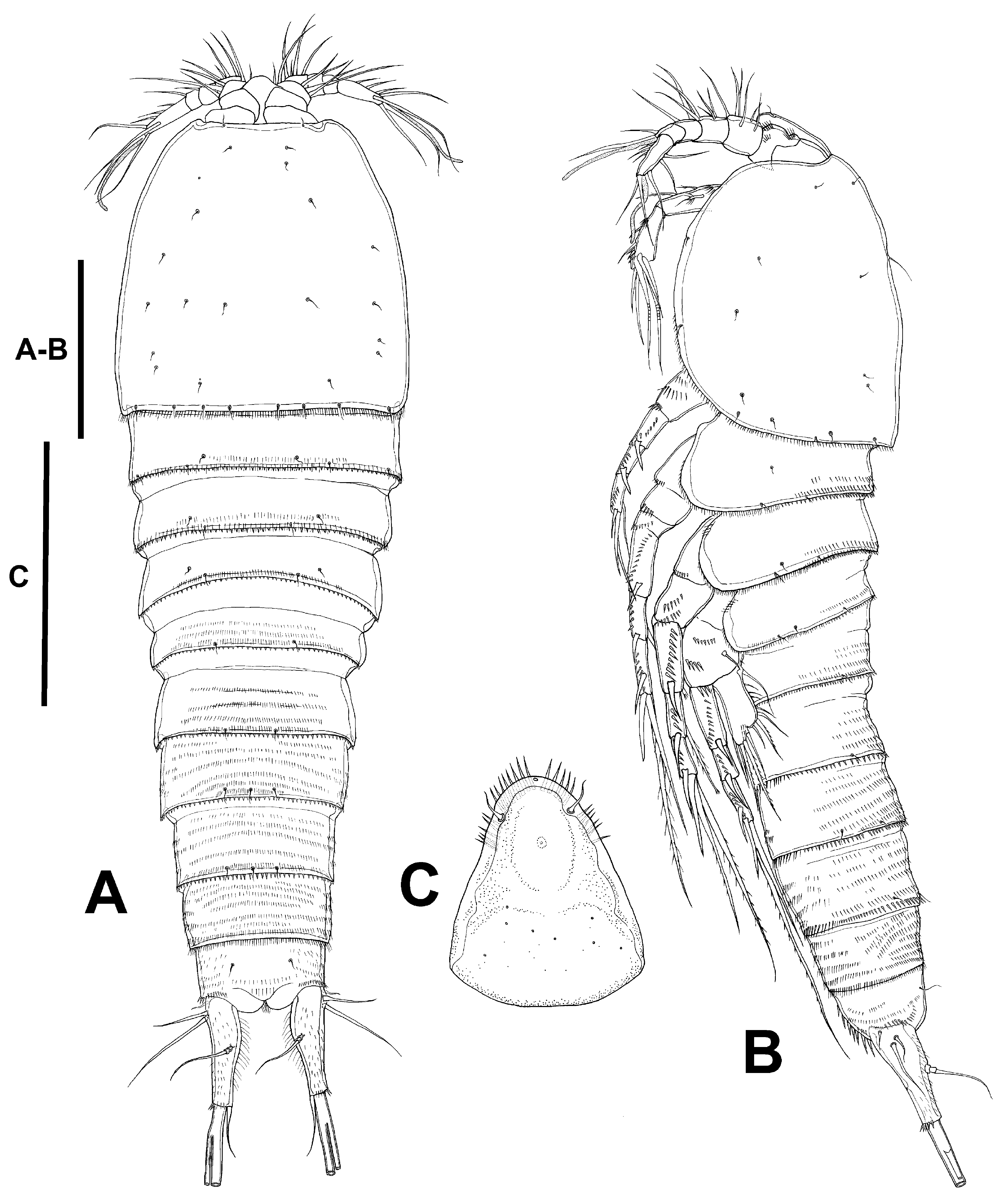
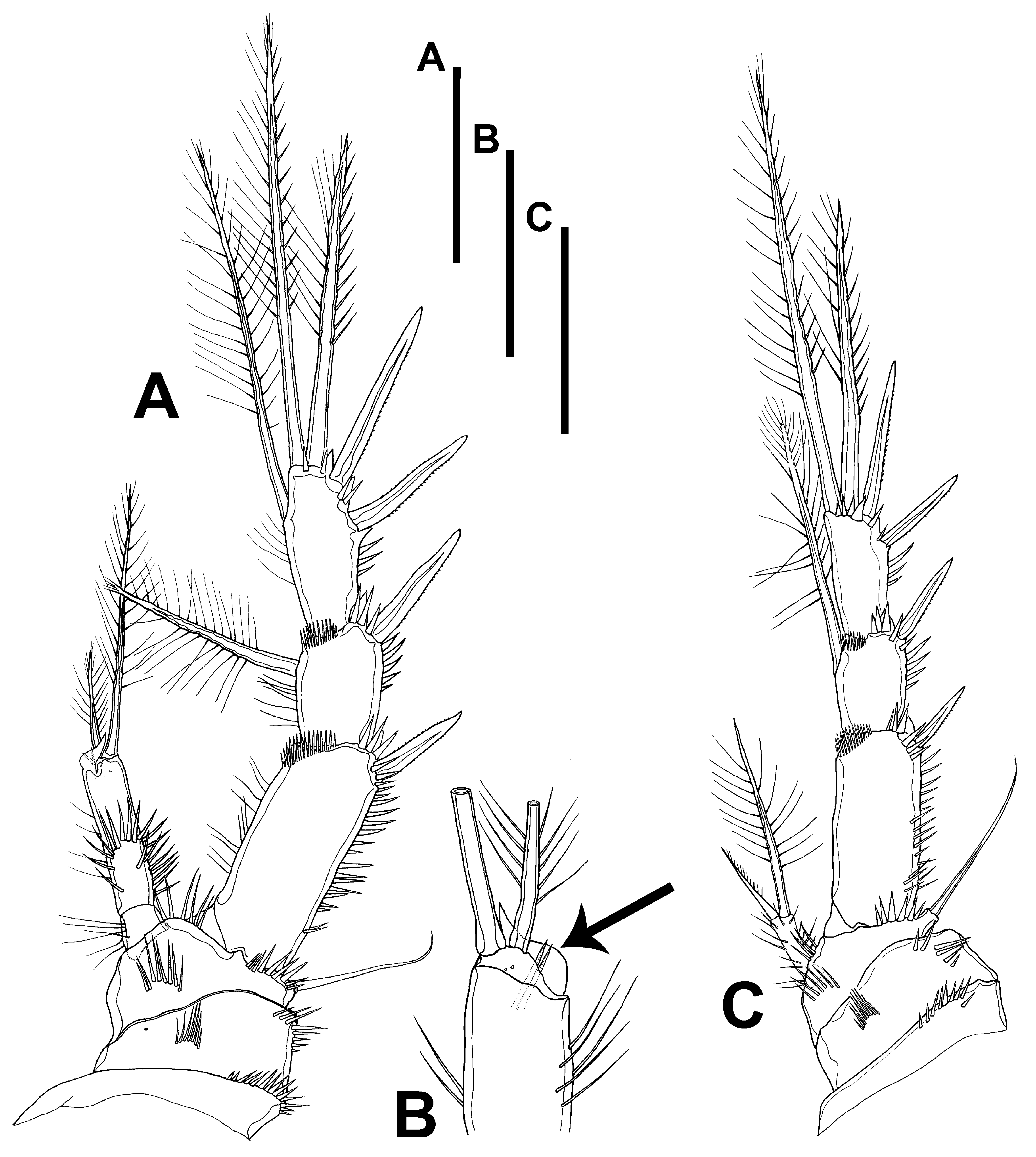
( Figs. 27–37 View FIGURE 27 View FIGURE 28 View FIGURE 29 View FIGURE 30 View FIGURE 31 View FIGURE 32 View FIGURE 33 View FIGURE 34 View FIGURE 35 View FIGURE 36 View FIGURE 37 )
Etymology. The species is named in honor and memory of the Taíno people who were the original inhabitants of St. John Island (as early as 880 BC) and nearby Caribbean islands ( Wild 1999).
Material examined. One female holotype ( USNM No. 1418184), one male allotype ( USNM No. 1418185), and four female and four male paratypes ( USNM No. 1418186) preserved in alcohol; 6 January, 2007; col. Ray Gerber .
Type locality. A small (0.36 ha) and shallow (less than 0.5 m depth) salt pond located near the shore of Privateer Bay , on the east end of St. John Island, US Virgin Islands (18˚20’16.96” N and 64˚39’58.40” W).
Distribution. US Virgin Islands: Near Privateer Bay on St. John Island (present study).
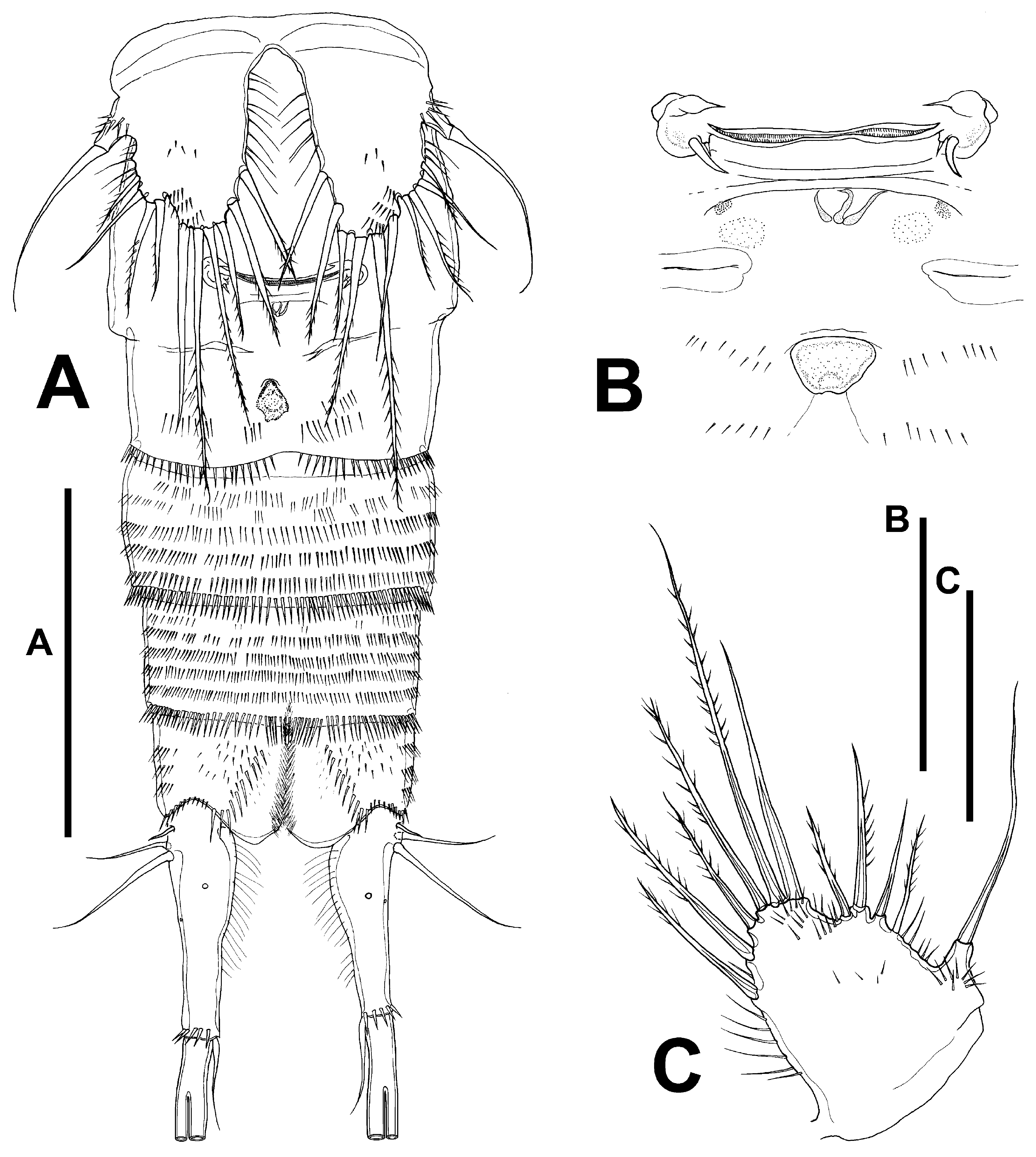
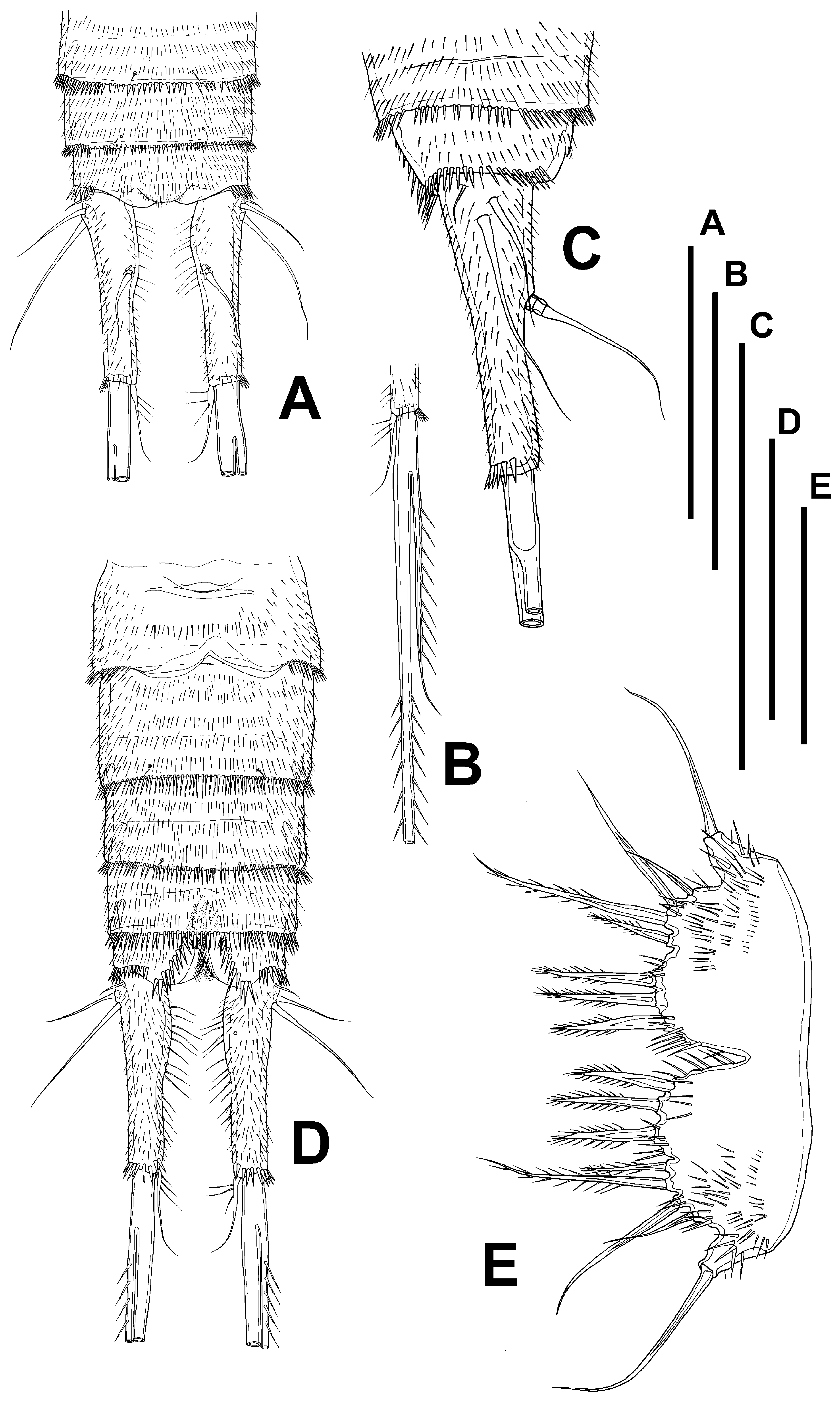
Description. Female. Habitus ( Fig. 27 View FIGURE 27 A, B) tapering posteriorly; total body length measured from tip of rostrum to posterior margin of caudal rami ranging from 526 µm to 610 µm (mean= 568 µm; n= 8). Rostrum set off, triangular, with pair of setules subapically and with row of spinules distally ( Fig. 27 View FIGURE 27 C). Cephalic shield with fine and short spinules along margin dorsolaterally ( Fig. 27 View FIGURE 27 A, B). Dorsal and lateral surface of free thoracic somites (P2–P4 bearing-somites) with transverse rows of minute spinules as shown, with short spinules along posterior margin. Dorsal and lateral surface of first urosomite (P5 bearing-somite) ( Fig. 27 View FIGURE 27 A, B) with transverse rows of minute spinules, with row of small spinules along posterior margin. Second and third urosomites distinct dorsally and laterally ( Fig. 27 View FIGURE 27 A, B), completely fused ventrally forming genital double-somite ( Fig. 28 View FIGURE 28 A); dorsal and lateral surface of first and second half of genital double-somite with transverse rows of minute spinules, with row of larger spinules along posterior margin; ventrally with longer spinules as shown ( Fig. 28 View FIGURE 28 A). Fourth and fifth urosomites as in second half of genital double-somite dorsally ( Fig. 27 View FIGURE 27 A), ventral surface with transverse rows of minute spinules, with larger spinules along posterior margin ( Fig. 28 View FIGURE 28 A). Dorsal surface of anal somite ( Fig. 27 View FIGURE 27 A) with transverse rows of minute spinules and with dorsolateral strong spinules close to joint with caudal rami; anal operculum crescentic medially, with spinules as shown ( Fig. 27 View FIGURE 27 A). Caudal rami ( Figs. 27 View FIGURE 27 A, B, 28A) about 3.6 times as long as wide; dorsal surface with sparse small spinules dorsally, smooth ventrally; with ventral row of larger spinules close to insertion of caudal setae distally; with seven elements in all ( Fig. 27 View FIGURE 27 A, B, 28A); seta I very small, situated proximally on lateral surface of ramus, close to setae II and III, the latter setae longer; setae IV and V fused basally, 16% and 49% of total body length, respectively; seta VI situated on distal inner corner; seta VII situated dorsally midway length of ramus on inner edge.
Antennule ( Fig. 29 View FIGURE 29 A) six-segmented; surface of segments smooth except for two rows of spinules on first and third segments. Armature formula, 1-(1), 2-(9), 3-(6), 4-(1+[1+ae]), 5-(1), 6-(9+[1+ae]).
Antenna ( Fig. 29 View FIGURE 29 B, C) with small coxa. Allobasis with two abexopodal setae. Free endopodal segment with small inner spinules proximally, with stronger spinules subdistally; with two lateral inner spines and a slender seta, and five distal elements, two of them geniculate. Exopod elongate, one-segmented, with few spinules, with two lateral and one apical seta (the two lateral setae are somewhat rigid and could actually be spinules (see Fig. 29 View FIGURE 29 C)).
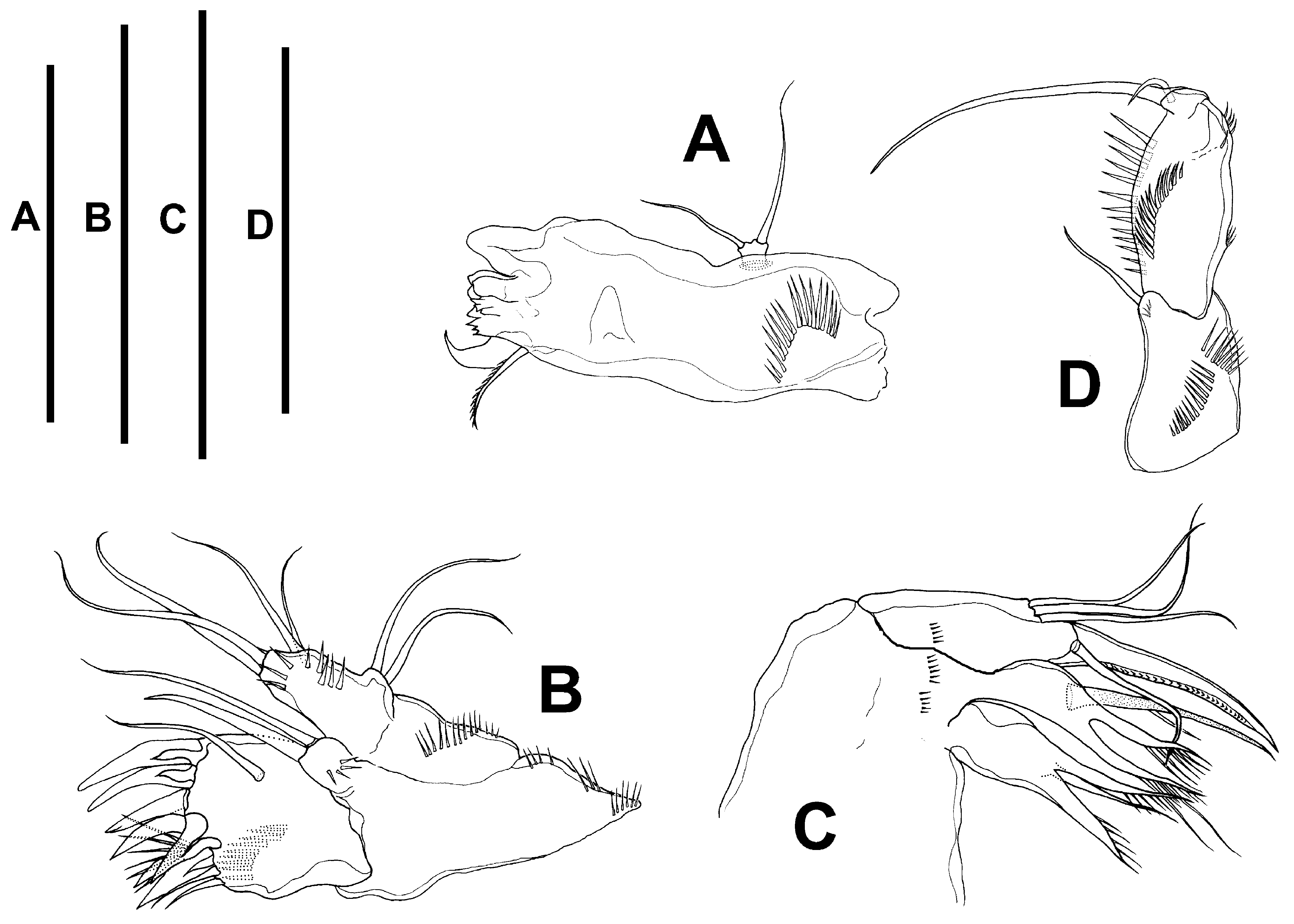
Mandible ( Fig. 30 View FIGURE 30 A) robust, with rows of spinules proximally; chewing edge with teeth as figured, with one pyriform element and one lateral pinnate seta. Palp one-segmented, with two long setae unequal in length.
Maxillule ( Fig. 30 View FIGURE 30 B) robust; arthrite of praecoxa with few spinules, with one surface seta, and seven spines and two slender setae distally. Coxa with some spinules, with two slender setae. Basis with spinules as figured, with two apical setae. Exopod and endopod incorporated to basis, represented by two setae each.
Maxilla ( Fig. 30 View FIGURE 30 C): syncoxa with spinules as shown; with two endites, each bearing three setae. Allobasis drawn into strong claw with one accompanying seta. Endopod represented by three setae.
Maxilliped ( Fig. 30 View FIGURE 30 D) subchelate. Syncoxa with row of spinules, with one seta on inner distal corner. Basis without armature, with spinules as shown. Endopod drawn into long and slender claw with one accompanying small seta.
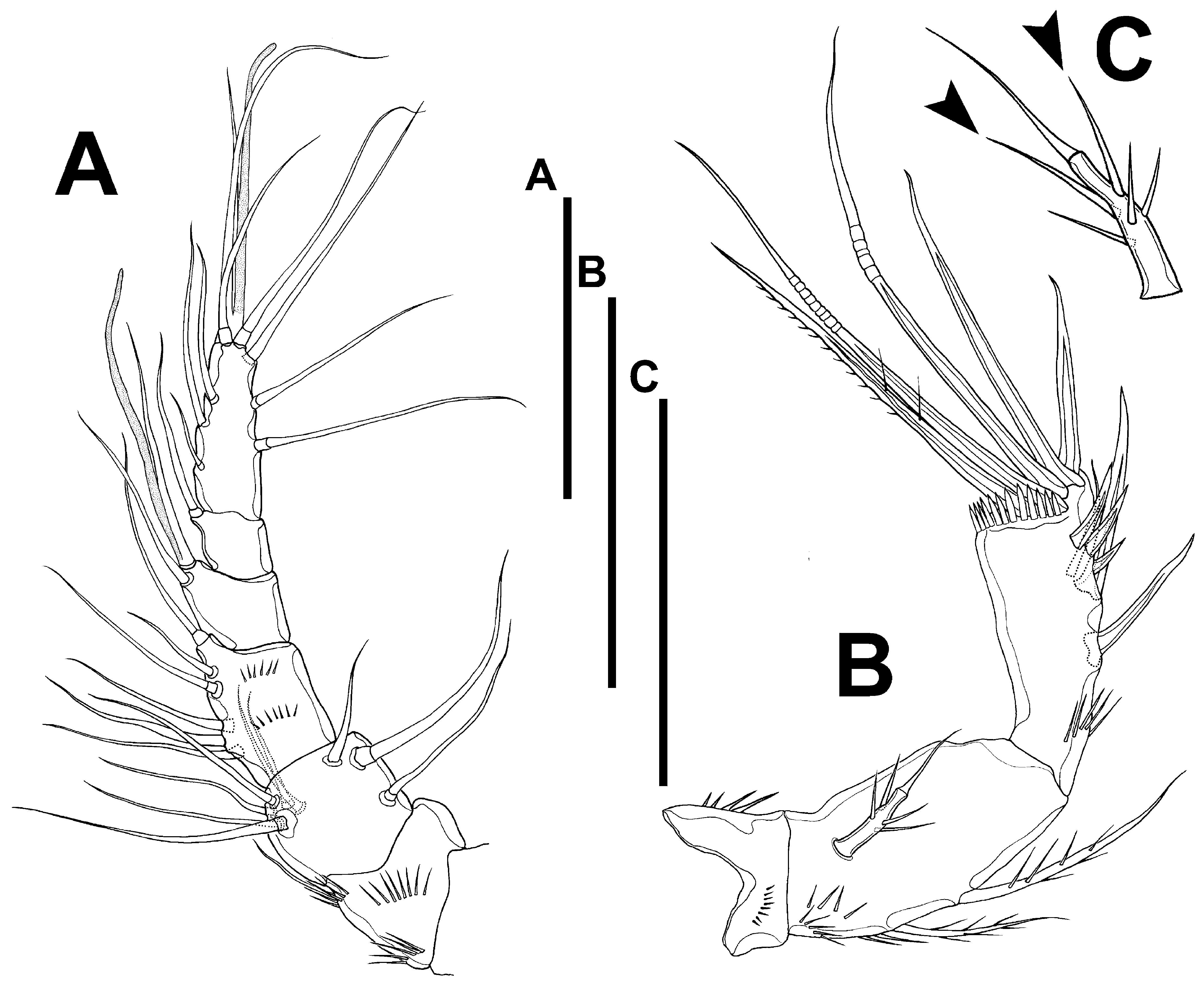
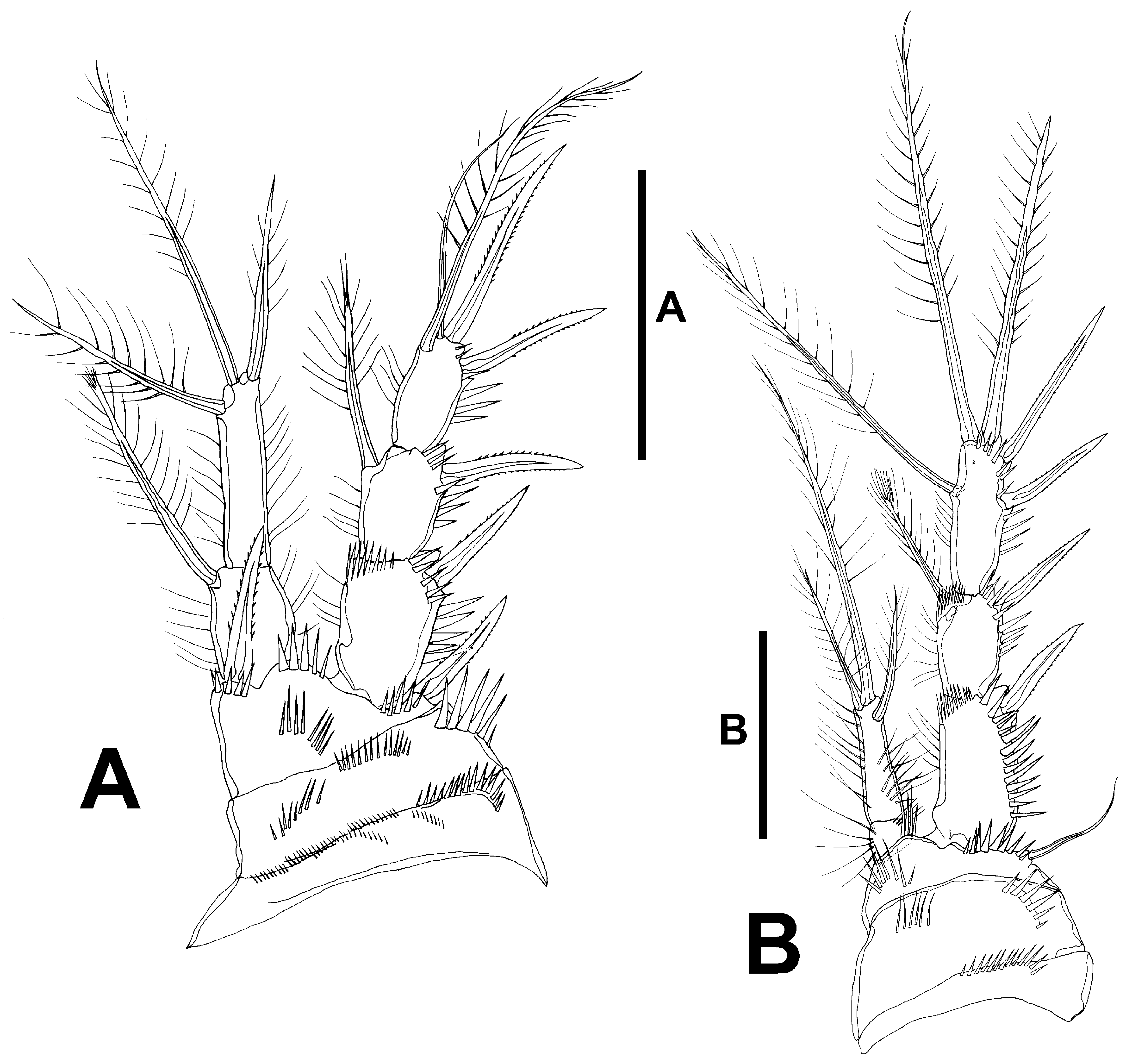
P1 ( Fig. 31 View FIGURE 31 A): praecoxa with spinules close to joint with coxa. The latter with anterior transverse rows of spinules and outer row of strong spinules. Basis with inner and outer spine; with median rows of spinules, with stronger spinules at base of exopod, between rami and at base of inner and outer spines. Exopod three-segmented, slightly longer than endopod; EXP1 without, EXP2 with inner seta; EXP3 with four elements. Endopod twosegmented, reaching proximal third of EXP3; ENP1 about 1.4 times as long as wide, barely reaching tip of EXP1, inner seta shorter than both endopodal segments combined, with brush tip; ENP2 elongate, about 4.6 times as long as wide, with three elements.
P2 ( Fig. 31 View FIGURE 31 B): praecoxa and coxa ornamented as figured. Basis with spinules between rami and medially close to base of endopod, and with stronger spinules at base of exopod; outer element setiform. Exopod three-segmented and ornamented as shown; EXP1 without inner seta; inner seta of EXP2 about 0.4 times as long as outer apical seta of EXP3, with brush tip; EXP3 with five elements, of which inner seta about 1.1 times as long as outer apical seta, without brush tip. Endopod two segmented, reaching distal margin of EXP1; ENP1 small, slightly wider than long, with outer and inner spinules; ENP2 elongate, about 3.5 times as long as wide, with long spinules as shown, with one outer spine-like and two apical elements; outer element shortest, apical seta longest reaching beyond EXP3.
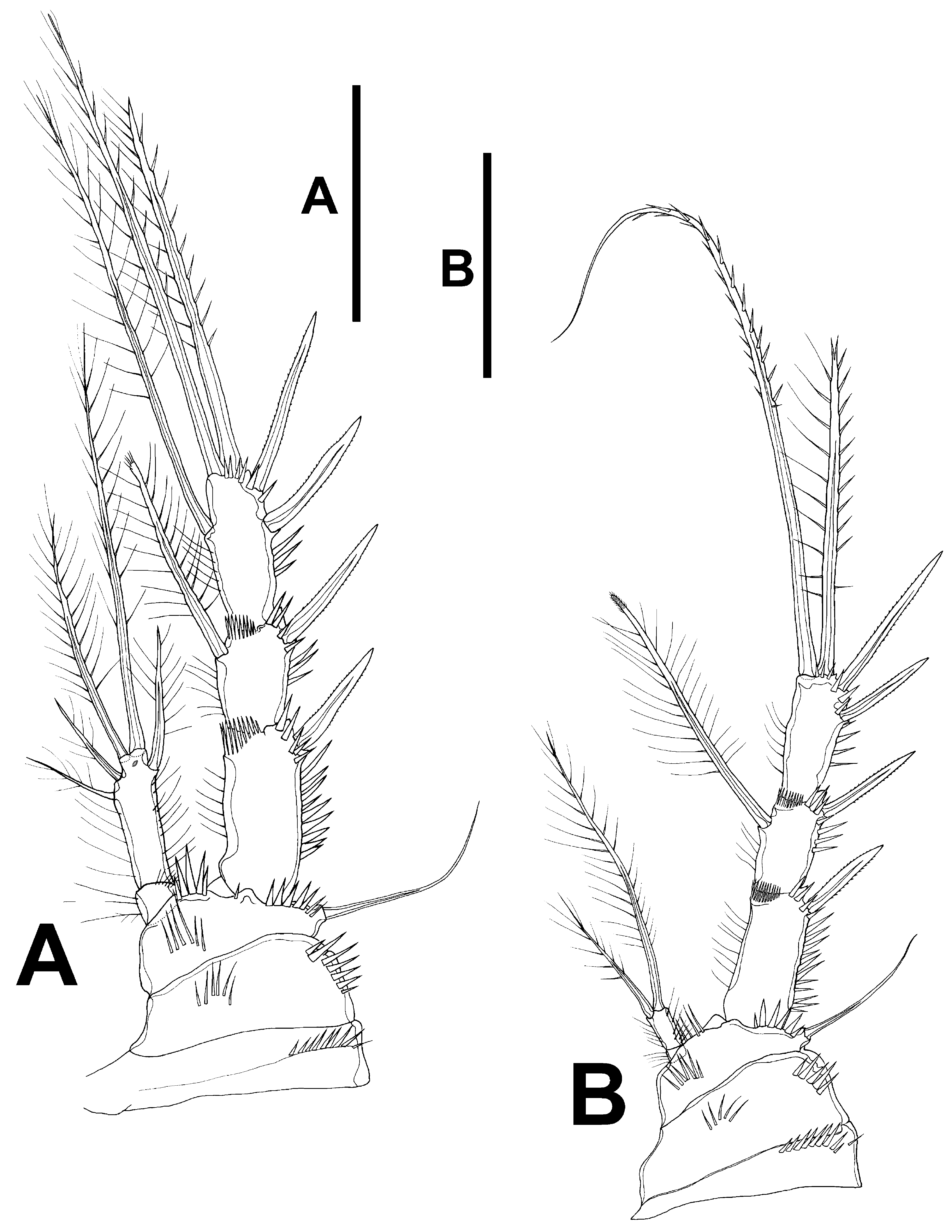
P3 ( Fig. 32 View FIGURE 32 A): praecoxa and coxa as in P2; basis with outer seta. Exopod as in P2; EXP1 without inner armature; inner seta of EXP2 about 0.5 times as long as outer apical seta of EXP3, with brush tip; EXP3 with five elements, of which inner seta about 1.1 times as long as outer apical seta, without brush tip. Endopod twosegmented, barely reaching tip of EXP1; ENP1 nearly as long as wide, with long spinules as shown; ENP2 elongate, about 3.3 times as long as wide, with two inner and two apical setae, and one outer spine-like element; inner setae shortest, apical setae longest (outer apical seta reaching beyond, inner apical seta barely reaching tip of EXP3).
P4 ( Fig. 32 View FIGURE 32 B): praecoxa, coxa and basis as in P3. Exopod as in P3, except for armature formula of EXP3 (without inner seta); EXP1 without inner armature; inner seta of EXP2 slightly shorter than outer apical seta of EXP3, with brush tip; EXP3 with four elements. Endopod very small, two-segmented; ENP1 minute, about as long as wide; ENP2 elongate, about twice as long as wide, with slender spinules, with two apical setae, of which inner shorter, outer reaching middle of EXP3.
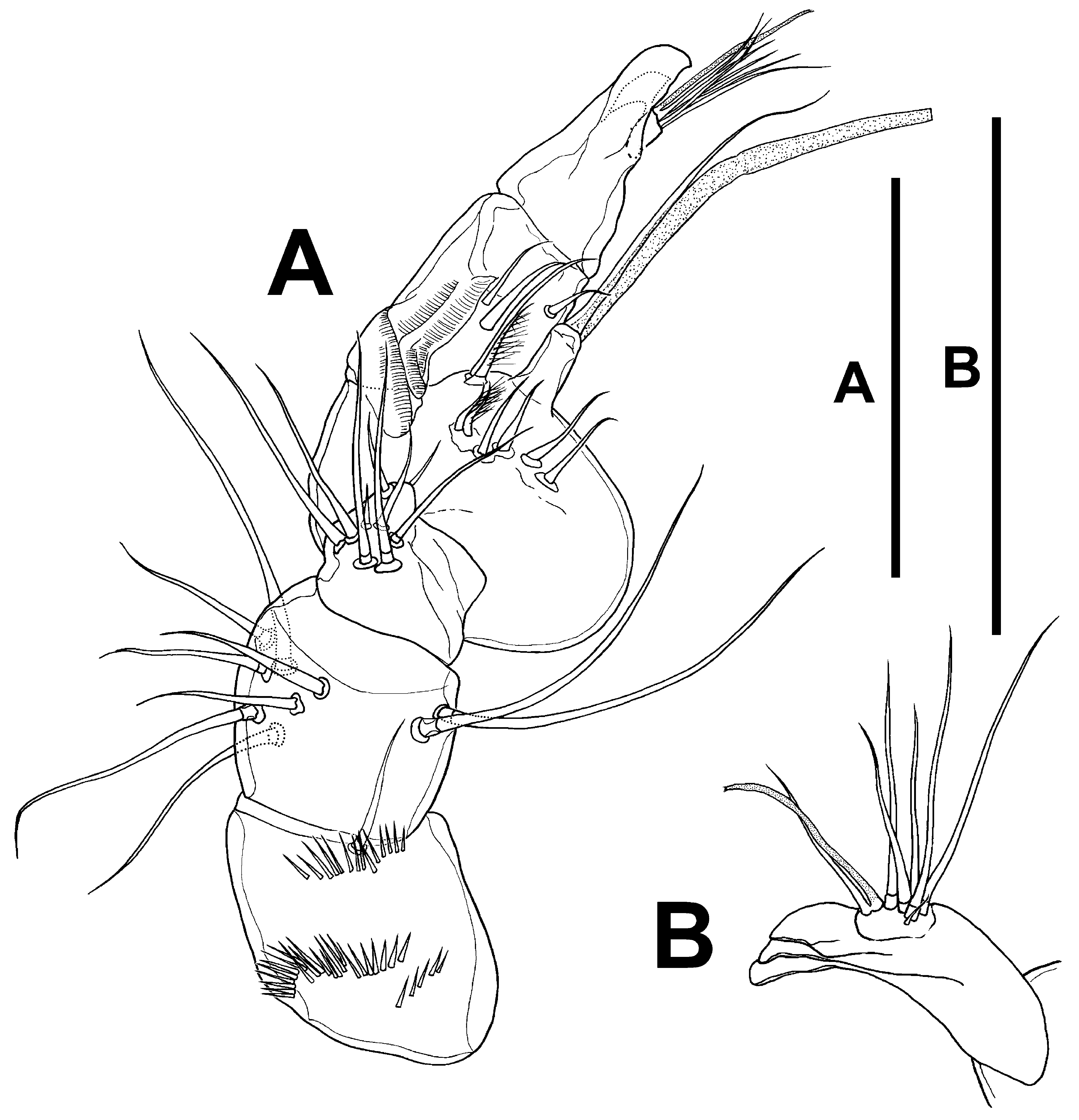
P5 ( Fig. 28 View FIGURE 28 A, C): exopod and baseoendopod fused, barely separated by gap between rami. Baseoendopod with outer seta of basis; endopodal lobe longer than exopod, with spinules as figured, with two inner and four apical setae; relative length of setae as shown. Exopod with outer spinules, with four setae in all.
P6 ( Fig. 28 View FIGURE 28 B) represented by median plate, each vestigial leg represented by two small setae. Copulatory pore in the middle of genital somite.
Male. Total body length measured from tip of rostrum to posterior margin of caudal rami, ranging from 486 µm to 628 µm (mean= 556 µm; n= 8). Habitus as in female except for clearer distinction between prosome and urosome, and for separate second and third urosomites ( Fig. 33 View FIGURE 33 A, B); anal somite ( Figs. 33 View FIGURE 33 A, B, 34A, C, D) as in female except for denser spinular ornamentation in the male; caudal rami as in female; caudal setae IV and V as in female, except for length relative to total body length of 17% and 61%, respectively. Ventral ornamentation of third, fourth and fifth urosomites ( Fig. 34 View FIGURE 34 D) denser than in female.
Rostrum ( Fig. 33 View FIGURE 33 C): sexually dimorphic, elongate.
Antennule ( Fig. 35 View FIGURE 35 A, B): six-segmented; subchirocer; last segment with two acute teeth ( Fig. 35 View FIGURE 35 B). Armature formula difficult to define; most probably as follows: 1-(1), 2-(9), 3-(9), 4-(8+[1+ae]), 5-(3), 6-(6+[1+ae]). The armature on the last segment arises from a plate-like swelling, it seems not to be a true segment. The nature of this structure as well as an in-depth analysis of the segmentation of the antennule deserves further investigation.
Antenna, mandible, maxillule, maxilla and maxilliped (not shown) as in female.
P1 ( Fig. 36 View FIGURE 36 A): as in female except for dimorphic projection on inner distal corner of basis.
P2 ( Fig. 36 View FIGURE 36 B) as in female except for relatively stouter outer spines of the exopod, for relatively longer EXP1, and for relatively shorter setae of ENP 2 in the male, of which apical seta barely reaching tip of EXP3.
P3 ( Fig. 37 View FIGURE 37 A, B): as in female except for relatively stouter outer spines, and for longer EXP1. Endopod clearly two-segmented; ENP1 nearly as long as wide, with long inner spinules, unarmed; ENP2 with inner distal apophysis bent outwards, very short, with two apical setae relatively shorter than in female (outer apical seta barely reaching tip of EXP3, inner apical seta barely reaching middle of EXP2); with paired asprothekes ( Fig. 37 View FIGURE 37 B).
P4 ( Fig. 37 View FIGURE 37 C) as in female, except for stouter outer spines of exopod, relatively longer EXP1, and relatively shorter setae of ENP2 (outer seta reaching slightly beyond EXP1).
Both P5 fused ( Fig. 34 View FIGURE 34 E); exopod and baseoendopod fused; division between rami indicated by slight notch. Exopod with spinules as shown; with four elements. Baseoendopod with outer seta of basis; endopodal lobe with outer and inner spinules as shown; with three elements in all.
P6 ( Fig. 34 View FIGURE 34 D) represented by plate; without armature. Armature formula in Table 4.
Leg P1 P2 P3 P4 P5 Female EXP I-0; I-1;I,I1,1 I-0; I-1;II,2,1 I-0; I-1;II,2,1 I-0; I-1;II,2,0 4 ENP 0-1;0,I1,1 0-0;I,2,0 0-0;I,2,2 0,0;0,2,0 6 Male EXP I-0; I-1;I,I1,1 I-0; I-1;II,2,1 I-0; I-1;II,2,1 I-0; I-1;II,2,0 4 ENP 0-1;0,I1,1 0-0;I,2,0 0-0;0,2,Apophysis 0,0;0,2,0 3 Variability. The number of spinules/setae on the antennary exopod can vary from four to five in females and males. The number of seta on the second and last antennular segment of the female can vary from 8 to 9. The number of setae on the fourth antennular segment of the male can vary from 7 to 8.


| USNM |
Smithsonian Institution, National Museum of Natural History |
No known copyright restrictions apply. See Agosti, D., Egloff, W., 2009. Taxonomic information exchange and copyright: the Plazi approach. BMC Research Notes 2009, 2:53 for further explanation.
|
Kingdom |
|
|
Phylum |
|
|
Class |
|
|
Order |
|
|
Family |
|
|
Genus |