
Obama tribalis, Marques & Rossi & Valiati & Leal-Zanchet, 2018
|
publication ID |
https://doi.org/10.11646/zootaxa.4455.1.4 |
|
publication LSID |
lsid:zoobank.org:pub:92D6B362-047F-4BA0-91F1-4CF3F1D104A6 |
|
DOI |
https://doi.org/10.5281/zenodo.5959077 |
|
persistent identifier |
https://treatment.plazi.org/id/74A2CF86-A564-4630-8E72-74339F68508D |
|
taxon LSID |
lsid:zoobank.org:act:74A2CF86-A564-4630-8E72-74339F68508D |
|
treatment provided by |
Plazi |
|
scientific name |
Obama tribalis |
| status |
sp. nov. |
Obama tribalis sp. nov.
Etymology: The specific name, frOm latin tribālis (tribal), refers tO the dOrsal pigmentatiOn which resembles the crOssed stripes pattern Of the tribal style drawings.
Type-material. Holotype: MZUSP PL.2140: leg. E. FederOlf, 30 June 2013, Gravataí, state Of RiO Grande dO Sul, Brazil—anteriOr tip: transverse sectiOns On 7 slides; anteriOr regiOn at the level Of the Ovaries : sagittal sectiOns On 42 slides; pre-pharyngeal regiOn: transverse sectiOns On 26 slides; cOpulatOry apparatus: sagittal sectiOns On 43 slides.
Paratypes: MZU PL.00287: leg. E. FederOlf, 30 June 2013, Gravataí, state Of RiO Grande dO Sul, Brazil—anteriOr tip: transverse sectiOns On 13 slides; anteriOr regiOn at the level Of the Ovaries: sagittal sectiOns On 10 slides; pre-pharyngeal regiOn: transverse sectiOns On 4 slides; pharynx and cOpulatOry apparatus: sagittal sectiOns On 23 slides. MZU PL.00288: leg. E. FederOlf, 30 June 2013, Gravataí, state Of RiO Grande dO Sul, Brazil—cOpulatOry apparatus: hOrizOntal sectiOns On 12 slides.
Diagnosis: species Of Obama with dOrsal grOund cOlOur yellOwish cOvered by irregular flecks, mOre cOncentrated laterally; pharynx cylindrical; prOstatic vesicle with twO pOrtiOns, prOximal pOrtiOn fOrked and glObOse and distal pOrtiOn funnel-shaped; penis papilla lOng, cOnical and symmetrical; male and female atria with slightly fOlded walls, and with ample cOmmunicatiOn.
Molecular diagnosis: this species includes all pOpulatiOns that cluster with sequences Of specimens included in this study (GenBank accessiOn numbers MH378885 View Materials – MH378886 View Materials ) with significant suppOrt in an adequate mOlecular delimitatiOn mOdel.
Type-locality. Gravataí, RiO Grande dO Sul (RS), Brazil.
Distribution: knOwn Only frOm the type-lOcality, Gravataí, RS, Brazil.
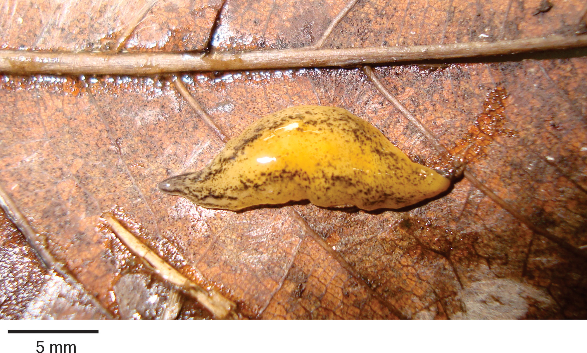
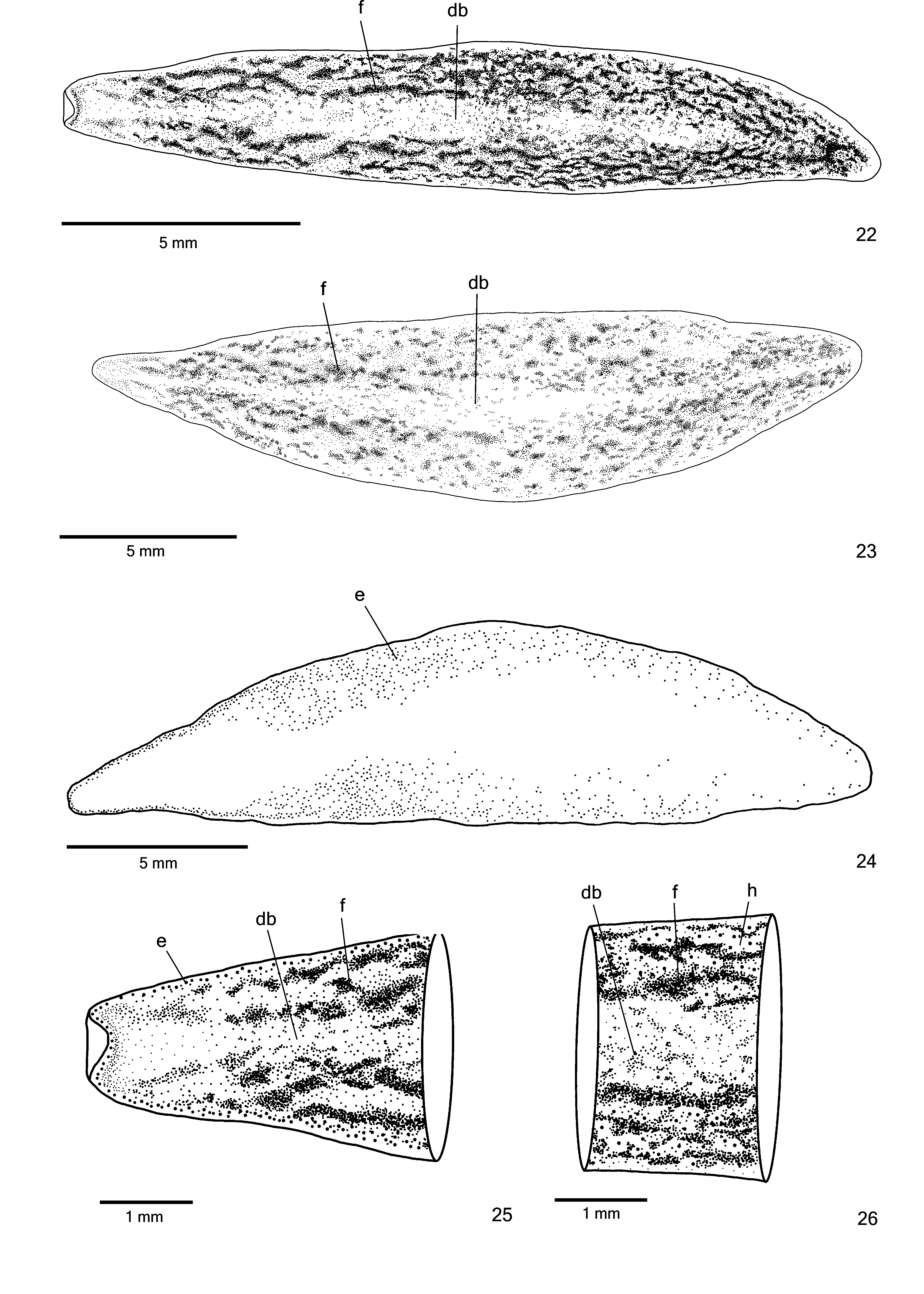
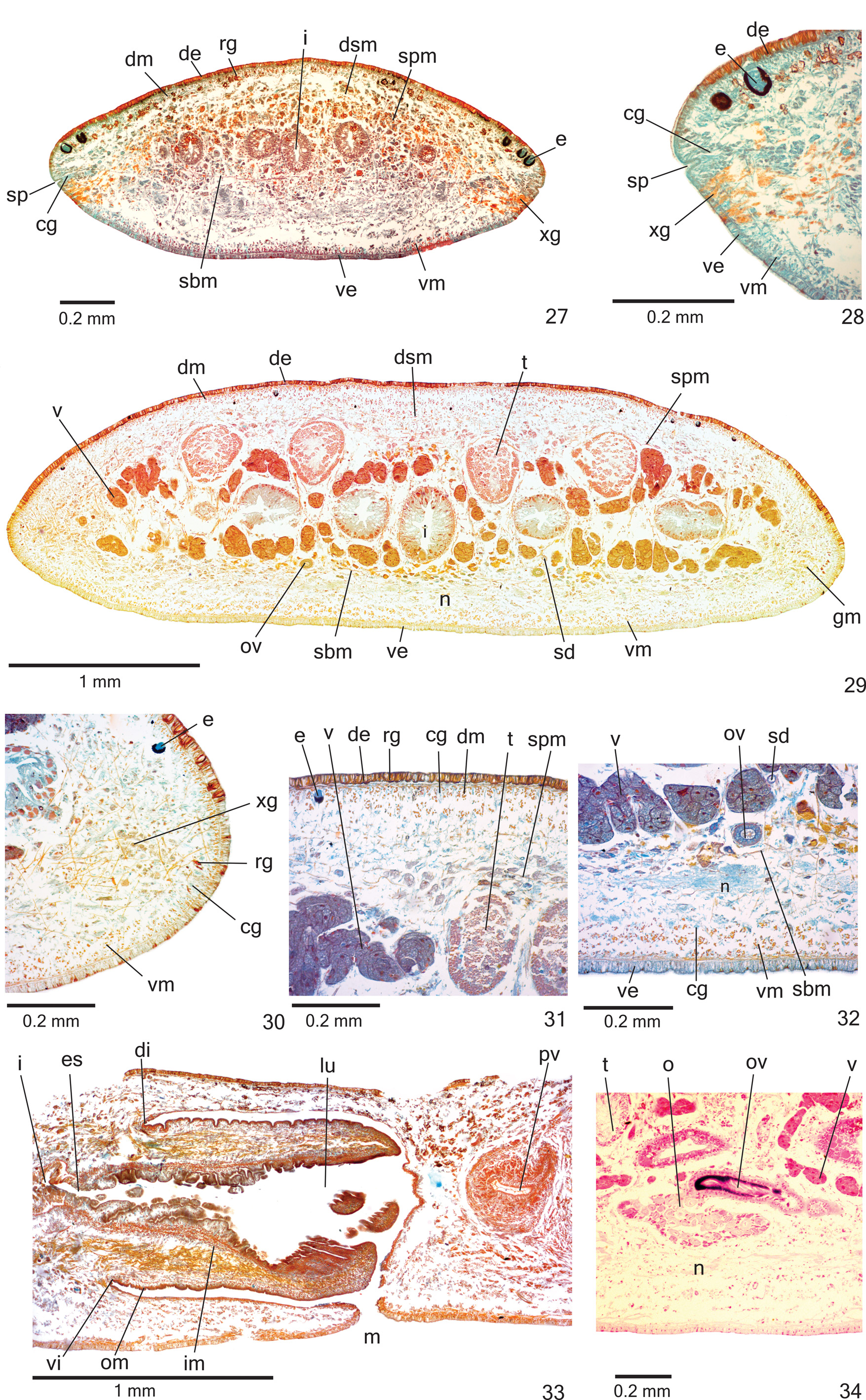
Description. External features. BOdy fOliaceOus and dOrsal surface slightly cOnvex; anteriOr and pOsteriOr tips rOunded ( Fig. 21 View FIGURE 21 ). When creeping, maximum length 33 mm. After fixatiOn, maximum length 23 mm. MOuth at third-fOurth Of bOdy and gOnOpOre at pOsteriOr fOurth Of bOdy, in mOst specimens ( Table 3).
Live specimens with dOrsal grOund cOlOur yellOwish, cOvered by greyish pigmentatiOn in cephalic regiOn ( Fig. 21 View FIGURE 21 ). Blackish pigmentatiOn cOntOurs cephalic regiOn and cOnstitutes irregular flecks Over dOrsal surface, mOre cOncentrated laterally, sOmetimes fOrming a pattern that resembles crOssed stripes at sOme pOints ( Figs. 21–23 View FIGURE 21 View FIGURES 22–26 ). Median regiOn Of dOrsal surface almOst withOut flecks. Paratype MZU PL. 0 0 287 with mOre abundant Overlaying black pigmentatiOn than hOlOtype ( Fig. 22 View FIGURES 22–26 ); paratype MZU PL. 0 0 288 with very few irregular flecks Over dOrsal surface ( Fig. 21 View FIGURE 21 ). Ventral surface greyish with yellOwish margins. After fixatiOn, dOrsal grOund cOlOur fades.
Eyes, initially mOnOlObate and uniserial, surrOund anteriOr tip ( Figs. 24–25 View FIGURES 22–26 ). After first millimetre Of bOdy, eyes becOme larger and spread OntO dOrsal surface, Occupying maximum width Of abOut One-third Of bOdy width On either side Of bOdy. Eyes becOme trilObate after One-fifth Of bOdy length and remain dOrsal, but less numerOus tOwards pOsteriOr tip ( Fig. 24 View FIGURES 22–26 ). IncOnspicuOus clear halOs may Occur arOund dOrsal eyes ( Fig. 26 View FIGURES 22–26 ). Diameter Of pigment cups abOut 30 µm.
Sensory organs, epidermis and body musculature. SensOry pits ( Figs. 27–28 View FIGURES 27–34 ), as simple invaginatiOns (20–40 µm deep), cOntOur anteriOr tip and Occur ventrOmarginally in irregular, single rOw in anteriOr third Of bOdy.
Glands Of three types discharge thrOugh whOle epidermis Of pre-pharyngeal regiOn: rhabditOgen cells with xanthOphil secretiOn (scarcer ventrally and with smaller rhabdites), cyanOphil glands with finely granular secretiOn ( Figs. 31–32 View FIGURES 27–34 ) and sparser xanthOphil glands with finely granular secretiOn. Creeping sOle Occupies whOle bOdy width. Glands Of at least fOur types cOnstitute the incOnspicuOus glandular margin ( Figs. 29, 30 View FIGURES 27–34 ), which is nOticeable after first millimetre Of bOdy: numerOus xanthOphil glands and sparser cyanOphil glands, bOth with cOarse granular secretiOns, besides xanthOphil and cyanOphil glands with fine granules. Glands discharging thrOugh anteriOr tip Of bOdy similar tO thOse Of pre-pharyngeal regiOn ( Figs. 27–28 View FIGURES 27–34 ).
CutaneOus musculature with usual three layers (circular, Oblique and lOngitudinal layers); lOngitudinal layer with thick bundles ( Figs. 29–32 View FIGURES 27–34 ). Thickness Of cutaneOus musculature twO times that Of epidermis ( Table 4). Ventral musculature slightly thicker than dOrsal at sagittal plane in pre-pharyngeal regiOn ( Table 4). Thickness Of cutaneOus musculature gradually diminishes tOwards bOdy margins and anteriOr tip.
Mesenchymal musculature ( Figs. 29, 31–32 View FIGURES 27–34 ) well develOped, mainly cOmpOsed Of three layers: (1) dOrsal subcutaneOus, with decussate fibres (abOut 5–7 fibres thick), (2) supra-intestinal transverse (abOut 7–9 fibres thick) and (3) sub-intestinal transverse (abOut 7–13 fibres thick), besides fibres in variOus directiOns. Mesenchymal musculature less develOped in anteriOr regiOn than in pre-pharyngeal regiOn ( Fig. 27 View FIGURES 27–34 ).
Pharynx. Pharynx cylindrical, nearly 7% Of bOdy length, Occupies mOst Of the pharyngeal pOuch ( Fig. 33 View FIGURES 27–34 ). Pharyngeal insertiOns almOst at same transversal level. MOuth lOcated clOse tO end Of pharyngeal pOuch ( Fig. 33 View FIGURES 27–34 ). OesOphagus shOrt, with fOlded walls. OesOphagus: pharynx ratiO 11–13%.
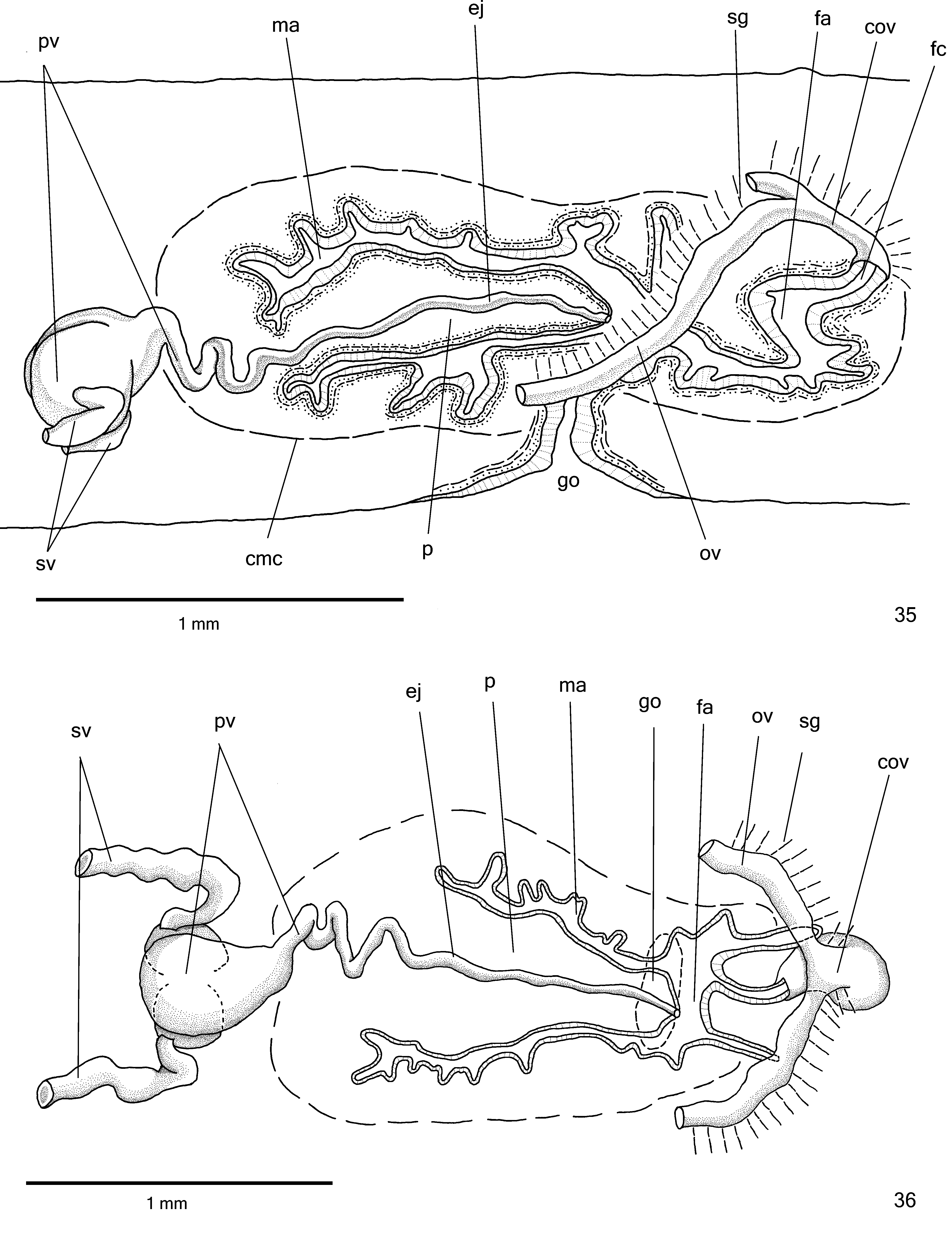
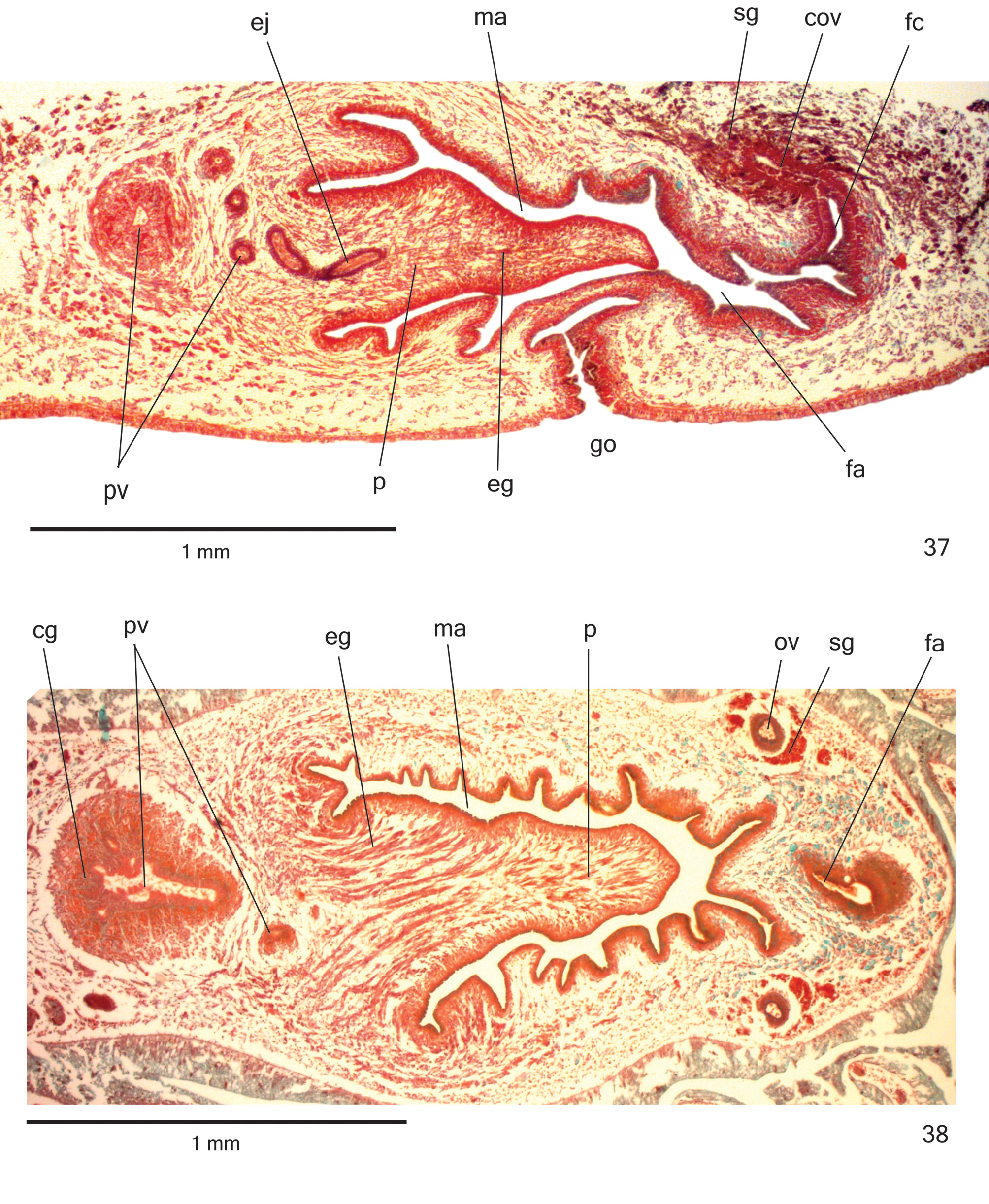
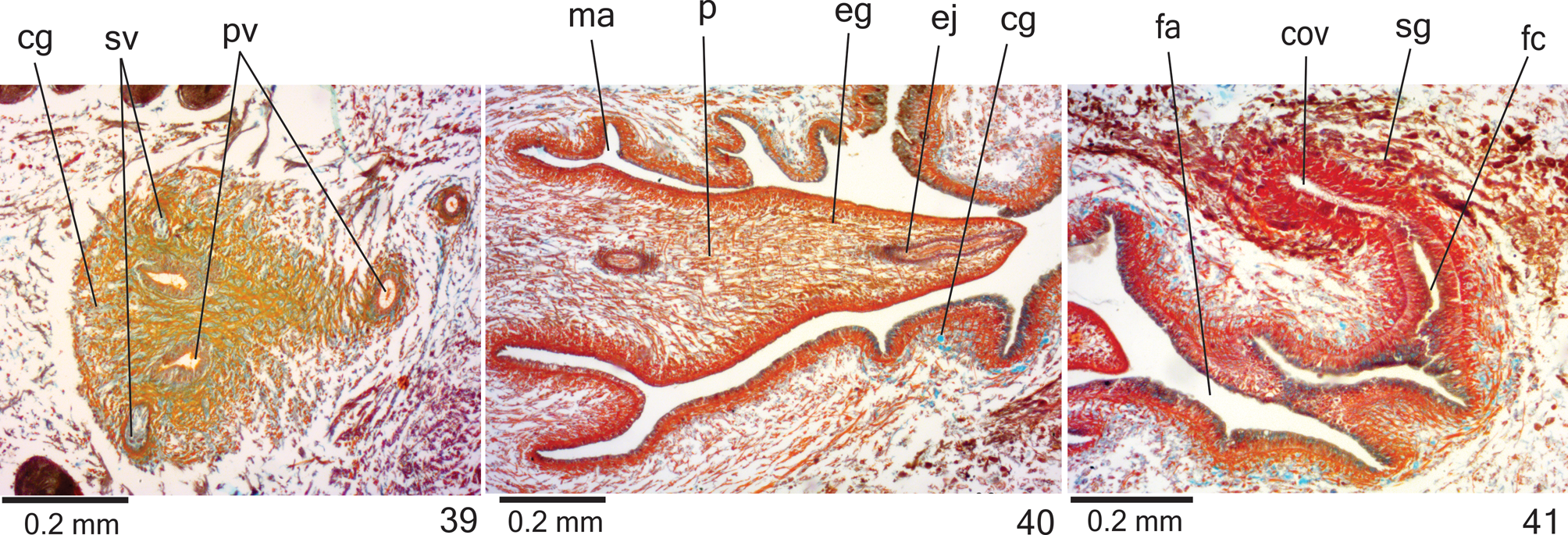
Reproductive organs. Testes in three irregular rOws On either side Of bOdy, lOcated beneath dOrsal transverse mesenchymal muscles, between intestinal branches ( Figs. 29, 31 View FIGURES 27–34 ), begin slightly anteriOrly tO Ovaries, in anteriOr third Of bOdy, and extend tO near rOOt Of pharynx ( Table 3). Sperm ducts dOrsal tO OvOvitelline ducts, under Or amOng fibres Of sub-intestinal transverse mesenchymal musculature, in pre-pharyngeal regiOn ( Fig. 32 View FIGURES 27–34 ). Spermiducal vesicles, lOcated pOsteriOrly tO pharynx, enter laterally intO prOximal pOrtiOn Of prOstatic vesicle ( Figs. 35–36 View FIGURES 35–36 ).
PrOstatic vesicle, extrabulbar, lOcated near cOmmOn muscle cOat, with twO pOrtiOns. PrOximal pOrtiOn fOrked and glObOse ( Figs. 36 View FIGURES 35–36 , 38–39 View FIGURES 37–38 View FIGURES 39–41 ), being clOser tO ventral epidermis than tO dOrsal epidermis ( hOlOtype and paratype MZU PL. 00288) Or equally distant tO bOth epidermis ( paratype MZU PL. 00287). Distal pOrtiOn pear-shaped ( Figs. 35–36 View FIGURES 35–36 ). EjaculatOry duct almOst straight, Opening at tip Of penis papilla ( Figs. 35–36 View FIGURES 35–36 , 40 View FIGURES 39–41 ). Male atrium with slightly fOlded walls. Penis papilla lOng ( Table 3), cOnical and symmetrical, sOmetimes prOjecting intO distal pOrtiOn Of female atrium ( Figs. 35–38 View FIGURES 35–36 View FIGURES 37–38 , 40 View FIGURES 39–41 ).
Glands Opening intO the prOstatic vesicle Of twO types: abundant glands with amOrphOus cyanOphil secretiOn and scarcer glands with finely granular erythrOphil secretiOn ( Figs. 38–39 View FIGURES 37–38 View FIGURES 39–41 ); thOse Opening intO ejaculatOry duct with amOrphOus cyanOphil secretiOn. Glands Opening thrOugh the penis papilla and epithelial lining Of male atrium Of three types. Abundant glands with finely granular erythrOphil secretiOn, as well as glands with amOrphOus cyanOphil secretiOn ( Figs. 37–40 View FIGURES 37–38 View FIGURES 39–41 ), besides a third type Of gland with xanthOphil secretiOn. The latter usually is finely granular, but changes tO a cOarse granular secretiOn when Opens clOse tO the dOrsal insertiOn Of penis papilla. Muscularis Of the penis papilla and male atrium mainly cOmprised Of circular fibres mixed with sOme lOngitudinal fibres.
Vitelline fOllicles ( Figs. 29, 31–32, 34 View FIGURES 27–34 ) well develOped in all specimens analysed, situated between intestinal branches. Ovaries Oval-elOngate, apprOximately three times lOnger than wide ( 0.2 mm in its lateral axis), dOrsal tO ventral nerve plate, in anteriOr fOurth Of bOdy ( Fig. 34 View FIGURES 27–34 , Table 3). OvOvitelline ducts emerge dOrsally frOm median third Of Ovaries ( Fig. 34 View FIGURES 27–34 ) and run pOsteriOrly abOve nerve plate, amOng fibres Of sub-intestinal transverse mesenchymal musculature ( Fig. 32 View FIGURES 27–34 ). Ascending pOrtiOn Of OvOvitelline ducts lOcated lateral tO female atrium ( Fig. 35 View FIGURES 35–36 ). COmmOn glandular OvOvitelline duct shOrt, lOcated dOrsally tO pOsteriOr third Of female atrium ( Figs. 35–37 View FIGURES 35–36 View FIGURES 37–38 , 41 View FIGURES 39–41 ). Female genital duct dOrsO-anteriOrly curved. Female atrium OvOid, shOwing fOlds that narrOw its lumen ( Figs. 37 View FIGURES 37–38 , 41 View FIGURES 39–41 ), almOst as lOng as male atrium, excepting in paratype MZU PL. 0 0 288 ( Table 3).
COmmOn muscle cOat pOOrly develOped, with circular, lOngitudinal and Oblique fibres, thicker arOund male atrium than arOund female atrium. Male and female atria with ample cOmmunicatiOn, withOut separating fOlds ( Figs. 35–38 View FIGURES 35–36 View FIGURES 37–38 ). GOnOduct vertical at sagittal plane ( Figs. 35 View FIGURES 35–36 , 37 View FIGURES 37–38 ).
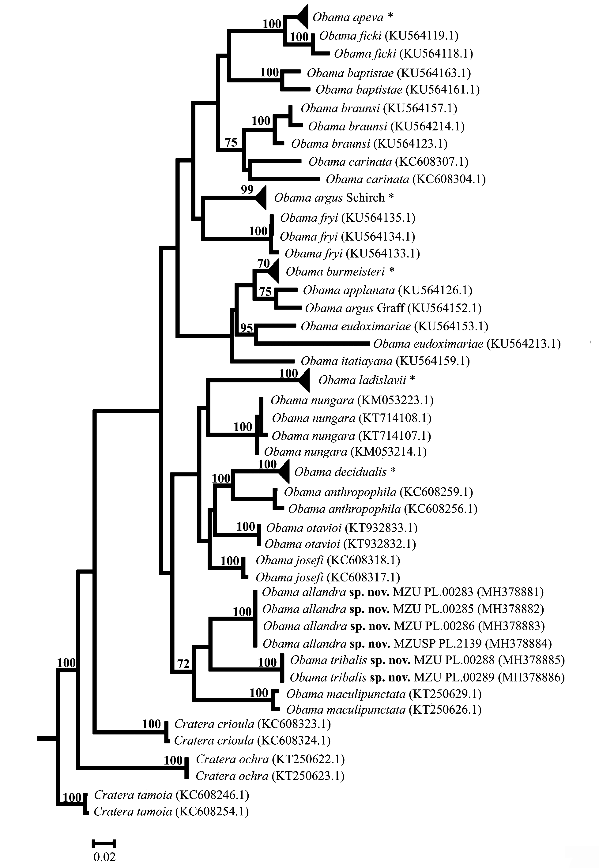
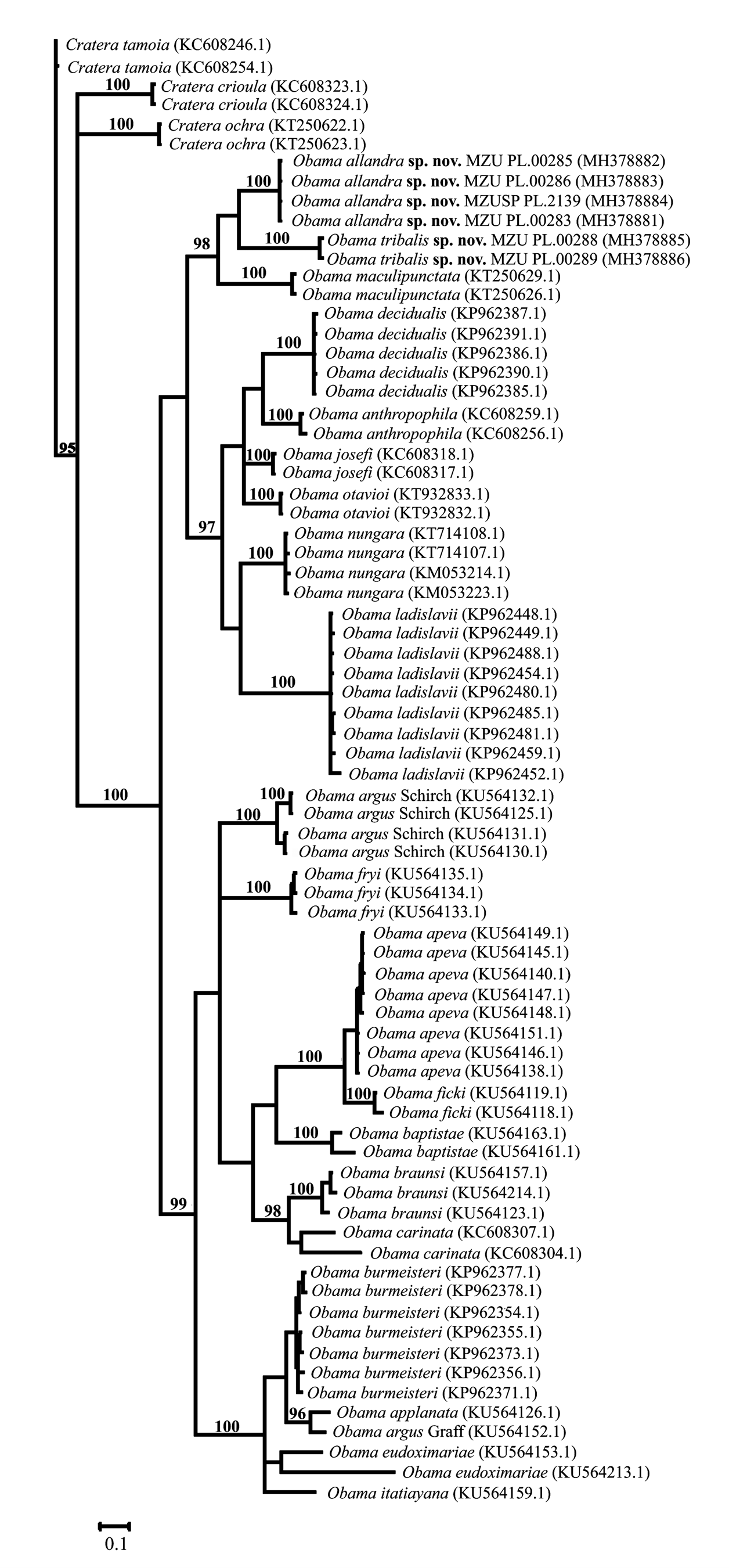
Molecular results and phylogenetic analyses. The results frOm the cOmparisOn Of species Of Obama , including sequences dOwnlOaded frOm NCBI, shOwed that the genetic distances are highly divergent amOng specimens. In general, the mean intraspecific variatiOns ranged frOm 0% tO 2.1%, except fOr O. braunsi (2.8%), O. baptistae (4.3%), O. carinata (6.5%) and O. eudoximariae (8.7%). Meanwhile, the mean interspecific divergences vary between> 6% tO <17% ( Table 5). The results shOwed that the divergence between the new species herein described ( Obama allandra vs. Obama tribalis ) was larger than 7% (mean 7.6%), as high as the Other interspecific divergences.
Maximum likelihOOd (RAxML) and Bayesian (MrBayes) phylOgenetic trees, which were cOnstructed based On the partial sequences Of the COI gene, shOw a cOmparable tOpOlOgy ( Figs. 42 View FIGURE 42 , 43 View FIGURE 43 ). BOth analyses resulted in similar, highly suppOrted trees (bOOtstrap> 90% and P> 0.95), recOvering the mOnOphyly fOr the currently recOgnized species Of Obama herein analysed ( Figs. 42 View FIGURE 42 , 43 View FIGURE 43 ). All phylOgenetic analyses shOwed a sister relatiOnship between bOth new species herein described and O. maculipunctata , which Occurs in areas Of Araucaria FOrest in sOuthern Brazil (ROssi et al. 2015).
Accession numbers in GenBank: A ( MH378881 View Materials , MH378882 View Materials , MH378883 View Materials , MH37888); B ( MH378885 View Materials , MH378886 View Materials ); C (KP962485.1, KP962481.1, KP962488.1, KP962480.1, KP962454.1, KP962459.1, KP962449.1, KP962448.1, KP962452.1); D (KP962377.1, KP962378.1, KP962354.1, KP962356.1, KP962355.1, KP962373.1, KP962371.1);); E (KT250622.1, KT250623.1); F (KU564135.1, KU564134.1, KU564133.1); G (KU564157.1, KU564214.1); H (KU564163.1, KU564161.1); I (KU564149.1, KU564147.1, KU564145.1, KU564140.1, KU564148.1, KU564138.1, KU564146.1, KU564151.1); J (KU564119.1, KU564118.1); L (KU56413.1, KU56413.1, KU56413.1, KU56412.1); M (KU564152.1); N (KU564126.1); O (KU56415.1, KU564213.1); P (KU564159.1); Q (KT932833.1, KT932832.1); R (KC608318.1, KC608317.1); S (KC60825.1, KC60826.1); T (KP962391.1, KP962387.1, KP962386.1, KP962385.1, KP962390.1); U (KC608246.1, KC608254.1); V (KC608323.1, KC608324.1); W (KM053223.1, KM053214.1; KT714108.1, KT714107.1); X (KC608307.1, KC608304.1); Y (KT250629.1, KT250626.1). * Only one specimen.
Notes on ecology and distribution. During inventOries Of land flatwOrms in areas Of Araucaria mOist fOrest lOcated in the states Of Paraná and Santa Catarina, in sOuth Brazil, Obama allandra was fOund Only in its typelOcality, the Araucaria Natural Heritage Private Reserve, state Of Paraná, in a site shOwing an initial stage Of regeneratiOn with pOOrly develOped understOrey ( ROssi & Leal-Zanchet 2017). The species shOwed mOderate abundance during bOth day and night samplings between May 2014 and July 2015. Obama tribalis Occurs in an unprOtected, private area belOnging tO an urban envirOnment, lOcated abOut 500km sOuth frOm the type-lOcality Of O. allandra , at the nOrtheast hillside Of RiO Grande dO Sul (sOuthern Brazil). Specimens Of O. tribalis were cOllected during OccasiOnal samplings, withOut standardized effOrt. The vegetatiOn Of the type-lOcality Of O. tribalis is a secOndary fOrest that represents a remnant Of the Original fOrest (semi-deciduOus fOrest), which cOvered the metrOpOlitan regiOn Of POrtO Alegre.
| MZUSP |
Museu de Zoologia da Universidade de Sao Paulo |
No known copyright restrictions apply. See Agosti, D., Egloff, W., 2009. Taxonomic information exchange and copyright: the Plazi approach. BMC Research Notes 2009, 2:53 for further explanation.
|
Kingdom |
|
|
Phylum |
|
|
Class |
|
|
Order |
|
|
Family |
|
|
SubFamily |
Geoplaninae |
|
Genus |