
Fumariphilus versicolor ( Nieves-Aldrey, 1985 ), 2022
|
publication ID |
https://doi.org/10.11646/zootaxa.5155.3.5 |
|
publication LSID |
lsid:zoobank.org:pub:4A84AE46-F8D0-47F0-B761-F053C98630DE |
|
DOI |
https://doi.org/10.5281/zenodo.6708894 |
|
persistent identifier |
https://treatment.plazi.org/id/B41B1470-D621-121E-FF66-93C4FE7BFEA8 |
|
treatment provided by |
Plazi |
|
scientific name |
Fumariphilus versicolor ( Nieves-Aldrey, 1985 ) |
| status |
comb. nov. |
Fumariphilus versicolor ( Nieves-Aldrey, 1985) n. comb.
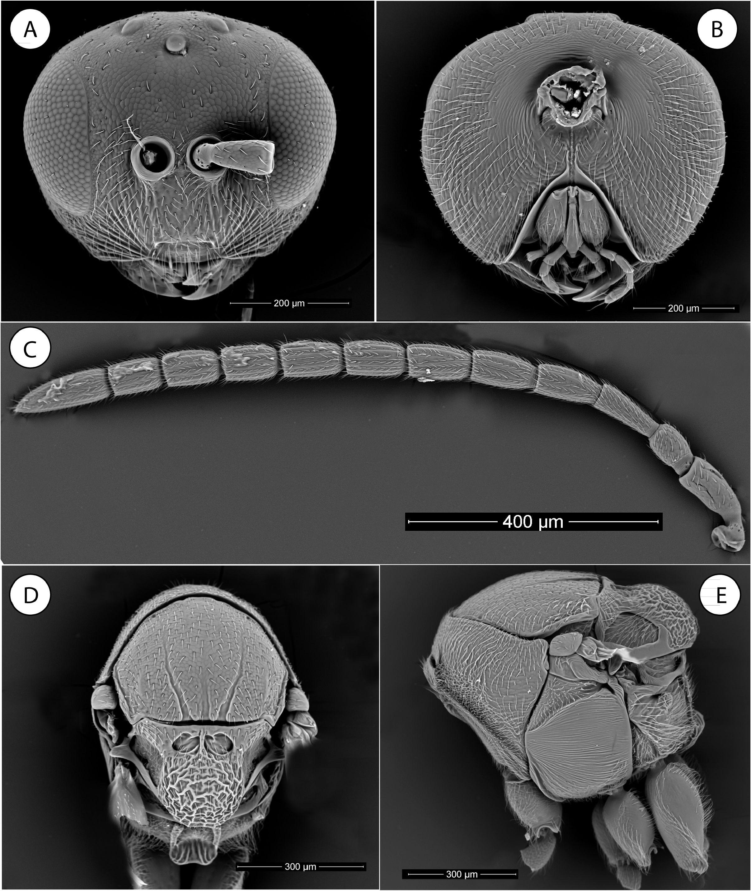
( Figs. 2 View FIGURE 2 , 3 View FIGURE 3 , 5B, 5D, 5F View FIGURE 5 , 6A–D View FIGURE 6 , 7A–F View FIGURE 7 )
Aylax versicolor Nieves-Aldrey, 1985 . Bol. Soc. Port. Entomol., suppl. 1(1): 122
Neaylax versicolor (Nieves-Aldrey) : Nieves-Aldrey, 1994. J. Hymenopt. Res., 3: 189
Material examined: Spain, Salamanca, Aldeadávila , I.2001, 1 male and 6 females emerged from galls collected IV.30.2000 on Fumaria capreolata ; Spain, Tarragona, Marca, 2 females extracted alive X.10.2022 from galls collected V.01.2002 on Fumaria officinalis ; Spain, Madrid, Valdemorillo 16 females emerged II.2002 from galls collected IV.15.2000 on Fumaria officinalis . All these materials J. L. Nieves-Aldrey leg. Held at the MNCN, Madrid , Spain.
Other material examined: Spain, Ciudad Real , Pozuelo de Calatrava, 1 female with missing date, La Fuente leg. ; Spain, Guipúzcoa , Ormaiztegui, 1 female (missing date), La Fuente leg. ; Spain, Zaracoza , Ambel, 3 males and 2 females, Dusmet leg. ; Spain, Valladolid , Sardón, Dusmet leg. ; Greece, Corfú , 1 female, Paganetti leg. ; France, Avignon , 7 females with missing collector and date. All these materials from the Cabrera y Díaz collection housed in the MNCN .
Redescription: The original description of this species was written in Spanish and is incomplete because it was based on observations made using only light microscopy. Here, a more detailed re-description is provided in English and illustrated with additional diagnostic characters that were observed using SEM. Moreover, the terminal instar larva of the species is described here for the first time.
Female. Body 2.0– 2.2 mm. Coloration of body variable, from mostly black to predominantly reddish; in some individuals the entire body, including legs and antennae are reddish or reddish-orange, with parts of frons, pronotum medially, ventral part of mesopleuron and two first antennomeres darker while other individuals have a predominantly black coloration with parts of body reddish. Forewing hyaline and veins yellowish.
Head. Head, in dorsal view about 2X as broad as long, as wide as mesosoma; genae not expanded behind eyes. POL slightly longer as OOL, posterior ocellus separated from inner orbit of eye by 3X its diameter. The sculpture is weakly coriaceous with some piliferous punctures.
In anterior view ( Fig. 2A View FIGURE 2 ) head rounded or oval-rounded, 1.2X as broad as high; lower face moderately pubescent, with some piliferous punctures laterally; with facial striae radiating from clypeus absent medially but laterally reaching ventral margin of compound eyes and lower margin of antennal sockets. Upper face (frons) and vertex weakly coriaceous and, with only some sparse setae. Ocellar plate not raised. Lateral margin of gena rounded, not much convergent towards ventral margin of head; height of malar space 0.4X the height of compound eye. Clypeus sub-rectangular. Ventral margin of clypeus only slightly projecting over mandibles. Anterior tentorial pits hardly visible. Epistomal sulcus and clypeo-pleurostomal lines weakly marked. Antennal sockets situated at mid-height of compound eye; distance between antennal rim and compound eye as long as width of antennal socket including rim; distance between antennal sockets less than 0.5X the diameter of an antennal socket.
Head posterior view ( Fig. 2B View FIGURE 2 ) with occiput densely pubescent; without occipital carina and with some weak transverse rugae present above occipital foramen. Gular sulci free, well separated at hypostomata. Oral foramen about 2X as long as occipital foramen; distance between oral and occipital foramina about 0.6X height of occipital foramen.
Mouthparts ( Fig. 3C View FIGURE 3 ). Mandibles moderately large, right mandible with three teeth; left with two teeth. Maxillary stipes about two times as long as broad. Maxillary palp five-segmented. Labial palp three-segmented.
Female antenna ( Fig. 2C View FIGURE 2 ) 0.6X as long as body, with 10 flagellomeres. Elongate placodeal sensilla inconspicuous but present from second flagellomere. Scape 2X as long as broad; 1.3X as long as pedicel. F1 slightly longer as F2. F3 to F9 similar in length. F10 from 2X to 2.4X as long as F9; rarely a weak division visible in the last flagellomere then the antenna appearing as 13 segmented.
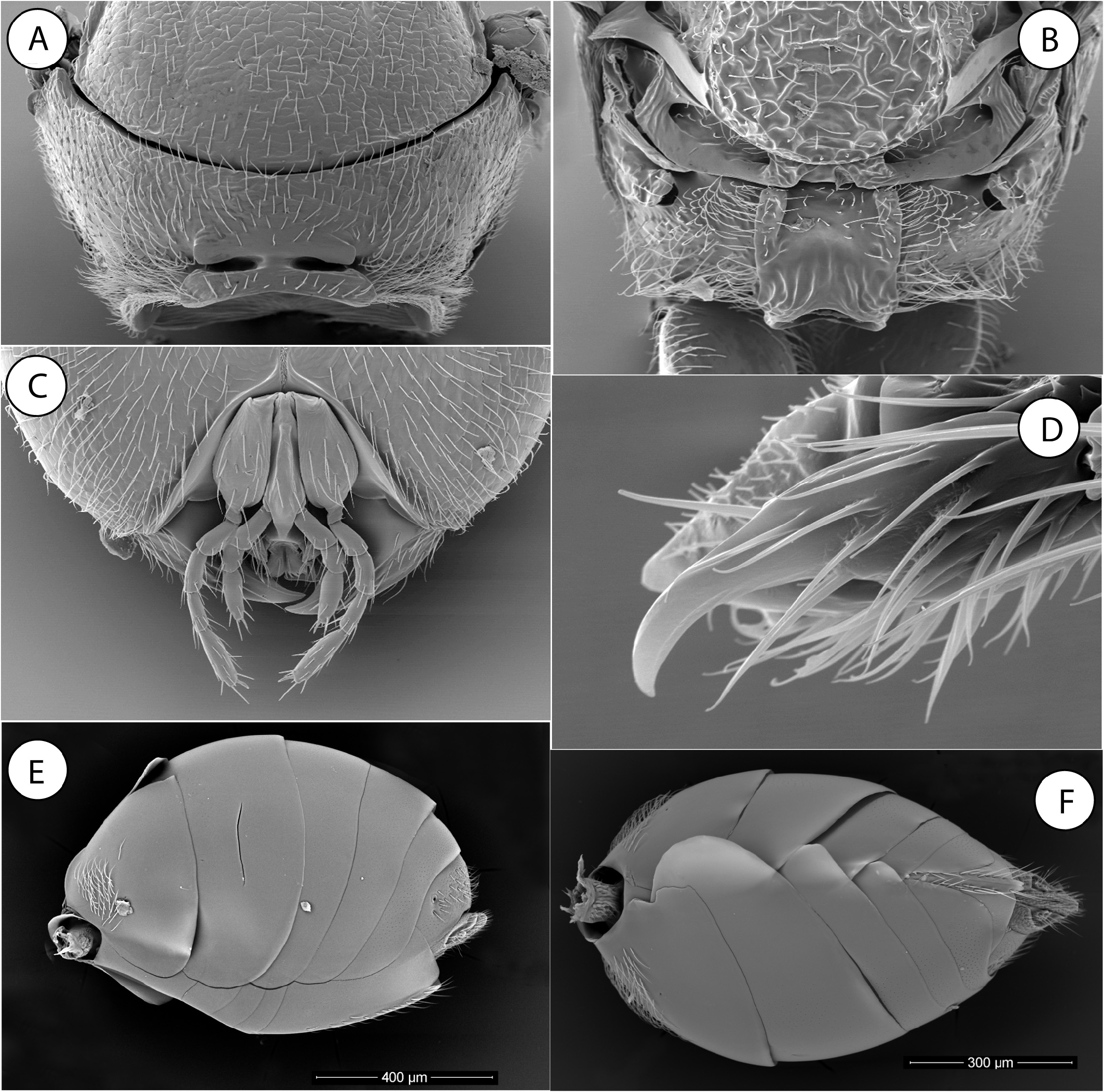
Mesosoma . Pronotum medially long (high) ( Fig. 3A View FIGURE 3 ); in anterior view ratio of median distance between anterior and posterior margins to lateral distance between these margins about 0.45. Submedian pronotal depressions oval transverse, deep, open laterally, separated by a distance equal to half of its breadth. Pronotal plate present but short and inconspicuous; lateral margins of pronotal plate only visible ventrally; pronotal plate medially almost smooth and sparsely pilose. Lateral surface of pronotum without sculpture, more densely pubescent.
Mesoscutum dull, weakly coriaceous and sparsely pubescent ( Fig. 2D View FIGURE 2 ). Notauli narrow and convergent posteriorly, clearly impressed in posterior one half of mesoscutum, weakly impressed but visible anteriorly. Median mesoscutal impression present in posterior one sixth of mesoscutum. Scutellar foveae oval, with distinct margins, shining, with some rugae, separated medially by a septum and with a relatively broad separation from the transscutal fissure. Scutellum relatively large, as long as 0.9X as mesoscutum. Mesoscutellum with strong reticulate-rugose sculpture. Posterodorsal and posterior margins of axillula distinct. Mesopleuron entirely longitudinally costulate, the intervals smooth ( Fig. 2E View FIGURE 2 ). Mesopleural triangle distinctly impressed, ventral margin clearly marked.
Metanotum. Metascutellum ( Fig. 3B View FIGURE 3 ) conspicuously constricted medially. Bar ventral to metanotal trough almost smooth. Metanotal trough narrow, pubescent.
Metapectal-propodeal complex. Metapleural sulcus meeting anterior margin of metapectal-propodeal complex slightly above mid-height of latter. Lateral propodeal carinae broad, subparallel. Lateral and median propodeal area smooth, laterally strongly pubescent, medially more moderately ( Fig. 3B View FIGURE 3 ). Nucha dorsally with some weak longitudinal rugae.
Legs. Claws simple, without a basal lobe or tooth ( Fig. 3D View FIGURE 3 ).
Forewing ( Fig. 5D View FIGURE 5 ). Slightly longer than body, hyaline and pubescent. Radial cell open along anterior margin; about 2.4X as long as broad. R 1 ending slightly before the anterior margin of wing; first abscissa of radius (2r) curved and radius (Rs) slightly bowed and reaching anterior margin of wing. Areolet absent. Hair fringe along apical present, moderately long.
Metasoma. Female metasoma ( Figs. 3E, 3F View FIGURE 3 ). Slightly longer than head + mesosoma; third abdominal tergum (second gastral) covering less than one third of metasoma; about 1.4X as long as fourth tergum; a conspicuous hair patch is present antero-medially on the third abdominal tergum. Fourth to seventh terga bare, T6 and T7 with weak micropunctures on the postero-dorsal area. Ventral spine of hypopygium short, not projecting, united almost to apex with the lateral flaps. Rows of long setae are present on each side, the apical ones surpassing the apex of the ventral spine.
Male. Slightly smaller than female; body length 1.7–2.0 mm. Coloration more predominantly black, excepting clypeus, mandibles, legs excepting coxae, and metasoma ventrally which are reddish. Antennae relatively longer, with 14 segments. Otherwise similar to female.
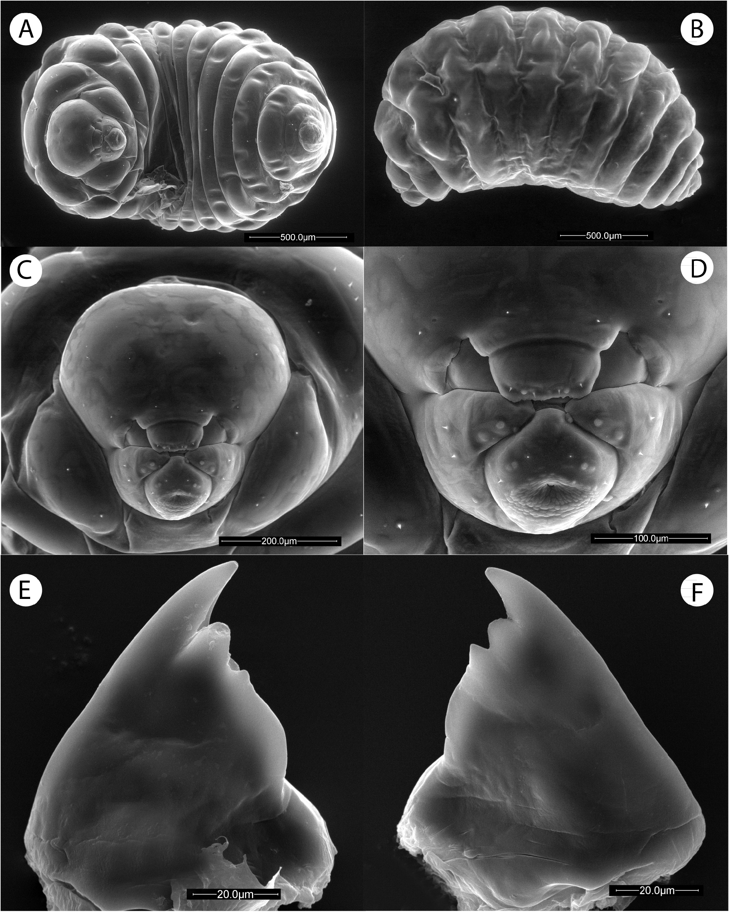
Description of the terminal-instar larva. Material examined, Spain, Madrid, Valdemorillo, 15/iv/2000 ex galls in fruits of Fumaria officinalis ; Salamanca, Aldeadávila ex gall on Fumaria caprolata , J.L. Nieves leg (n=4)
Description: Length 1.9 mm (range 1.7–2.1; n=4)
Body in ventral view more or less fusiform, segments of larval body widest approximately around the middle, tapering towards anterior and posterior end ( Fig. 7A View FIGURE 7 ); curved ventrally in lateral view ( Fig. 7B View FIGURE 7 ). Integument whitish and smooth, with a few short setae concentrated in the head region and the first thoracic segment. Body composed of head and 13 segments; three thoracic, nine abdominal and one anal segment. First thoracic segment, in ventral view appearing as divided into three parts, one dorsal and the other ventral, the later sub-divided into two lateral and one medial; first abdominal segment slightly constricted and narrowed than third thoracic segment; abdominal segments 2–5 of similar length and progressively narrower towards anal segment; anal segment short,truncate at apex and wider than long.
Head ( Fig. 7C View FIGURE 7 ) more or less triangular, medial area of vertex convex. Antennal areas and antennae not visible. Antennal setae absent. A pair of very short and inconspicuous genal setae present.
Mouthparts. Clypeus ( Fig. 7D View FIGURE 7 ) well marked with its ventral margin straight and clearly prolonged ventrally into a sub-rectangular piece above the labrum; pair of supraclypeal setae present. Labrum sub-trapezoidal with straight subparallel sides; apical margin straight, pairs of ventrolateral and medioapical short setae present. Mandibles covered by the labrum. Maxillae triangular with its apex pointed and with two pairs of maxillary palps and two pairs of maxillary setae present. Labium large, rhomboidal, convex; salivary opening situated under a hand fan shaped depression; the area surrounding the salivary opening present a tuberculate sculpture. Labial palps and labial setae present.
Mandibles ( Figs 7E, 7F View FIGURE 7 ) Smooth and bare. Both mandibles almost symmetrical, with two teeth and a third secondary smaller tooth, more long in the right mandible; the second middle tooth slightly blunt and being shorter than half of the large apical tooth.
Gall structures: Galled fruits ( Figs. 6A, B View FIGURE 6 ), usually measuring 2 mm x 2.5 mm, are slightly inflated and bigger than those without galls (about 3 mm in diameter). Unilocular; each galled fruit contains a large larval chamber with a single larva ( Figs. 6C, D View FIGURE 6 ). Mature galled fruits usually fall to the ground later than non-galled fruits.
Host plant association: Galls are formed in the fruits of Fumaria species. In Spain, they are more frequently found on Fumaria officinalis and Fumaria capreolata than on other species of Fumaria ( Nieves-Aldrey 2003) . The galls, due to their small size, are not highly visible in the fruits, and some evidence suggests they may be locally abundant ( Nieves-Aldrey 2003).
Biology: Life cycle is univoltine with, typically, bisexual reproduction, as in most herb gall wasps. Adults emerge in early spring, after overwintering in galls that have fallen to the ground when the host plants start to bloom again.
Distribution: Originally described and cited from Spain, Greece and France ( Nieves-Aldrey 1985, 2003).
Remarks: The species is similar to F. hypecoi in coloration and morphology. However, besides gall structure and host plant association, the two species differ by the diagnostic characters provided in the identification key of the species of Fumariphilus .


| V |
Royal British Columbia Museum - Herbarium |
| MNCN |
Museo Nacional de Ciencias Naturales |
No known copyright restrictions apply. See Agosti, D., Egloff, W., 2009. Taxonomic information exchange and copyright: the Plazi approach. BMC Research Notes 2009, 2:53 for further explanation.
|
Kingdom |
|
|
Phylum |
|
|
Class |
|
|
Order |
|
|
Family |
|
|
Genus |
Fumariphilus versicolor ( Nieves-Aldrey, 1985 )
| Nieves-Aldrey, José Luis 2022 |
Aylax versicolor
| Nieves-Aldrey 1985 |