
Patia orise orise
|
publication ID |
https://doi.org/10.11646/zootaxa.4559.3.2 |
|
publication LSID |
lsid:zoobank.org:pub:EDE68167-8CD0-4C99-82A8-8EAB1604E86F |
|
DOI |
https://doi.org/10.5281/zenodo.5934279 |
|
persistent identifier |
https://treatment.plazi.org/id/B66087B9-1B6A-A332-FF16-F92A4BF3FBBB |
|
treatment provided by |
Plazi |
|
scientific name |
Patia orise orise |
| status |
|
Patia orise orise View in CoL (male and female)
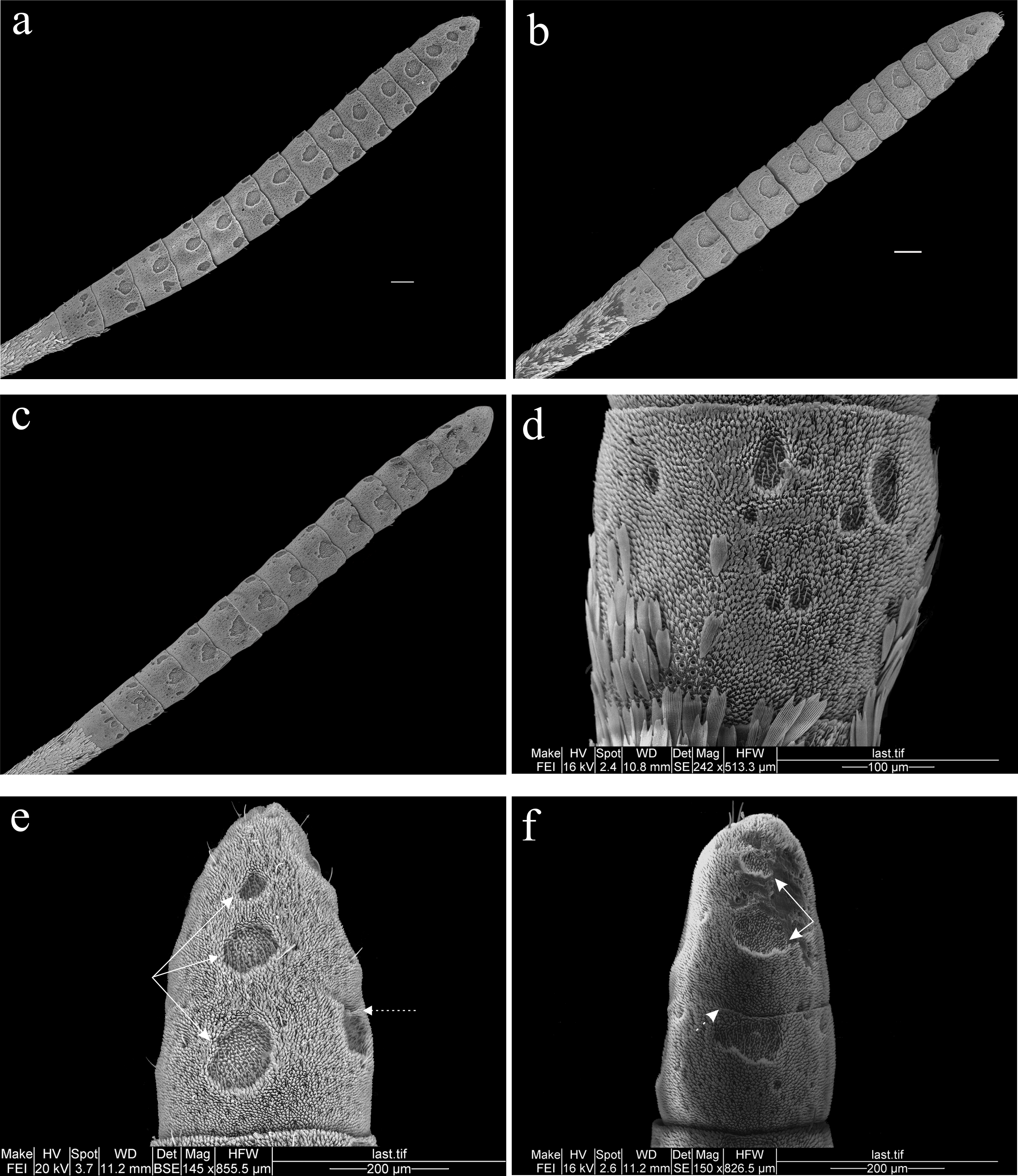
ANTENNAL CLUB ( Figs. 4a, b View FIGURE 4 ): The nudum measures around 4.5 mm.
ANTENNOMERES ( Figs. 4 View FIGURE 4 d–g): The female has 14 scaleless antennomeres, and the male has 15. The first basal is partially scaled on the lateral-dorsal side and the proximal margin on its ventral surface; in the male, the scaleless area occurs in the previous segment. The first segment of the nudum is compressed, and the second is almost isometric. From the third to the twelfth the antennomeres are depressed, and the most distal fused is very compressed. Sometimes, the division between the distal antennomere and the one that precedes it is incomplete. In the proximal antennomeres, the l/w ratio is 1.1–1.5 in the first, 0.9–1.1 in the second. From the third to the twelfth it is 0.6–0.9, and in the distal fused, it is 1.5–1.7. In the male the first three proximal antennomeres are cylindrical, from the fourth to the twelfth they are doliform (barrel-shaped), and the last one is cupuliform (dome-shaped) elongated with the blunt apex. In the female, all the antennomeres are doliforms, except for the distal.
SULCI AND PSEUDOSULCI ( Figs. 4 View FIGURE 4 e–h): It exhibits 12–15 central sulci and 27–30 laterals. The trisulcate configuration is noticed from the third antennomere. The central sulci are present in almost all segments and sometimes are absent in the first two and the second. These sulci occupy one-fourth to one-half of the length of the antennomere that contains them in the proximal ones and one-third to three-fourths in the medial and distal ones. In all cases, the central sulci are close to the distal margin of the antennomere, usually separated by two or seven rows of microtrichia m2; some of them have a truncated distal edge. The central sulci appear from the first or second antennomeres although they are disaggregated. In all segments, the sulci central have an irregular form with broken or discontinuous edges, although some are quasi-elliptical. The lateral sulci are presented from the first antennomere (both or only one of them) although they are disaggregated, from the second or third, they are complete (aggregates); its size is smaller than the central ones, or it is similar in the proximal segments. The lateral sulci are separated from the distal margin of the antennomeres by up to nine rows of microtrichia m2 and rarely are truncated by this margin. Some are very irregular, in such a way that portions of microtrichia m2 invaded the sulci, besides their edges are very discontinuous (female). The pseudosulci (ps), are scarce and they are present in the proximal segments of the club where they are part of the disaggregated sulci. They are located under the central sulcus or between it and the lateral ones, toward the middle of the antennomere or close to its distal margin; sometimes, they are under the lateral sulci.
MICROTRICHIA ( Figs. 4i, m, r View FIGURE 4 ): There are microtrichia types m1–m4. In the basal and medial antennomeres, the st:m1 ratio of the central sulci is from 1:4 to 1:5 and in the distal, from 1:4 to 1:6. The l/w ratio is from 1:3 to 1: 4 in all lateral sulci.
TRICHOID SENSILLA ( Fig. 4i View FIGURE 4 ): The average length of the trichoid sensilla is 21 µm (n = 7; 19.7–21.8 µm). In the pseudosulci, these sensilla have a similar measure. The number of trichoid sensilla in the central and lateral sulci along the nudum shows no pattern or trend. The largest number is in the sulcus of the fifth antennomere for the central sulci and the seventh for the lateral ones.
CHAETIC SENSILLA ( Fig. 4j View FIGURE 4 ): The chaetic sensilla show the characteristic distribution of the subfamily, and they are close to the distal margin of the antennomeres. The length of these sensilla is 37.5 µm on average (n = 3; 33.8–39.5 µm).
OTHER SENSILLA ( Figs. 4 View FIGURE 4 l–r). Without considering the trichoid sensilla, the auriculate are the most abundant (almost ten times more than the basiconic sensilla). In the mesial segments, the coeloconic sensilla are present under the lateral sulci; sometimes they are close to each other, separated by a single microtrichia m2. The squamiform sensilla are near the central sulci and measure 32.7 µm on average (n = 3; 30.4–34.7 µm). There is one campaniform sensillum in the fourth antennomere.
PORES: They are present within the sulci, between microtrichia m1.
No known copyright restrictions apply. See Agosti, D., Egloff, W., 2009. Taxonomic information exchange and copyright: the Plazi approach. BMC Research Notes 2009, 2:53 for further explanation.
|
Kingdom |
|
|
Phylum |
|
|
Class |
|
|
Order |
|
|
Family |
|
|
Genus |