
Myostenostomum Bulbocaudatum
|
publication ID |
https://doi.org/ 10.11646/zootaxa.4066.1.9 |
|
publication LSID |
lsid:zoobank.org:pub:208BBCEC-AECE-4961-8B7A-58A2CE0C6788 |
|
DOI |
https://doi.org/10.5281/zenodo.6056305 |
|
persistent identifier |
https://treatment.plazi.org/id/BD0ECC29-F77F-0A63-FF1F-FE8AFAB7709A |
|
treatment provided by |
Plazi |
|
scientific name |
Myostenostomum Bulbocaudatum |
| status |
|
Myostenostomum Bulbocaudatum View in CoL (Catenulida, Stenostomidae ): new for Ukraine, rare for the World
YULIIA KANANA
I.I. Schmalhausen Institute of Zoology NAS of Ukraine, B. Khmelnitsky str., 15, Kyiv, 0 1601 Ukraine. E-mail: y.kanana@izan.kiev.ua, y.kanana@gmail.com
At present, the stenostomid genus Myostenostomum Luther, 1960 according to Artois et al. (2013) contains 7 species: M. Bulbocaudatum Luther (type species), M. ilmenicum Rogozin , M. lutheri Rogozin , M. marcusi Rogozin , M. vanderlandi Rogozin , M. fasciatum (Veydovsky) and M. gigerium (Kepner & Carter) , although the two latter are thought to belong to the genus Stenostomum Schmidt (Tyler et al. 2006–2015). A remarkable feature of the representatives of the genus Myostenostomum is a muscular ring on their gut.
M. bulbocaudatum was first recorded in Finland and described by Luther in 1960 ( Luther 1960). Recently this species was recorded in the Russian Federation from the Southern Ural by Rogozin (2012). Unfortunately, drawings made by both authors do not have necessary details of the cerebral ganglia. Hence, this morphological character is useful and reliable even when the refractile bodies are absent ( Rogozin 1992). Here Myostenostomum bulbocaudatum is registered in Ukraine for the first time. The morphology description and illustrations are given.
The specimens were collected by the author on 10.08.2014 from a natural freshwater pool in the vicinity of the village Sulimovka (Boryspil raion, Kyiv Oblast; 50.395032, 31.186336).
Samples of vegetation were taken using a small net (mesh width 70 µm) or cut off and washed in sampled water, as well as collecting water from the sample site. The collected vegetation and material were kept in glass bottles. Water from the samples was extracted into a Petri dish and then examined using a dissecting microscope. Each worm that was found was picked up with a glass pipette and put on a slide. Before they were preserved, the collected worms were first studied alive using a dissecting and a light microscope. Slightly squeezed specimens were drawn freehand and photographed using a digital camera attached to a Zeiss Axio Imager M 1.
Results and discussion
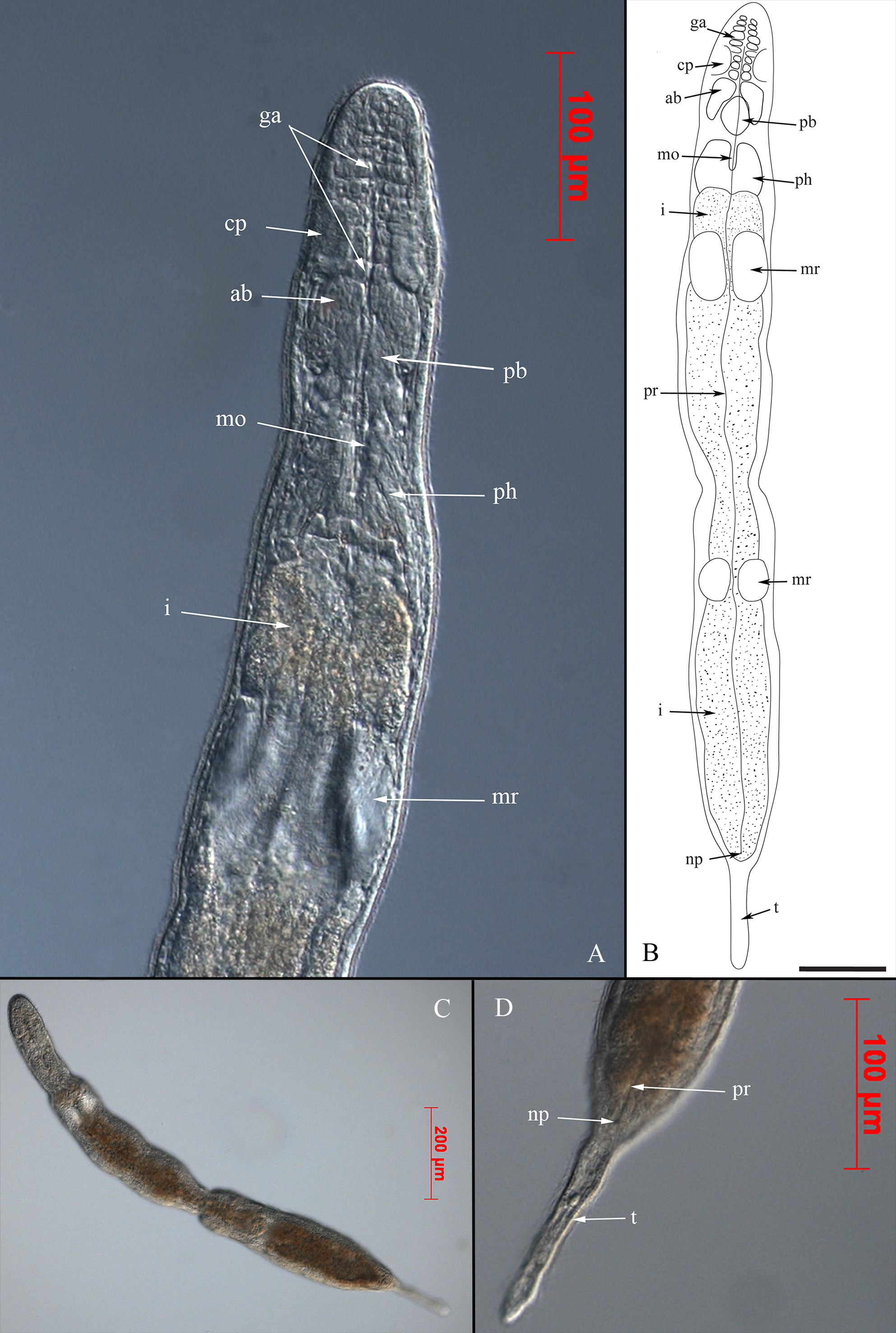
Specimens with two zooids were found ( Fig. 1 View FIGURE 1 , B and C). The animals are 1.2 mm long. The color is white in reflected light. The body is slender, evenly shaped and cylindrical with posterior part ending with the caudal appendage, or “tail” ( Fig. 1 View FIGURE 1 , D). The “tail” is somewhat elevated, dorsally situated. The epidermis is completely covered with short cilia. The ciliated pits are long and deep.
The cerebral ganglia (“brain”) consist of two lobes ( Fig. 1 View FIGURE 1 , A); the anterior lobes are more distinct, paired, beanshaped, and anteriorly have two chains of approximately 10 metamerically arranged ganglia, while posterior lobe is almost rhomb-like. Refractile organs are absent.
The mouth opening is more or less V-shaped. The pharynx is muscular, coniform, and its proximal part contacts the intestine. The length of the pharynx is approximately equal to its width. Pharyngeal glands are slightly visible. The muscular ring on a gut is short. Its length and width are almost equal.
The protonephridium is visible along the whole body and ends in a nephridiopore at the posterior end right before the base of the “tail”.
Individuals with a developed genital system were not found.
Known distribution. Finland ( Luther 1960); Southern Ural ( Russian Federation) ( Rogozin 2012); first record for Ukraine.
Body shape, dorsal “tail”, ciliated pits and posterior lobe shape correspond to Luther’s description ( Luther 1960). However, the pharynx’ length and width, which are almost equal in species from Ukraine, are similar to ones described in Myostenostomum marcusi ( Rogozin 1992) .
The body in species from Ukraine is not swollen in the region of muscular ring like in one from Southern Ural which in addition has two “tails” ( Rogozin 2012). At the same time, author does not present a cerebral ganglia description in the text as well as does not depict it in the illustration. Considering this, Rogozin’s finding needs to be checked whether it is M. bulbocaudatum or different Myostenostomum .
Therefore, our detection of M. bulbocaudatum is quite natural to complement the records of this species, but for adequate distribution analysis of M. bulbocaudatum more information from different geographical regions is needed.
No known copyright restrictions apply. See Agosti, D., Egloff, W., 2009. Taxonomic information exchange and copyright: the Plazi approach. BMC Research Notes 2009, 2:53 for further explanation.
|
Kingdom |
|
|
Phylum |
|
|
SubPhylum |
Catenulida |
|
Class |
|
|
Family |