
Olmeccyclops hondo, Fiers, Frank & Jocque, Merlijn, 2013
|
publication ID |
https://doi.org/ 10.11646/zootaxa.3630.2.4 |
|
publication LSID |
lsid:zoobank.org:pub:41C065DF-D429-4120-A5A7-CE72860D2E21 |
|
DOI |
https://doi.org/10.5281/zenodo.5632091 |
|
persistent identifier |
https://treatment.plazi.org/id/BD3787D0-FFAF-FFEA-3AC8-FB97FE58E0C4 |
|
treatment provided by |
Plazi |
|
scientific name |
Olmeccyclops hondo |
| status |
sp. nov. |
Olmeccyclops hondo sp. nov.
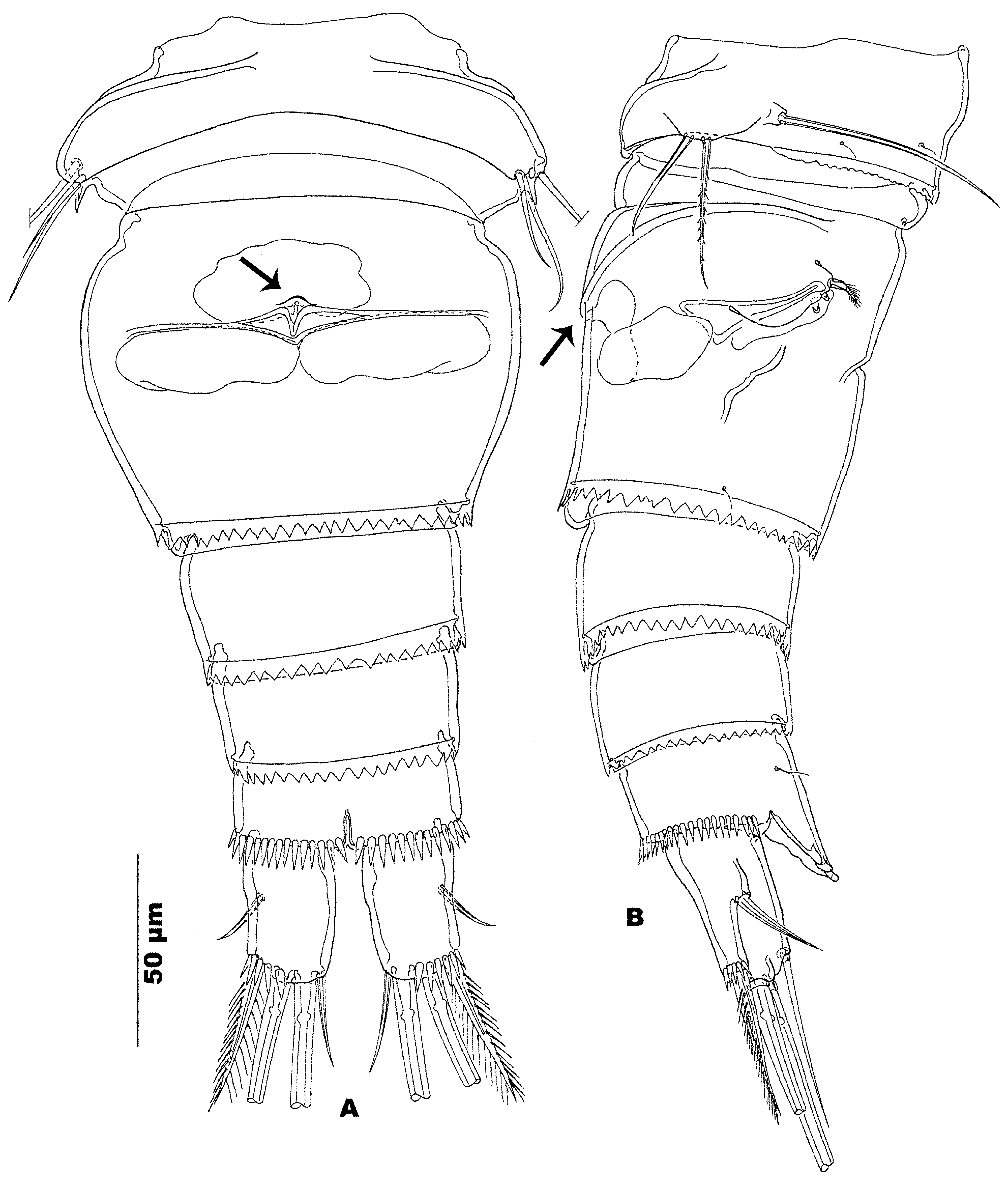
( Figs. 1–4 View FIGURE 1 View FIGURE 2 View FIGURE 3 View FIGURE 4 )
Material examined. Holotype, female, dissected and mounted on 4 slides, registered RBINSc COP 9936A-D; paratype female, preserved, registered RBINSc COP 9937.
Type locality. Honduras, El Cusuco National Park, Elfin forest floor (see: Material and methods for details).
Etymology. From the Spanish word hondo , meaning “mysterious, deep” and traditionally said to be used by Christopher Colombus in 1502 to mark the deep waters off the present day Honduras, proposed herein as specific epithet to mark the occurrence of the species deep in the cloud forests of El Cusuco National Park. Gender masculine.
Description. Female. Habitus ( Fig. 1 View FIGURE 1 A) cyclopid-shaped. Body depressed, widest along posterior margin of cephalothorax. Metasome gently tapering caudally, shorter that cephalothorax (ratio 1/1.2). Transition between prosome and urosome indistinct in dorsal view. Intersomal arthrodial membrane between leg 5-bearing pediger and genital double-somite wide, without particularly reinforced integument ( Fig. 2 View FIGURE 2 A, B). Genital double-somite vaseshaped in dorsal view ( Fig. 1 View FIGURE 1 A), widest in anterior third (length:width ratio: 1/1.15). Leg 6 vestiges positioned dorsolaterally in anterior third of double-somite ( Fig. 2 View FIGURE 2 b). Urosomites 4 and 5 parallel-sided. Ratio urosome/body length: 1:2.6. Body length (holotype) 555 μm and (paratype) 750 μm.
Integument of body somites smooth, integument structure with refractile punctuations (not illustrated). Posterior margin of cephalothorax and metasomites straight. Posterior margin of genital double-somite and urosomites 4 and 5 with wide, transparent, with serrate fringe. Posterior margin of anal somite with uninterrupted girdle of robust spinules ( Fig. 1 View FIGURE 1 A, 2A, B). Anal operculum wide and prominently expanded caudally, either linguiform with crescentic apex ( Fig. 1 View FIGURE 1 C: holotype) or irregularly undulate ( Fig. 1 View FIGURE 1 D: paratype).
Caudal rami ( Fig. 1 View FIGURE 1 C, D, 2A, B) twice as long as wide, cylindrical, with large triangular depression along medial margin. Anterolateral seta long, about 3/4 of ramal length, without spinules near insertion. Posterolateral element as long as ramus, rigid, serrate along outer, pinnate along inner margin. Outer terminal seta half as long as inner one, both pinnate and with breaking plane near insertion. Medial seta stout, pinnate, shorter than ramus (2/ 3–3/4 of ramal length). Dorsal seta located near to inner distal corner of ramus, articulating on single basal part, and slightly longer than ramus. Surface of rami smooth except for row of spinules extending from insertion of distolateral setae to halfway posteroventral margin ( Fig. 2 View FIGURE 2 A, B).
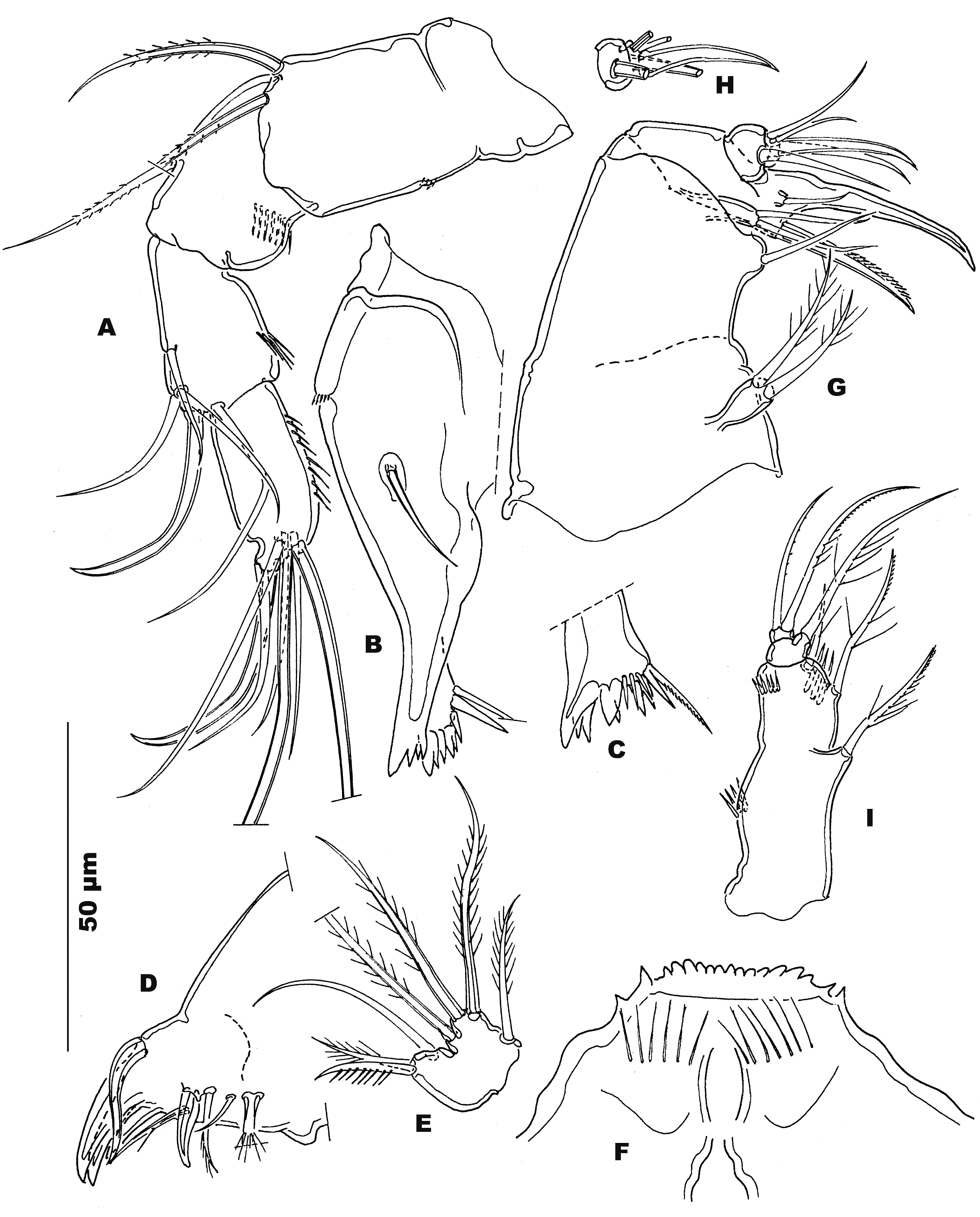
Rostrum ( Fig. 1 View FIGURE 1 E) roughly triangular with pointed apex and pair of sensilla near transition between rostrum and cephalothorax. Sieve plates not observed.
Antennule ( Fig. 1 View FIGURE 1 F) 10-segmented, reaching halfway along cephalothorax when deflected backwards. Armature formula (from proximal to distal segment; Aesth=aesthetasc):1(7)-2(7)-3(3)-4(1)-5(2)-6(3)-7(2+Aesth)- 8(2)-9(2+Aesth)-10(7+Aesth). Aesthetasc on segment 7 linguiform, on segment 9 filiform, and on segment 10 tubiform. First segment with proximal comb of long and slender spinules, subsequent segments without ornamentation. Conical element on segment 4 short.
Antenna ( Fig. 3 View FIGURE 3 A) 4-segmented with 2 abexopodal setae but without exopodite vestige on coxobasis; spinule pattern on coxobasis limited to small cluster of minute spinules near middle of outer margin; endopodite segments (from proximal to distal) with 1, 5 and 7 setae, respectively. Outer margin of endopodite segments with spinules, surfaces smooth.
Mandible ( Figs. 3 View FIGURE 3 B, C) with robust multi-cuspidate medial margin; palp obsolete, represented by single stout seta. Accessory element on cutting edge associated with large prominent spinule. Surface of gnathobasis smooth, except for few minute spinules on outer half of frontal margin and near the medial margin.
Labrum ( Fig. 3 View FIGURE 3 F) with multicuspidate margin and two combs of long and widely spaced slender spinules. Maxillulary arthrite ( Fig. 3 View FIGURE 3 D) with 3 medial claw-shaped naked elements, confluent with segment. Ventral subdistal element naked and long. Dorsal margin bearing 5 elements with outermost one (broken in Fig. 3 View FIGURE 3 D) long and densely pinnate. Maxillulary endopodite fused with basis ( Fig. 3 View FIGURE 3 E), bearing 3 pinnate setae. Basis with seta representing exopodite. Medial margin with 1 pinnate and 1 serrate element. Subdistal element naked.
Maxilla ( Fig. 3 View FIGURE 3 G) 4-segmented with incomplete remnant separation between precoxa and coxa. Proximal and distal endite with 2 terminal setae, median endite represented by single seta. Basis with smooth claw, serrate accessorial element, and short seta on posterior surface. Endopodite ( Fig. 3 View FIGURE 3 H) 2-segmented with, on proximal segment, 2 rigid elements, on distal segment, 2 slender lateral setae and a stout claw-shaped smooth terminal element. Surface of segments without ornament.
Maxilliped ( Fig. 3 View FIGURE 3 I) composed of 3 segments: syncoxa, basis and one-segmented endopodite. Syncoxa with 3 elements: 1 precoxal and 2 coxal. Basis with one seta, endopodite with 2. Two spinule rows along outer margin of syncoxa and one medial cluster near insertion of coxal setae.
Legs 1–4 ( Figs. 4 View FIGURE 4 A–D, respectively) intercoxal sclerites large, with crescentic distolateral edges and concave distal margin. Surface and margins devoid of spinular ornamentation. Precoxa smooth. Medial coxal seta present in legs 1–4, rather long and pinnate. Coxal surface smooth frontally and caudally in legs 1 and 2. Coxa of leg 3 frontally smooth, caudally with spinular comb near proximal outer corner. Coxa of leg 4 frontally smooth, with spinular comb near proximal outer corner and near distal margin on caudal surface. Medial spine on leg 1 basis robust, serrate along outer margin only. Medial basis margin of legs rounded and hairy. Distal margin of leg basis set with spinules midway (legs 1–4, not illustrated for legs 3 and 4) and near insertion of medial spine in leg 1. All rami distinctly 2-segmented, exopodites with solid appearance. Setal armature slender and pinnate. Distal spine of second leg 4 endopodite segment longer than segment (ratio 1/1.5) and inserted between 2 minute triangular expansions on distal segment margin. Exopodite spine formula: 2.3.3.2, exopodite seta formula 4.4.4.3.
Leg 5 ( Figs 2 View FIGURE 2 A, B) positioned on ventrolateral margin of pediger, segments undifferentiated. Basal segment vestige inserted on short cylindrical pedestal, sparsely pinnate. Distal segment vestige represented by narrow crescentic expansion bearing 2 elements: medial one shortest and smooth, outer one 1.3 times longer than median one, pinnate with short setules.
Leg 6 ( Fig. 2 View FIGURE 2 B) represented by 3 elements: outermost longest, setiform and pinnate, middle and inner one short and triangular, smooth, with hyaline appearance. Leg 6 valves naked. Copulatory pore orifice in middle of anterior third of double-somite, located in small depression (arrowed in Fig. 2 View FIGURE 2 A). Pore canal extending caudally, fitting in V-shaped caudal bend of lateral arms. Caudal seminal receptacula narrowly ovoid, laterally extended. Anterior part of receptacle irregularly ovate with lobate margin.
Male. Unknown.


No known copyright restrictions apply. See Agosti, D., Egloff, W., 2009. Taxonomic information exchange and copyright: the Plazi approach. BMC Research Notes 2009, 2:53 for further explanation.
|
Kingdom |
|
|
Phylum |
|
|
Class |
|
|
Order |
|
|
Family |
|
|
Genus |