
Chaetonotus (Primochaetus) heideri Brehm, 1917
|
publication ID |
https://dx.doi.org/10.11646/zootaxa.3701.5.3 |
|
publication LSID |
lsid:zoobank.org:pub:472882BF-6499-47D3-A242-A8D218BE2DFD |
|
persistent identifier |
https://treatment.plazi.org/id/C1146C7C-4C26-FF9A-02CD-C6AC1CA4FEF7 |
|
treatment provided by |
Plazi (2016-04-14 21:25:55, last updated 2017-01-16 11:40:15) |
|
scientific name |
Chaetonotus (Primochaetus) heideri Brehm, 1917 |
| status |
|
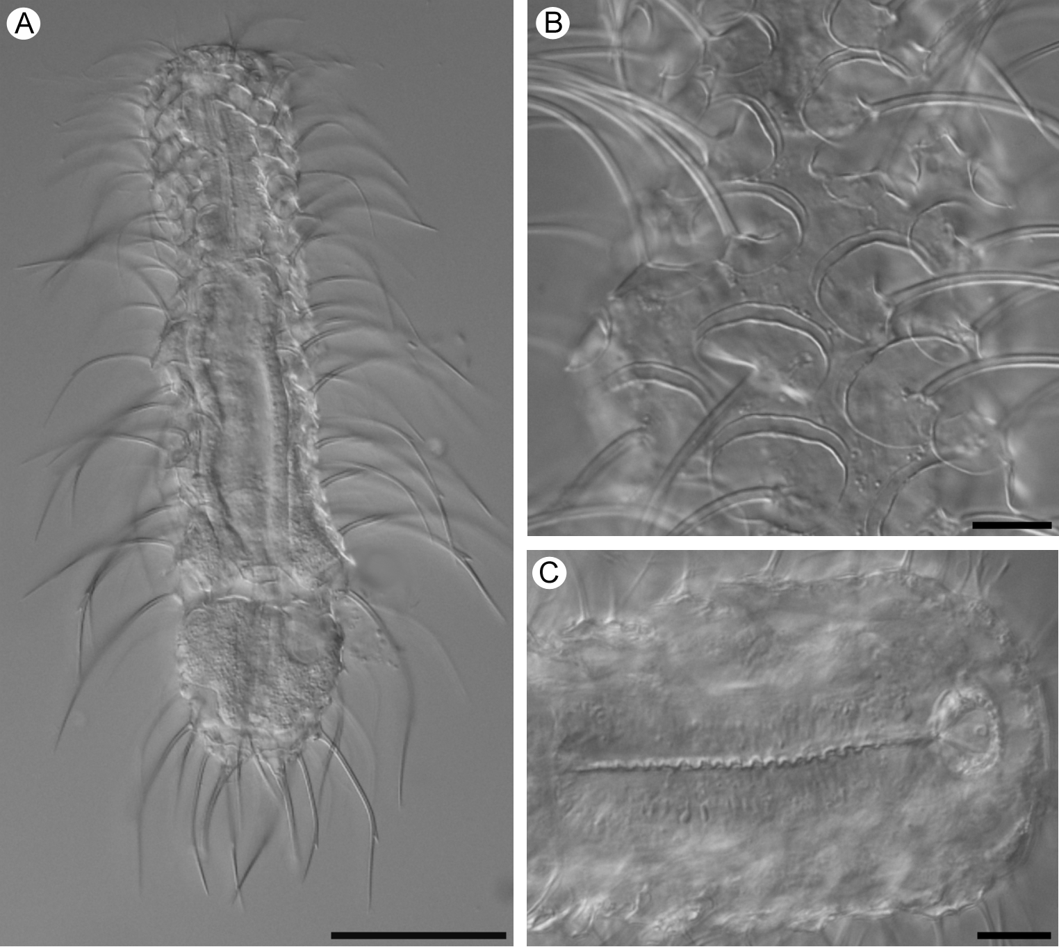
Chaetonotus (Primochaetus) heideri Brehm, 1917
( Fig. 18 View FIGURE 18 )
Localities: Lake Ånnsjön, Jämtland (N 63 º 15 ’ 65 ’’; E 12 º 27 ’ 03’’), July 8, 2008; Storlien E, Jämtland (N 63 º 18 ’ 42 ’’; E 12 º 06’ 43 ’’), July 10, 2008; Snasahögarna, Jämtland (N 63 º 12 ’ 39 ’’; E 12 º 18 ’ 19 ’’), July 7, 2008; Lake Gevsjön, Jämtland (N 63 º 22 ’ 14 ’’; E 12 º 42 ’ 10 ’’), July 10, 2008; Freshwater Rockpool among Sphagnum spp., Hållö, Bohuslän (N 58 º 20 ’ 03’’; E 11 º 12 ’ 50 ’’), July 14, 2009; Saltö, Bohuslän (N 58 º 52 ’ 21 ’’; E 11 º 07’ 34 ’’) September 13, 2008; Skaftö, Bohuslän (N 58 º 14 ’ 39 ’’; E 11 º 27 ’ 16 ’’), July 23, 2009; Bog at highway E 10, Lapland (N 68 º 25 ’ 58 ’’; E 18 º 23 ’ 00’’), July 1, 2010; A small pond close to lake Torneträsk at Abisko Scientific Research Station, Lapland (N 68 º 21 ’ 19 ’’; E 18 º 49 ’ 21 ’’), June 30, 2010.
Material: 10 specimens.
TL, 113–195 µm; FL, 20–25 µm; AL, 15–18 µm; PhL, 36–44 µm; MD, 8–10 µm; TNC, 16–17; DC, 7–9; DR, ~ 20; VLC, 8; HS, 3– 4 x 3–4 µm; NS, 3– 8 x 3–12 µm; DS, 7– 12 x 9–16 µm; HSp, 4–9 µm; NSp, 10–16 µm; DSp, 16–36 µm; VC, 5–7.
A very variable species (Balsamo 1980; Schwank 1990). Head rounded to weakly five-lobed with two pairs of cephalic sensory ciliary tufts. Cephalion and pleurae weakly developed. Hypostomium sometimes developed as a more or less concave bar (Schwank 1990). Two pairs of dorsal sensory bristles; anterior pair inserted between scales, posterior pair emerging from rounded pentagonal double-keeled scales. Furca relatively short with adhesive tubes constituting 2 / 3–4 / 5 of the total furca length. Dorsal surface covered with heart-shaped to pentagonal scales, sometimes with more or less well developed double anterior edge (Schwank 1990). Scales increase in size from the head region to mid-posterior trunk region with a subsequent decrease towards the caudal end. Barbed spines originate from approximately the center of each scale, increasing in length from anterior to posterior. The posteriormost spines can overshoot the furca.
Ventrolateral scales similar in appearance to those of the dorsal surface, decreasing in size medially. Ventral interciliary field covered with numerous keeled rounded scales or several columns (5–7) of rounded spined scales (Balsamo 1980). At the posterior end of the interciliary field 2–4 scales with very long simple spines sometimes present. The medial spines can be longer than the furca.
Mouth large. Pharynx with weak swellings at both ends. PhIJ at approximately U 26. Intestine straight with anus at approximately U 84.
The Swedish specimens were all adults in parthenogenetic phase. They showed a high variability in size of scales and length of spines. According to Balsamo (1980) C. (P.) heideri is a very variable species. The Swedish specimens agree more or less with form C and D (Balsamo 1980). It is very likely that C. (P.) heideri is a complex of closely related morphological species. The species is fairly common and could serve as a model organism to study species delimitation within freshwater chaetonotid gastrotrichs.
Previously reported from France (Grilli et al. 2008), Germany (Brehm 1917), Great Britain (Martin 1990?), Italy (Balsamo 1983), Poland (Kisielewski 1981), Romania (Rudescu 1967), Russia (Preobrajenskaja 1926), Sweden (Kånneby et al. 2009; 2013), Brazil (Kisielewski 1991), Canada (Schwank 1990) and USA (Emberton 1981).
No known copyright restrictions apply. See Agosti, D., Egloff, W., 2009. Taxonomic information exchange and copyright: the Plazi approach. BMC Research Notes 2009, 2:53 for further explanation.
|
Kingdom |
|
|
Phylum |
|
|
Class |
|
|
Order |
|
|
Family |
|
|
Genus |
1 (by plazi, 2016-04-14 21:25:55)
2 (by ImsDioSync, 2017-01-16 11:38:02)
3 (by ImsDioSync, 2017-01-16 11:40:15)
4 (by ImsDioSync, 2019-03-29 20:57:03)
5 (by ExternalLinkService, 2019-09-26 14:50:56)
6 (by ExternalLinkService, 2021-10-29 03:18:45)
7 (by ExternalLinkService, 2021-10-29 10:12:22)
8 (by ExternalLinkService, 2021-10-31 08:12:10)
9 (by plazi, 2023-10-26 19:20:28)