
Passalus ( Pertinax ) occidentalis Hincks, 1950
|
publication ID |
https://doi.org/10.1080/00222933.2020.1759721 |
|
persistent identifier |
https://treatment.plazi.org/id/C4008788-6906-9E0F-FF35-FD1F81F26E28 |
|
treatment provided by |
Carolina |
|
scientific name |
Passalus ( Pertinax ) occidentalis Hincks, 1950 |
| status |
stat. nov. |
Passalus ( Pertinax) occidentalis Hincks, 1950 n. stat. ( Figures 5 – 6 View Figure 5 View Figure 6 )
Passalus mancus occidentalis Hincks, 1950: 1041 ; Hincks and Dibb 1958: 16; Miles 2017:
120, fig. 18.
Diagnosis. Large size species; macropterous, with straight anterior frontal edge; mediofrontal area with coarse punctuations located mainly in the anterior region; tall and strong posterofrontal ridges; small laterofrontal + mediofrontal tubercles; reduced pronotal punctuations concentrated only near the lateral fossae; finely pubescent prepisternum; metatarsal with disc well delimited by a carina with group of coase punctuations located in the posterior region; conspicuous and fine metasternal pubescence; broad metasternal lateral grooves.
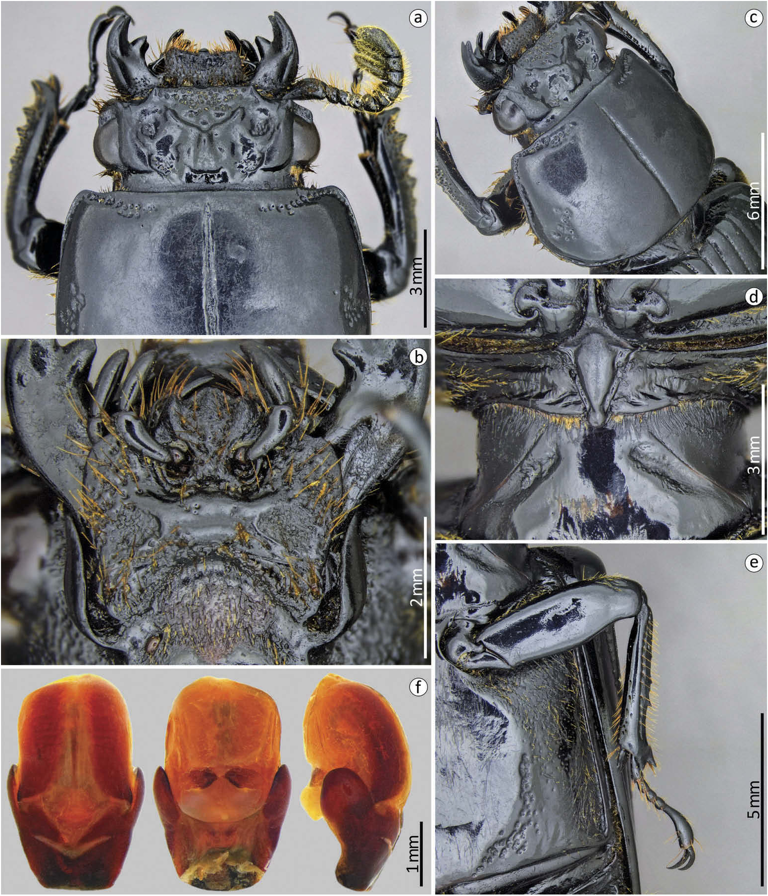
Redescription. Body ( Figure 5 View Figure 5 ): habitus: flat; size: big (< 30 mm in length). Head ( Figure 6 View Figure 6 (a,c)): Labrum: anterior border slight concave. Clypeus: hidden under the frons; with anterior angles under the lateral + mediofrontal tubercles. Anterior frontal edge: straight, without a notch in the median region. Secondary mediofrontal tubercles: absent. Laterofrontal + mediofrontal tubercles: small and obtuse. Mediofrontal area: transversal, 2.2x wider than long, flat, and shallow; with coarse punctuation scattered throughout the entire anterior region to the cephalic nodule. Cephalic nodule: small, well-marked and oval-shaped. Inner tubercles: small, conspicuous, slightly larger than the laterofrontal + mediofrontal tubercles, of which they are separated, but located closer to these than the apex of the central tubercle. Anterofrontal ridges: absent. Posterofrontal ridges: high, strong, and slightly sinuous, beginning at the base of the central tubercle. Laterofrontal areas: flat and smooth surface. Central tubercle: small, conical, low, with non-free apex, and back with slight groove. Lateroposterior tubercles: conspicuous, close to the central tubercle. Postfrontal area: flat, smooth surface. Postfrontal groove: well marked. Lateropostfrontal areas: deep, divided into two parts by a ‘ bridge ’ with punctuations in both areas. Epicranial sutures: poorly marked. Epicranial pits: shallow but clearly visible. Anterior angles of the head: undeveloped, with acute apex, smaller than the laterofrontal + mediofrontal tubercles. Canthus ocular: apexes rounded not reaching half eye. Antennas: trilamellate, with robust lamellae, the distal being wider than the other two. Mouthparts ( Figures 5 View Figure 5 (b), 6(b)): Ligula : tridentate with a larger middle tooth and narrower than the lateral teeth. Hypostomal process: wide, glabrous, and separate from mentum. Mentum mediobasal area: not dilated, with very few small punctuations in the posterior area; not protruded anterior region with a slight notch. Mentum lateral lobes: rounded apexes. Mentum lateral scars: large, shallow, and rounded. Mandibles: incisor lobe with three wellformed teeth at apex; robust suprainternal teeth; inconspicuous infrabasal pits. Maxilla: lacinia bidentate at the apex. Prothorax. Pronotum ( Figures 5 View Figure 5 (a,c), 6(a – c)): Anterior edge: straight. Anterior angles: straight, slightly obtuse. Marginal groove: well marked, shallow and with coarse punctuations in the anterior region, with dilated apex; reaching more than two-thirds of the pronotal width in the anterior margin. Lateral fossae: small, well marked and softly deep; irregular format. Pronotal punctuations: coarse, concentrated near the lateral fossae. Prosternum ( Figures 5 View Figure 5 (b), 6(d)): Prepisternum: not dilated in the anterior region; pubescence throughout the lateroposterior region. Prepimerum: glabrous. Prosternelum: rhomboid with narrow apex. Mesosternum ( Figures 5 View Figure 5 (b), 6(d)): smooth, shiny, and glabrous. Mesosternal scars: long, broad, and deep; without punctuations or pubescence. Metasternum ( Figures 5 View Figure 5 (b), 6(e)): Metasternal disc: well delimited by a carina; coarse and dense punctuations in the lateroposterior region. Metasternal punctuations: setigerous punctuations throughout the region adjacent to metasternum. Metasternal pubescence: fine and conspicuous in throughout the region adjacent to metasternum and in the metasternal lateral groove. Metasternal lateral groove: narrow, with broad apex as broad as mesotibiae; shallow, not punctuated. Elytra ( Figure 5 View Figure 5 (a)): Approximately 2.6x longer and 1.1x wider than pronotum. Striae: narrower than the interstriae, marked with round and thin punctuations, inconspicuous in the dorsal striae and well marked in the lateral striae. Epipleura : glabrous. Humeri: glabrous. Legs ( Figures 5 View Figure 5 (a,b), 6(e)): Profemur: ventral anterior border with well-marked groove, not reaching the apex of the profemur; ventral posterior border with long setae near the apex. Protibiae: not dilated. Mesotibiae: two small spines on the outer face. Metatibiae: without spine. Abdomen ( Figure 5 View Figure 5 (b)): sternite VII with well-marked and incomplete groove; slightly rough sides. Aedeagus ( Figure 6 View Figure 6 (f)): In ventral view: median lobe with smaller width than the parameres and the basal piece together and almost as long as these; with two sclerotised plaques on the lateral margin. Basal piece and parameres separate; parameres projections not reach 1/4 the length of the median lobe; arched anterior edges of the parameres, with a groove separating them medially; basal piece without re-enactment at anterior region. In side view, slightly rounded apex of the parameres. In dorsal view with separate parameres.
Dimensions. Total length: 32 mm; cephalic length: 3.1 mm; cephalic width: 6.5 mm; mediofrontal area length: 1.3 mm; mediofrontal area width: 2.7 mm; canthus ocular length: 0.7 mm; canthus ocular width: 0.3 mm; area of the mediofrontal area: 2.5 mm 2; mandibles external angle: 148º; antennal club length: 1.4 mm; antennal club width: 1.3 mm; distal lamella length: 0.5 mm; medial lamella length: 0.3 mm; width of the mentum at the lateral scars: 3.3 mm; mentum mediobasal area width: 1.3 mm; diameter of the mentum lateral scars: 0.8 mm; pronotal length: 7 mm; pronotal width: 9.4 mm; length of the pronotal anterior groove: 2.8 mm; elytral length: 18.5 mm; elytral width: 11 mm; humeral width: 9.6 mm; profemur length: 5 mm; length of the anterior ventral groove of the profemur: 2.7 mm; protibiae width: 1.2 mm; protarsal length: 3.6 mm; length of the last protarsomer: 1.4 mm; mesotibiae width: 0.7 mm; metasternal lateral groove width: 0.7 mm; metasternal disc length: 7.8 mm; abdomen length: 7.7 mm; aedeagus length: 3.2 mm; aedeagus width: 2.1 mm; median lobe length: 2.5 mm; paremeres projection lenght: 0.6 mm; basal piece lenght: 0.8 mm.
Material examined: type material. Holotype ( ♂) F2439.51. ECUADOR: San José de Chimbo// Passalus (Pertinax) / mancus Burm. ssp./ occidentalis Hincks /Type/det. W.D. Hincks//Manchester Museum/ HOLOTYPE //F2439.51 ( MMUE).
Passalus (Pertinax) mancus . Material examined. BRAZIL: Bahnhof Alto da Serra /S . [ São ] Paulo . Brasil . x. 26/leg . Luederwaldt /via Benesh // Passalus / mancus Burm ./det . Luederwaldt // Manchester Museum . 1♂ ( MMUE) . P. mancus Burm. / Bahnhof Alto da Serra /S . [ São ] Paulo . x. 26/leg. et det . Luederw ./via Benesh.//Manchester Museum. 1♂
( MMUE). Nova Teotonia/ Brasilien /27ºS – 52-53ºW/ 25 fev 1927 // Pertinax mancus /Burm. 1♀ ( INPA). Nova Teotonia/ Brasilien /27ºS – 52-53ºW/ 14 March 1927 // Pertinax mancus / Burm. 1♀ ( INPA). Itatiaya – 1100 m./km 5/13 – 7 – [1]933/J.F.Zikán// Pertinax / mancus Burm. //Coleção/J. F. Zikan//Nº 7.693/I. O. C. Coleoptera // Pertinax /3// Passalus mancus / Burmeister, 1847/M. Bevilaqua Det. 2019. 1♀ ( UFAM). Rio Grande/do Sul/N. [Nova] Petrópolis/P. buck leg./I. 28.// Passalus / mancus Burm. /Luederw. det. 28//Coleção/ J. F. Zikan//Nº 7.695/I. O. C. Coleoptera // Passalus mancus /Burmeister, 1847/M. Bevilaqua Det. 2019. 1♀ ( UFAM). Alto Itatiaya/ 2200 m./1. vii. 33/J.F.Zikan//Coleção/ J. F. Zikan// Passalus mancus /Burmeister, 1847/M. Bevilaqua Det. 2019//CEIOC/12333. 1♀ ( CEIOC). Itatiaia/E. [Estado] do Rio – Brasil /J.F.Zikan/21 – vii – 33//Coleção J. F. Zikan// Passalus mancus /Burmeister, 1847/M. Bevilaqua Det. 2019//CEIOC/12334. 1♀ ( CEIOC).
Remarks. Hincks originally described this species as a subspecies of Passalus mancus Burmeister as a ‘ geographic form ’ of this widely distributed species. However, the analysis of the specimen allowed us to verify that they are two distinct species, which differ by size, since P. occidentalis n. stat. is bigger ( 30 – 32 mm in length) while P. mancus is smaller ( 20 – 29 mm in length); by the posterofrontal ridges, which are stronger, taller and sinuous in P. occidentalis , while in P. mancus they are weaker, shorter and straighter; by the punctuations at mediofrontal area which in P. occidentalis are more abundant and coarse, while in P. mancus it ’ s smooth or poorly punctuated; by the cephalic nodule present in P. occidentalis and absent in P. mancus ; by the lateroposterior tubercles, conspicuous and prominent in P. occidentalis , while in P. mancus are inconspicuous and poorly detached; by the mesosternal scars that are elongated, wide and deep in P. occidentalis , while in P. mancus they are slightly triangular-shape, shallow with opaque surface; by the metasternal punctuations that in P. occidentalis are more abundant and reach the median region of the metasternal disc forming a carina, while in P. mancus these punctuations are concentrated only in the posterior region formed by a group of few, sometimes absent, thin punctures, and by the aedeagus that in P. occidentalis presents larger and wider median lobe, with smaller and more distant parameres in ventral view, separated from the basal piece only in the median region in ventral view, while in P. mancus the median lobe is smaller and narrower, with larger and closer parameres in ventral view, totally separated from the basal piece by a suture. P. occidentalis n. stat. also resembles P. matilei Boucher since both species are large, macropterous, have coarse punctuations in the mediofrontal area, wide and deep mesosternal scars, bristles in metasternum, wide metasternal lateral grooves and by the shape of aedeagus. However, they differ by the P. occidentalis n. stat. be a little smaller in body length, have coarser and deeper metasternal punctuations, reaching the median region of the metasternal disc and less dense metasternal pubescence, while P. matilei presents thinner metasternal punctuations that do not reach the median region of the metasternal disc, besides presenting denser mestasternal pubescence. It is possible that these species are synonymous, but without proper analysis of the holotype of P. matilei there is no way to take any position, but if they are distinct species, considering the descriptions of Passalus matilei , P. mancus and P. occidentalis , it is possible that they form a group of medium/large-sized species with developed wings that inhabit Andean and Atlantic forest regions.
Distribution. Ecuador (San José de Chimbo).

No known copyright restrictions apply. See Agosti, D., Egloff, W., 2009. Taxonomic information exchange and copyright: the Plazi approach. BMC Research Notes 2009, 2:53 for further explanation.
|
Kingdom |
|
|
Phylum |
|
|
Class |
|
|
Order |
|
|
Family |
|
|
Genus |
Passalus ( Pertinax ) occidentalis Hincks, 1950
| Bevilaqua, Marcus & Fonseca, Claudio Ruy Vasconcelos da 2020 |
Passalus mancus occidentalis
| Hincks WD & Dibb JR 1958: 16 |
| Hincks WD 1950: 1041 |