
Passalus ( Pertinax ) cuneatus Hincks, 1950
|
publication ID |
https://doi.org/10.1080/00222933.2020.1759721 |
|
persistent identifier |
https://treatment.plazi.org/id/C4008788-690A-9E14-FEF4-FCA881436A53 |
|
treatment provided by |
Carolina |
|
scientific name |
Passalus ( Pertinax ) cuneatus Hincks, 1950 |
| status |
|
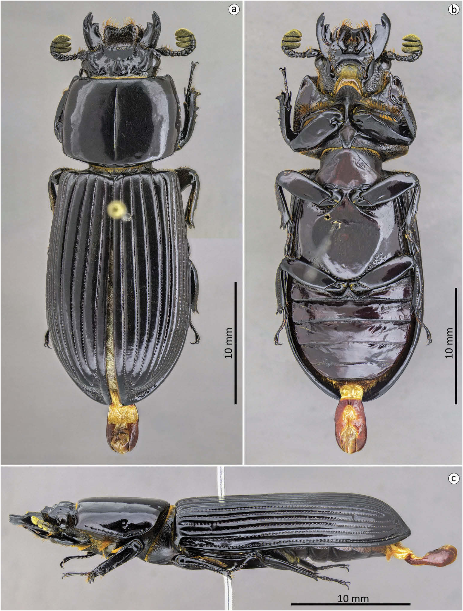
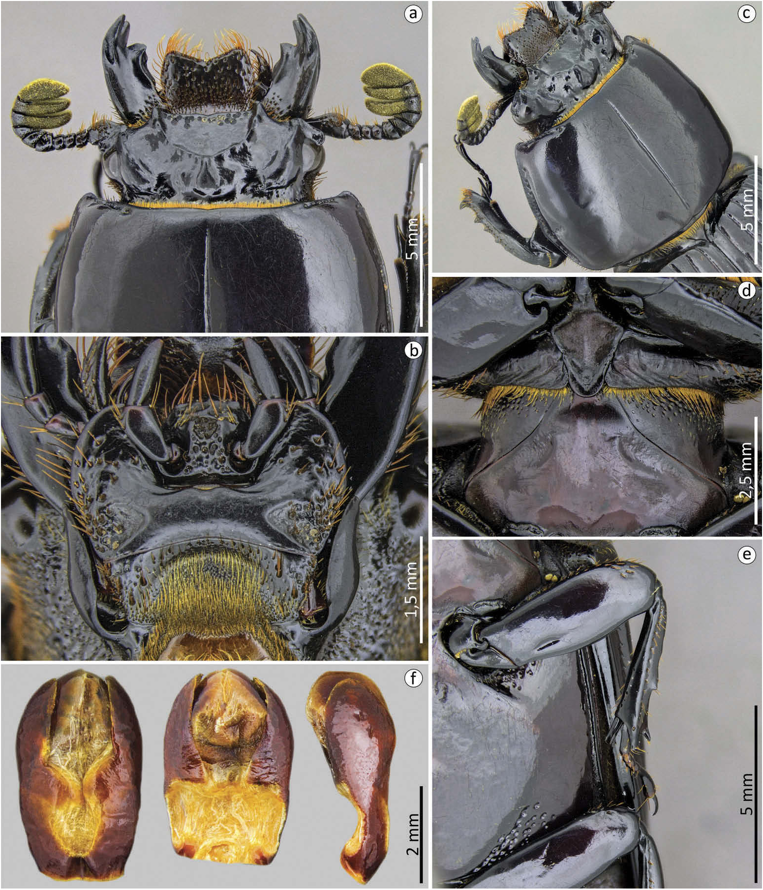
Passalus ( Pertinax) cuneatus Hincks, 1950 ( Figures 3 – 4 View Figure 3 View Figure 4 )
Hincks 1950: 1043, fig. 2; Hincks and Dibb 1958: 16; Reyes-Castillo 1970: 204; Reyes-
Castillo and Castillo 1992: 357; Miles 2017: 119, fig 12.
Diagnosis. Large size species; flat body; large and straight mandibles; straight anterior edge of the front; labrum with a notch in the ‘ V ’ shaped; large and smooth mediofrontal area with a row of punctures close to the anterior edge of the front; laterofrontal + mediofrontal tubercles practically attached to inner tubercles; posterofrontal ridges larger and lower than the anterofrontal ridges; practically absent pronotal punctuations; deep mesosternal scars; poorly delimited metasternal disc; metasternum glabrous with a small group of punctures in the posterior region; aedeagus with parameres reaching almost the apex of the median lobe and tegmen with wide opening in the median region in ventral view.
Redescription. Body ( Figure 3 View Figure 3 ): habitus: flat; size: big (< 30 mm in length). Head ( Figure 4 View Figure 4 (a,c)): Labrum: anterior border concave. Clypeus: hidden under the frons; with anterior angles under the lateral + mediofrontal tubercles. Anterior frontal edge: straight, with a notch in the median region. Secondary mediofrontal tubercles: absent. Laterofrontal + mediofrontal tubercles: conspicuous, acute, and projected forward. Mediofrontal area: caliciform 2.2x wider than long, flat and deep; with coarse punctuations only in the anterior region, near the anterior frontal edge. Cephalic nodule: large but not well marked and rounded shape. Inner tubercles: large, conspicuous with acute apexes, little smaller than the laterofrontal + mediofrontal tubercles, of which are practically attached by the anterofrontal ridges. Anterofrontal ridges: straight, smaller, and taller than the posterofrontal ridges. Posterofrontal ridges: low, strong, and straight, beginning at the apex of the central tubercle. Laterofrontal areas: slightly convex and smooth surface. Central tubercle: conical, small, low, with non-free apex and dilated back. Lateroposterior tubercles: inconspicuous, far from the central tubercle, with rounded shape. Postfrontal area: flat, smooth surface. Postfrontal groove: well marked and with a slight notch located medially. Lateropostfrontal areas: shallow, smooth, and glabrous surface. Epicranial sutures: well marked. Epicranial pits: deep and very visible. Anterior angles of the head: small, with obtuse apices, smaller than the laterofrontal + mediofrontal tubercles. Canthus ocular: apexes rounded not reaching half eye. Antennas: trilamellate, with robust lamellae, the distal being wider than the other two. Mouthparts ( Figures 3 View Figure 3 (b), 4(b)): Ligula : tridentate with a larger middle tooth and narrower than the lateral teeth, which are inconspicuous; punctuations mainly concentrated in the median region. Hypostomal process: wide, glabrous and near the mentum. Mentum mediobasal area: not dilated, smooth and glabrous, with non-protruding anterior region, notched in the middle. Mentum lateral lobes: rounded apexes. Mentum lateral scars: deep and oval-shape. Mandibles: incisor lobe with three well-formed teeth at apex; robust suprainternal teeth; infrabasal pits absent. Maxilla: lacinia bidentate at the apex. Prothorax. Pronotum ( Figures 3 View Figure 3 (a,c), 4(a – c)): Anterior edge: slightly sinuous. Anterior angles: straight and slightly protruding. Marginal groove: well-marked, deep in the lateral region, with coarse punctures throughout its extension, without dilation at the apex; reaching almost two-thirds of the pronotal width in the anterior margin. Lateral fossae: small, well marked and slighty deep; slightly rounded shape. Pronotal punctuations: practically absent, formed by a group of two to five punctures concentrated only in the lateral fossae. Prosternum ( Figures 3 View Figure 3 (b), 4(d)): Prepisternum: not dilated in the anterior region; fine pubescence throughout the lateroposterior region, being almost inconspicuously present on the margins of the anterior region. Prepimerum: glabrous. Prosternelum: rhomboid with broad and rounded apex. Mesosternum ( Figures 3 View Figure 3 (b), 4(d)): smooth, shiny and glabrous. Mesosternal scars: long, broad and deep; without punctuations or pubescence. Metasternum ( Figures 3 View Figure 3 (b), 4(e)): Metasternal disc: poorly delimited, but more dilated than the lateral region of the metasternum. Metasternal punctuations: group of punctures only in the lateroposterior region. Metasternal pubescence: absent. Metasternal lateral groove: narrow, thinner than mesotibiae, deep, without punctures or bristles. Elytra ( Figure 3 View Figure 3 (a)): Approximately 2.6x longer and 1.2x wider than pronotum. Striae: narrower than the interstriae, marked with round and inconspicuous punctuations on the dorsal striae and well defined in lateral striae. Epipleura : glabrous. Humeri: finely pubescent. Legs ( Figures 3 View Figure 3 (a,b), 4(e)): Profemur: ventral anterior border with poorly marked groove, not reaching the apex of the profemur; ventral posterior border with few setae only near the apex. Protibiae: not dilated. Mesotibiae: one or two small spines on the outer face. Metatibiae: one small spine on the outer face. Abdomen ( Figure 3 View Figure 3 (b)): sternite VII with well-marked and incomplete groove; smooth sides. Aedeagus ( Figure 4 View Figure 4 (f)): Median lobe with smaller width than the parameres and the basal piece together and almost as long as these. In ventral view with two sclerotised plaques on the lateral margin; basal piece and parameres fused forming a tegmen; parameres projected upwardly encapsulating the median lobe; anterior margin of parameres straight in the ‘ V ’ -shape. In lateral view, the apex of the parameres are slightly rounded. In dorsal view with widely separated parameres.
Dimensions. Total length: 34 mm; cephalic length: 2.8 mm; cephalic width: 6.8 mm; mediofrontal area length: 1.2 mm; mediofrontal area width: 2.7 mm; canthus ocular length: 0.9 mm; canthus ocular width: 0.3 mm; area of the mediofrontal area: 2.5 mm 2; mandibles external angle: 158º; antennal club length: 1.5 mm; antennal club width: 1.6 mm; distal lamella length: 0.6 mm; medial lamella length: 0.3 mm; width of the mentum at the lateral scars: 3.5 mm; mentum mediobasal area width: 1.6 mm; diameter of the mentum lateral scars: 0.7 mm; pronotal length: 7.5 mm; pronotal width: 10 mm; length of the pronotal anterior groove: 1.3 mm; elytral length: 19.8 mm; elytral width: 12.5 mm; humeral width: 9.7 mm; profemur length: 4.1 mm; length of the anterior ventral groove of the profemur: 1.2 mm; protibiae width: 1 mm; protarsal length: 3.4 mm; length of the last protarsomer: 1.1 mm; mesotibiae width: 0.8 mm; metasternal lateral groove width: 0.2 mm; metasternal disc length: 7.7 mm; abdomen length: 7.7 mm; aedeagus length: 4.4 mm; aedeagus width: 2.6 mm; median lobe length: 2.3 mm; paremeres projection lenght: 1.8 mm.
Material examined: type material. Holotype ( ♂) F2439.50. PANAMA: Chiriquí / Panama // Ex Staudinger &/Bang Haas, Dresden// Passalus (Pertinax) / cuneatus Hincks /Type/det. W.D. Hincks//Manchester Museum/ HOLOTYPE // ♂ dissect/S. Boucher det. 2012//F2439.50 ( MMUE).
Remarks. Hincks comments that this species is similar to Passalus punctatostriatus Percheron ; however, we believe it ’ s more similar to P. aequatorialis (Kirsch) , because both species have large body size (< 30 mm), macropterous, smooth frontal area with few punctures and pronotal and metasternal punctuations practically absent. However, the set of diagnostic characters of P. cuneatus distinguishes it from the other species of Passalus .
Distribution. Panama ( Chiriquí).
| MMUE |
Museum of Manchester University |
No known copyright restrictions apply. See Agosti, D., Egloff, W., 2009. Taxonomic information exchange and copyright: the Plazi approach. BMC Research Notes 2009, 2:53 for further explanation.