
Aspidiophorus lamellophorus Balsamo, Hummon, Todaro et Tongiorgi, 1997
|
publication ID |
https://doi.org/10.11646/zootaxa.4027.4.2 |
|
publication LSID |
lsid:zoobank.org:pub:17C4DA79-3D4A-4E35-ACDC-2BF7B1708263 |
|
DOI |
https://doi.org/10.5281/zenodo.6113451 |
|
persistent identifier |
https://treatment.plazi.org/id/C52AAB2D-FFAA-FFE3-FF41-47A325757C58 |
|
treatment provided by |
Plazi |
|
scientific name |
Aspidiophorus lamellophorus Balsamo, Hummon, Todaro et Tongiorgi, 1997 |
| status |
|
Aspidiophorus lamellophorus Balsamo, Hummon, Todaro et Tongiorgi, 1997 View in CoL
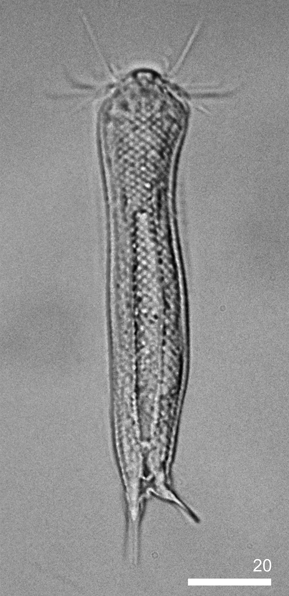
( Figs. 7–8 View FIGURE 7 View FIGURE 8 ; Table 3)
Locality. Southern Baltic Sea, outer Puck Bay, Jastarnia (N54° 41.4 E 18°40.9)
Material. Four specimens (all adult), 2 of which were photographed. The microphotographs are available in the Natural History Collections at Adam Mickiewicz University in Poznań under access number NHC-GAL- 11-1- 10 and in the collection of the first author.
Short description. Aspidiophorus lamellophorus is a species with a slender body, weakly marked neck constriction and narrow furca base. The head is short, semi-circular and five-lobed. The cephalion (U1–U3) is narrow and short and adheres to the head along its entire length. Epipleuria (U3–U4) are small and almost Character Ranges on adults specimens SD
Body length 112.78–117.35 3.231
Pharynx length 30.70–31.03 0.233
Width of anterior pharynx thickening (a) 8.16–9.09 0.658
Width of pharynx narrowing that follows anterior thickening (n) 5.72–6.09 0.262
Width of pharynx at its middle length (m) 6.66–6.74 0.057
Width of posterior pharynx thickening (p) 7.88–8.88 0.707
Length of cephalic cilia (anterior tuft) (4.08–4.18)–(18.75–18.81) 0.071; 0.042 Length of cephalic cilia (posterior tuft) (6.25–6.46)–(18.36–18.71) 0.148; 0.247 Hypostomium length 3.19–3.54 0.247
Hypostomium width 8.27–8.53 0.184
Cephalion length 6.41–6.67 0.184
Cephalion width 10.16–10.68 0.367; 0.696 Diameter of mouth ring 4.95–5.23 0.198
Furca length 18.60–18.77 0.120
Length of adhesive tube 10.40–10.49 0.064
Head scale length (1.67–1.73)–(2.17–2.39) 0.042; 0.156 Head scale width (2.13–2.17)–(2.69–2.72) 0.028; 0.021 Neck scale length (2.03–2.27)–(2.43–2.59) 0.170; 0.113 Neck scale width (2.50–2.64)–(2.88–3.01) 0.099; 0.092 Trunk scale length (2.10–2.35)–(3.01–3.53) 0.177; 0.368 Trunk scale width (2.70–2.89)–(3.64–4.03) 0.134; 0.276 Length of head ventral lamella (5.23–5.39)–(6.74–6.93) 0.113; 0.134 Width of head ventral lamella (2.16–2.52)–(2.38–2.88) 0.255; 0.354
......continued on the next page Second pair of posteriormost interciliary field scale length 9.15–9.25 0.071 Second pair of posteriormost interciliary field scale width 2.53–2.72 0.134 Number of ventral scales witch lamella in single longitudinal 35–39 2.828 row
Total number of longitudinal alternating rows of scales 31–32 0.707 Total number of longitudinal alternating rows of scales on 11 0 interciliary field
Pharynx formula a 26.58–29.29 1.919 Pharynx formula n 18.433–19.837 0.993 Pharynx formula m 21.463–21.954 0.347 Pharynx formula p 25.667–28.617 2.086 Ratio of scale distribution 60.78–71.43 7.531
unmarked in the body outline. Hypopleuria (U5–U8) are approximately three times larger than the epipleuria, flat and weakly marked in the head outline. The epipleuria are located dorsally and dorsolaterally, and the hypopleuria are located ventrolaterally. Ocellar granules are not present. The hypostomium (U5–U7) is wide and shaped like a square with rounded edges. It does not have reinforcements. Two pairs of cephalic ciliary tufts are present on the head. Each anterior tuft has four cilia that emerge around the dorsal edges of the epipleuria on U4/5. The first cilium in both anterior tufts is the shortest. The second cilium is very long (the longest of all cilia in the tuft). Two subsequent cilia are shorter than the second cilium but longer than the first one. Each posterior tuft has six cilia. The posterior tufts are located dorsally and emerge in a line directly beyond the ventral edges of epipleuria on U4/ 5. The first cilia emerge close to the cilia in the anterior tufts. The cilia gradually lengthen from the first cilium to the last one (see Table 3). The mouth ring is small and narrow and located subterminally on U2–U4. It has short cuticular reinforcements and is surrounded by suboral bristles. The pharynx (U4–U31) is narrow. Its anterior dilatation is clearly marked and stronger than the weakly marked posterior dilatation ( Fig. 8 View FIGURE 8 B). A weak reinforcement composed of a thin, strongly curved single cuticular rod is present inside the anterior dilatation on U6–U7. The pharynx connects to a straight intestine (U32–U88) through the pharyngeal-intestinal junction (U32). The junction is narrow and short. The intestine does not have a distinct anterior separate section different in form of morphology.
The head is similar in width to the trunk and separated from it by an indistinctly narrower neck ( Fig. 7 View FIGURE 7 ). The trunk slightly and gradually widens to its widest point located about its middle (U57). The trunk then gradually narrows up to a narrow, weakly marked furca base on U87. Furcal branches are close to each other. The furcal indentation is parabolic, and the ends of adhesive tubes point outwards. Adhesive tubes are straight, long and thin and do not taper towards their ends. The ends of the adhesive tubes are blunt.
The entire body, except for the ventral interciliary field, is covered with small pedunculated scales ( Figs. 7 View FIGURE 7 , 8 View FIGURE 8 A). The scales form 31–35 longitudinal alternating rows of 49–51 scales in each. The scales have a keel and are rhomboidal with a clearly marked peduncle base. They are regularly spaced, and their edges meet. Scales with lamellae form longitudinal rows located closest to ciliary bands, with 35–39 scales in each row. The lamellae are shaped like narrow rectangles and reach beyond the body edges.
Scales on the body vary slightly in size (see Table 3). They slightly and gradually become smaller from the dorsal side, through the dorsolateral, lateral, ventrolateral and ventral sides, to the ciliary bands. Furthermore, the scales become larger from the beginning of the head to the widest point of the trunk. Scales in the last longitudinal rows adjoining the ciliary bands are aligned diagonally at an angle of around 20° towards the bands.
On the ventral body side, locomotor cilia form longitudinal rows that run from U8 to U81 ( Fig. 8 View FIGURE 8 C). The longitudinal rows of cilia on the head and neck are wider than on the other body parts. The ventral interciliary field is covered with narrow, spineless scales with a clearly marked keel starting from about the beginning of the anterior pharynx dilatation (from U11). The scales of the ventral interciliary field are located close to one another, and their edges do not overlap. These scales are shaped like rectangles with slightly rounded edges. Scales on the ventral interciliary field gradually become larger from the front of the body to the widest part of the trunk, beyond which they slightly and gradually become smaller up to the furca base. The species has three pairs of terminal scales of the ventral interciliary field. The first, central pair of terminal scales is located on U86–U88. These scales are shaped like elongated ovals with a strong, long keel that runs along their entire length, and do not have spines. The second pair of scales is located on U84–U87, above and laterally in relation to the first pair. Scales in the second pair are similar in shape to those in the first one but are larger. The third pair (U87–U92) is located laterally in relation to the other two pairs on furcal appendages and is smaller than the others pairs. Scales in the third pair are shaped like narrow ovals. They are spineless and have a long, straight keel that runs along their entire length.
The species has three pairs of dorsal sensory bristles ( Fig. 8 View FIGURE 8 A). The first pair is located on the head, directly beyond the epipleuria (U5), and emerges from small, round papillae. The second pair is located dorsolaterally on the neck (U32) and emerges from small, round papillae as well. The third, posterior pair emerges from rhomboidal scales with two rectangular keels, located dorsolaterally on the posterior part of the trunk (U85–U86).
Taxonomic remarks. All the specimens of Aspidiophorus lamellophorus found in the Puck Bay correspond well with the original description. However, the adult individuals are slightly larger than the Italian specimens (112.8–117.4 Μm, compared to 109 Μm) and have a five-lobed, rather than three-lobed head (two pairs of pleuria are present, however, the epipleuria are small and very weakly marked), a hypostomium and three pairs of dorsal sensory bristles, instead of one pair. Furthermore, a weak cuticular reinforcement is present inside the anterior pharynx dilatation. The differences in body size may result from different environmental conditions ( e.g. lower water temperature, lower salinity and different sources of food). The observed differences of other traits may also represent phenotypic variability.
Emended differential diagnosis. Aspidiophorus lamellophorus is one of two species belonging to Aspidiophorus that has lamellae. In original description of A. lamellophorus authors ( Balsamo et al. 1997) not considered first species with lamellae from genus Aspidiophorus i. e. marine A. bisquamosus Mock, 1979 , therefore the comparison contained below is necessary.
A. lamellophorus shares with A. bisquamosus the following traits: the presence of ventral lamellae, pedunculated scales with clearly marked peduncle base and keel, body shape (no clearly marked neck narrowing) and a similar body size. However, it differs from A. bisquamosus in terms of the type and shape of lamellae (in A. bisquamosus , lamellae develop from spines and a keels. Spines and keels ends arise beyond lamellae posterior edges. Lamellae are smaller, shorter and shaped like an elongated tear and do not reach beyond the lateral body edges), the type, shape and alignment of scales (in A. bisquamosus , the scales are shaped like shields with rounded lower edges, are ornamented with three small dots on each scale and their edges overlap), the presence of scales of a different type on the furca base (on the furca base of A. bisquamosus , a pair of large scales with a long keel are present; these scales adhere to the cuticle with their entire surface) and the number, shape and alignment of terminal scales of the ventral interciliary field (in A. bisquamosus , four pairs of terminal scales of the ventral interciliary field are present, the second and third pairs are located one on top of the other, the fourth pair located on the inner side of the furcal branches and the second, third and fourth pairs possess spines).
Distribution. Previously, the species was recorded only at its locus typicus in the Adriatic Sea ( Balsamo et al. 1997; Todaro et al. 2003). The species was found in the mouth of the Isonzo River.

No known copyright restrictions apply. See Agosti, D., Egloff, W., 2009. Taxonomic information exchange and copyright: the Plazi approach. BMC Research Notes 2009, 2:53 for further explanation.
|
Kingdom |
|
|
Phylum |
|
|
Order |
|
|
SubOrder |
Paucitubulatina |
|
Family |
|
|
Genus |