
Gonatodes tapajonicus Rodrigues, 1980
|
publication ID |
https://doi.org/10.5281/zenodo.277513 |
|
DOI |
https://doi.org/10.5281/zenodo.5618486 |
|
persistent identifier |
https://treatment.plazi.org/id/C708878C-FFFD-995F-FF75-FE4FFA0DEC26 |
|
treatment provided by |
Plazi |
|
scientific name |
Gonatodes tapajonicus Rodrigues, 1980 |
| status |
|
Gonatodes tapajonicus Rodrigues, 1980
( Figs. 1 View FIGURE 1 C, 9, 10)
Gonatodes tapajonicus Rodrigues, 1980: 309 ( holotype MZUSP 53676, type-locality: Cachoeira do Limão, Rio Tapajós, Pará, Brazil); Vanzolini, 1986: 10; Avila-Pires, 1995: 283.
Material examined (numbers in bold between parentheses refer to localities in Fig. 3 View FIGURE 3 ): BRAZIL. PARÁ. Município Itaituba. Comunidade Aldeia Nova ("Aldêia Akay Muuybu"), anteriorly known as Comunidade Boa Fé, right margen of Rio Tapajós ( 04o 41' 58.2"S and 56o 22' 37.0"W) ( 45): MPEG 27708–27718, 2 juveniles, 5 males and 4 females, leg. M.J. Sturaro, P.L.V. Peloso and J. O. Gomes. Cachoeira do Limão, right margin of Rio Tapajós (~ 4o41’S and 56o21’W) ( 46): MZUSP 53676 ( holotype), male; MZUSP 53669, 53671–74, 53677 ( paratypes), a male and 5 female, leg. M.T. Rodrigues. Floresta Nacional de Altamira, Distrito de Moraes de Almeida ( 06º 1’8”S and 55º 18’17”W) ( 47): MPEG 25095, female, leg. C. Lima and R.R. Silva.
Diagnosis. A relatively large Gonatodes , with maximum SVL of 55 mm. Scales around midbody 118–133. Ventral scales in a longitudinal row 53–63. Proximal subdigital lamellae as wide as digits, in total 18–22 under fourth finger, 22–27 under fourth toe. Three, occasionally four, lateral rows of scales on distal portion of fingers and toes. Tail with midventral scales distinctly wider than long, forming a repetitive sequence of two single midventrals (one after the other), each in contact laterodistally with one scale per side, followed by a divided (only on proximal portion of tail, when present) or single midventral in contact laterodistally with two laterodistal scales per side (respectively 1’1’2” and 1’1’1”). A white suprahumeral ocellus, bordered by black, present both in males and females, although more conspicuous in males. Males with a vermiculated pattern of light and dark (vivid yellow and reddish brown in life) spots both on head and body dorsally; gular region with dark oblique streaks (yellow with reddish brown streaks in life).
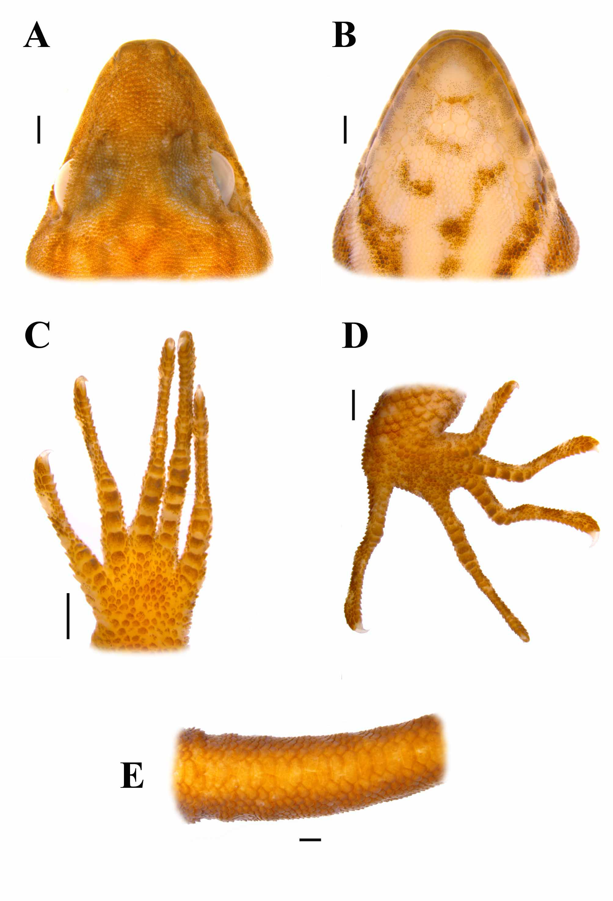
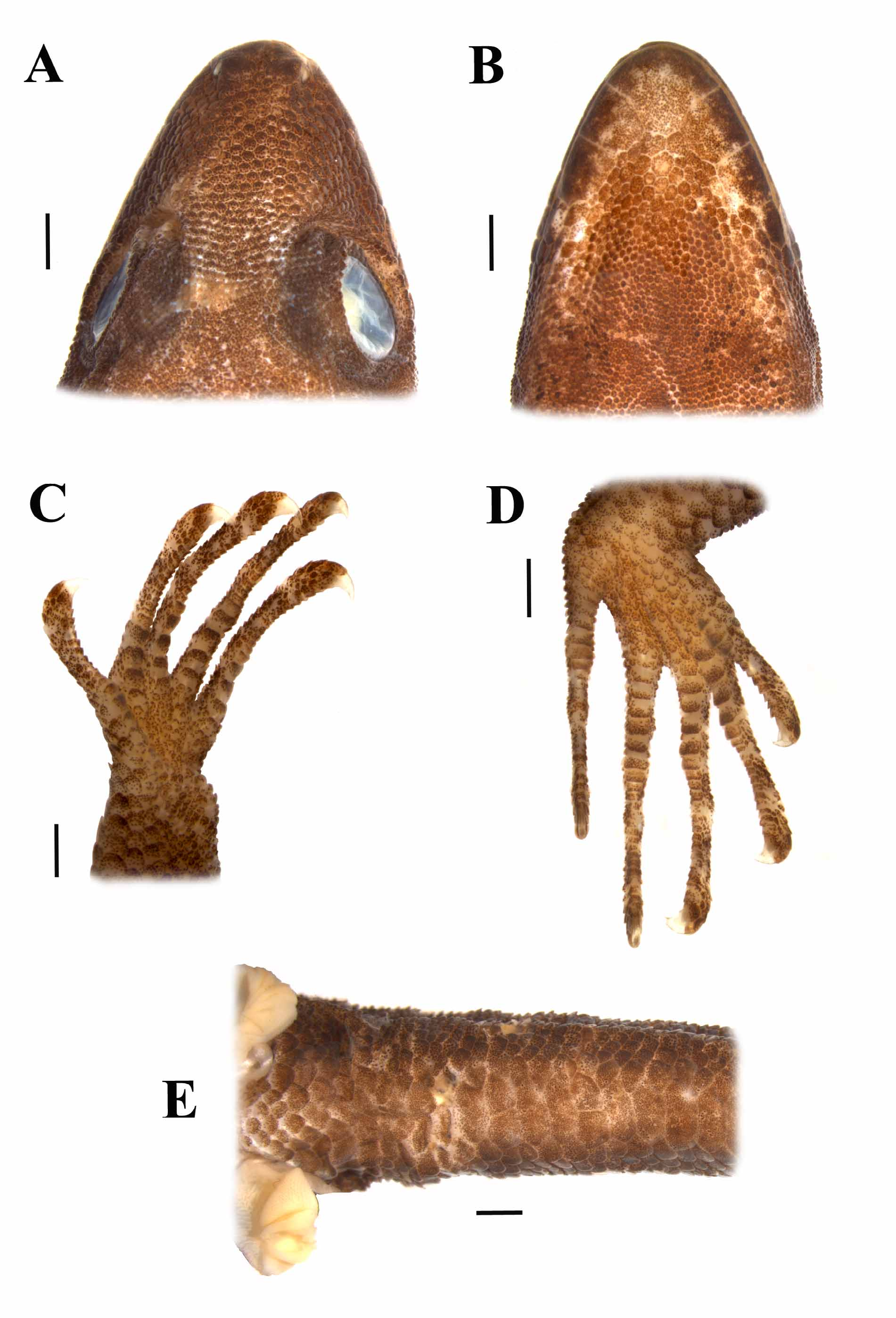
Description. Maximum SVL in males of 53 mm (MZUSP 53676), in females of 55 mm (MZUSP 53671) ( Rodrigues 1980). Tail round in cross section, tapering toward tip, 1.1–1.3 (1.21 ± 0.10, N =6) times SLV. Head length 0.22–0.27 (0.23 ± 0.01, N =19) times SLV, 1.4–1.6 (1.47 ± 0.06, N =19) times as long as wide, 1.3–1.5 (1.41 ± 0.06, N =19) times as wide as high. Snout round, moderately elongate ( Fig 9 View FIGURE 9 A), gently sloping toward top of head. Neck slightly narrower than head and anterior portion of body. Body cylindrical. Limbs well developed, lower arm 0.13– 0.15 (0.14 ± 0.01, N =19) times SVL, lower leg 0.12–0.16 (0.15, ± 0.01, N =19) times SVL.
Rostral convex, 1.8–2.4 (2.11 ± 0.17, N =19) times as wide as high; posterior margin with a shallow depression medially and slightly indented by 1–3 medial postrostrals, with or without a medial cleft extending anteriorly. Postrostrals 3 (79%), 4 (10.5%), or 5 (10.5%), laterals ones (supranasals) distinctly larger than medial ones ( N =19) ( Fig 9 View FIGURE 9 A). Nasal bordered by rostral, first supralabial (only a narrow contact in some specimens), 3 (68.4%) or 4 (31.6%) postnasals ( N =19), and supranasal; internostril distance 0.2–0.3 (0.25 ± 0.02, N =19) times head width. Supranasal roughly oval or circular, 1.0–1.5 (1.20 ± 0.14; N =19) times as wide as long. Postnasals slightly larger than, or similar in size to, adjacent loreals. Scales on snout convex, hexagonal to round, juxtaposed, relatively uniform in size. Canthus rostralis rounded. Loreal region with scales slightly more elongate than those on snout, largest on row adjacent to supralabials, 10–13 (11.1 ± 0.8, N =19) loreals on a line between postnasals and anterior corner of eye. Top and posterior portion of head, as well as supraorbital region, with granular scales. A short supraciliary flap present, anteriorly with 7–10 (8.6 ± 1.0, N =19) enlarged and flattened scales, among which 0–4 (2.6 ± 1.1; N =19) small, conical spines. Pupil round, eye diameter 0.20–0.27 (0.23 ± 0.01, N =19) times head length. Scales on temporal region similar to those on top of head. Ear-opening much smaller than eye, oval, posterior to, and at same level of, commissure of mouth. Supralabials 5–8 (6.7 ± 0.7; N =19), distinctly enlarged anteriorly and decreasing in size posteriorly, one or two of them posterior to centre of eye, followed to corner of mouth by small scales.
Mental large, distinctly wider anteriorly than posteriorly, with posterior margin forming a wide angle, 1.1–1.4 (1.25 ± 0.11; N =19) times as wide as long; 1 (10.5%), 2 (63.2%) or 3 (26.3%) postmentals ( Fig. 9 View FIGURE 9 B). Scales on chin flat, smooth, polygonal, juxtaposted, larger anteriorly, decreasing in size posteriorly. Infralabials 5–7 (5.8 ± 0.7; N =19), distinctly enlarged anteriorly and decreasing in size posteriorly; 1–3 of them posterior to centre of eye, followed to corner of mouth by small scales.
Scales on nape small and granular, becoming slightly larger on sides of neck. Scales on throat anteriorly granular, posteriorly flat, smooth, hexagonal or round, increasing in size toward the posterior region, with a short transitional zone between the anterior and posterior parts.
Dorsals granular, increasing in size toward the flanks. Ventrals larger than dorsals, roughly hexagonal, flat, smooth, imbricate, in oblique rows; 53–63 (57.4 ± 3.3; N =19) scales along the midventral line between anterior margin of forelimbs and vent; 18–21 (19.2 ± 0.9; N =18) scales in a transverse line at midbody, with a short transitional zone between ventrals and scales on flanks. Scales around midbody 118–133 (124.2 ± 3.2; N =18). Scales on preanal plate similar to ventrals, except for those bordering vent, which are very small. Escutcheon present in males on posterior portion of belly and on four rows (body-knee direction) on ventral surface of thighs.
Scales on anterodorsal surface of forelimbs flat, smooth, roundish or rhomboidal, imbricate, largest close to the wrist; on posterodorsal and ventral surface of forelimbs convex, smooth, rhomboidal, juxtaposed, relatively small. Scales on anterodorsal surface of thighs and ventral surface of hind limbs flat, smooth, roundish or rhomboidal, imbricate and relatively larger; on posterodorsal surface of thighs and dorsal surface of lower legs smaller, convex, smooth, rhomboidal, juxtaposed.
Lamellae under second ( II) through fourth ( IV) fingers (proximal lamellae in parentheses): II: 15–18 (5–6), III: 18–20 (5–7), and IV: 18–22 (6–8) (fig 9C). Lamellae under second through fourth toes (proximal lamellae in parentheses): II: 14–18 (5–7), III: 16–22 (5–8), and IV: 22–27 (10–12) ( Fig. 9 View FIGURE 9 D). Claws exposed, non-retractile, between two basal scales. Fingers and toes with three, occasionally four, lateral scales distally, between the fourth (counted from the claw towards the hand) subdigital lamella, and the dorsal scale.
Scales on tail dorsally and laterally relatively small, rhomboidal, flat, smooth, imbricate. On ventral surface of tail scales smooth, flat, imbricate, increasing in size toward midventral line; midventral scales, except close to the base of the tail, distinctly wider than long, forming a repetitive sequence of two single midventrals (one after the other), each in contact laterodistally with one scale per side, followed by a divided (only on proximal portion of tail, when present) or single midventral scale in contact laterodistally with two scales per side – respectively 1’1’2” and 1’1’1” in the codification of Avila-Pires (1995: Figure 2 View FIGURE 2 ) ( Fig. 9 View FIGURE 9 E).
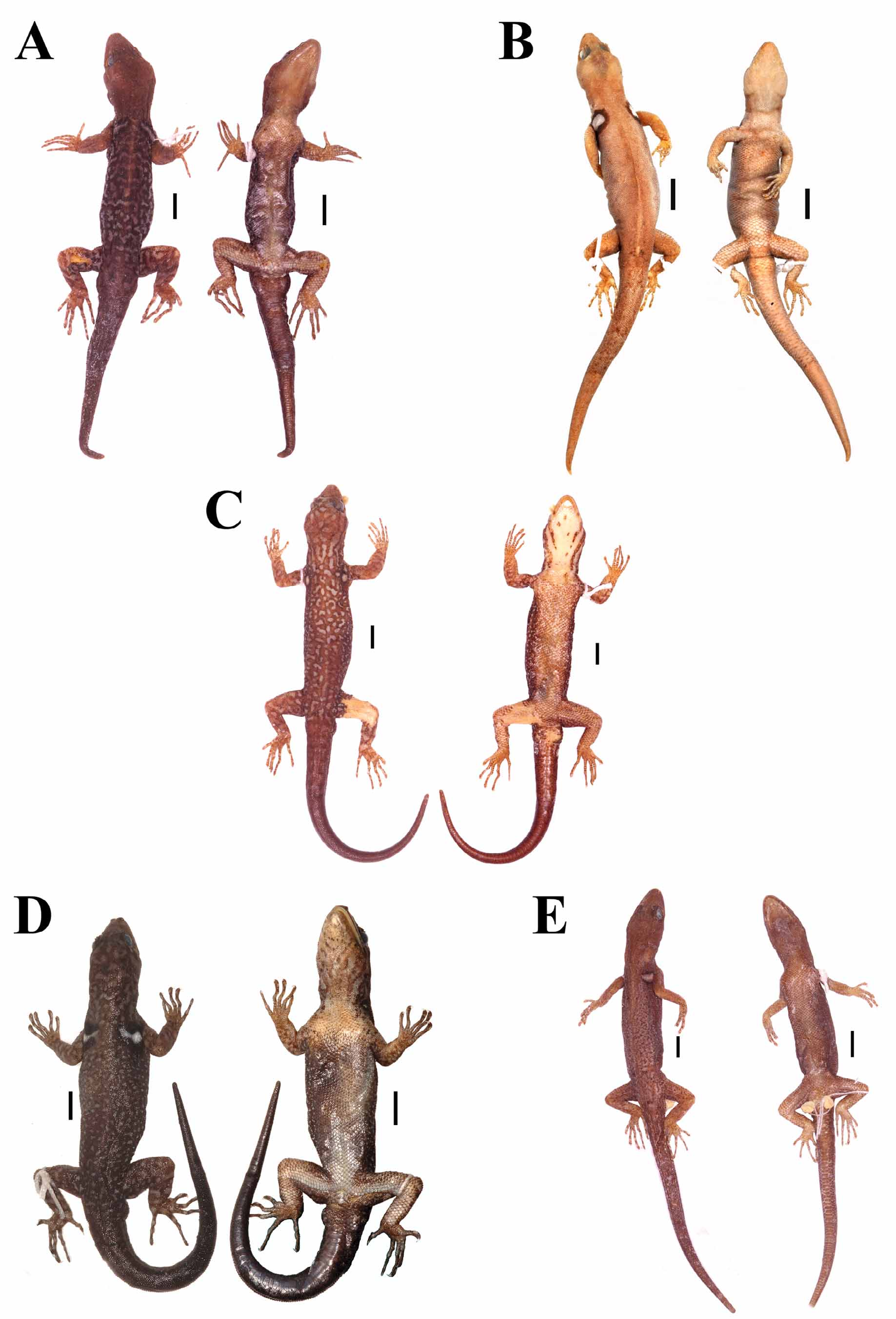
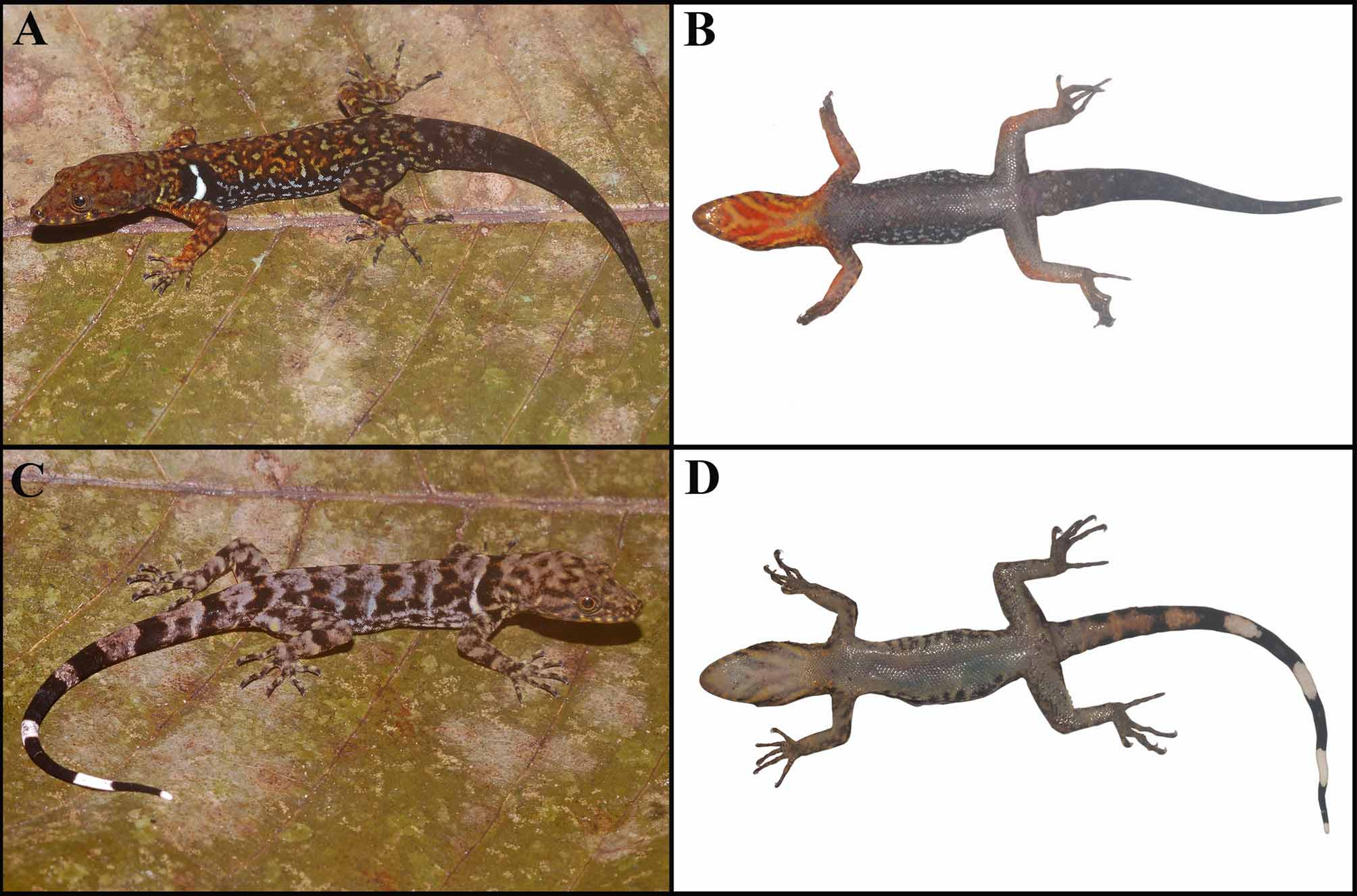
Color in preservative. In males ( Fig 1 View FIGURE 1 C), dorsal surface of head with relatively large reddish-brown and beige vermiculations. Sides of head with oblique and/or longitudinal, beige and brown bands. Back and flanks, base of tail and limbs with beige and brown vermiculations. A large, conspicuous, white suprahumeral ocellus with black margin. Ventral surface of head and gular region beige with oblique black streaks, in contact or not at midventral line; chest beige; belly dark-brown; underside of limbs and base of tail brown. Dorsal surface of tail brown, ventral surface reddish-brown; distal part in one specimen with a beige ring around the tail. Escutcheon area (belly and thighs) light gray.
In females, dorsal surface of head and limbs with brown and beige irregular spots. Sides of head with beige and brown oblique and/or longitudinal bands. Back gray with dorsolateral pairs of black and white spots; flanks gray with black and brown irregular spots. A moderately large, conspicuous, white suprahumeral ocellus, with black margin, smaller than that of males. Ventral surface of head and gular region white with dark oblique streaks, in contact or not at midventral line; belly and underside of limbs light gray. Tail brown and/or black dorsally, white and/or brown ventrally; original tail distally with white bands that form complete rings around the tail.
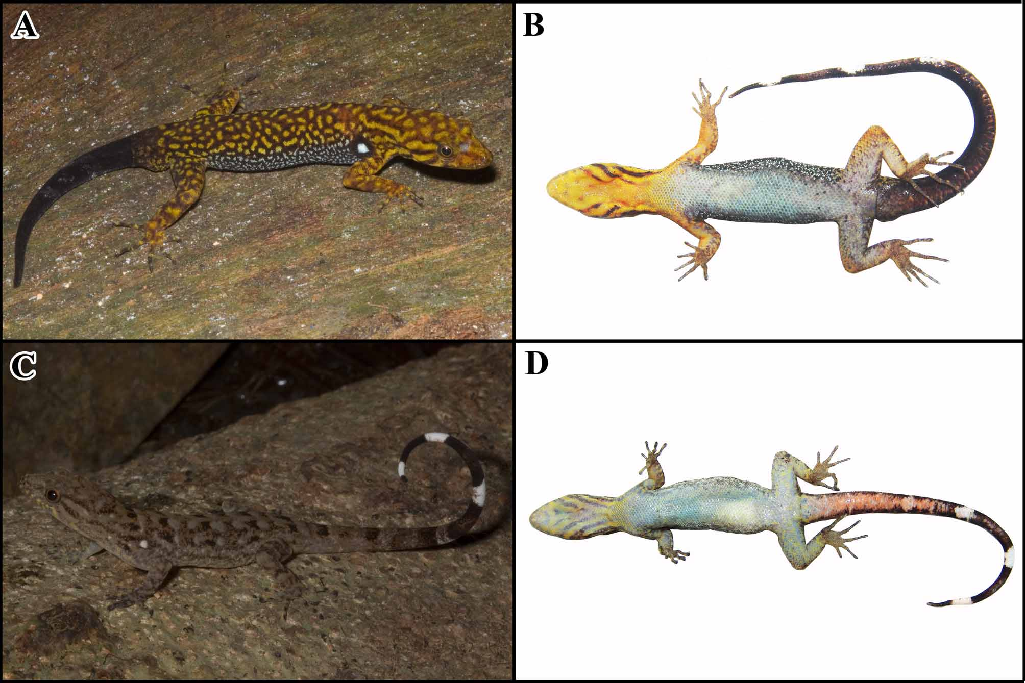
Color in life. In males ( Fig. 10 View FIGURE 10 A–B), head dorsally and laterally reddish-brown and vivid yellow. Suprahumeral ocellus white with black margin. Vermiculations on back brown and yellow, smaller vermiculations on flanks bluish-white and black. Head ventrally and gular region vivid yellow with reddish-brown streaks, chest vivid yellow. Remaining ventral region dark gray, lighter on escutcheon areas (belly and thighs). Tail dark gray to black all around, except near the base, where it is similar to the back.
In females ( Fig. 10 View FIGURE 10 C–D), head dorsally and lateraly gray and brown. Suprahumeral ocellus white with black margin. Back brown with pairs of black and gray spots. Head ventrally and gular region beige with light-yellow and gray streaks, chest white. Remaining ventral region white. Tail anteriorly brown with black spots dorsally, orange ventrally; posteriorly with black and white rings.
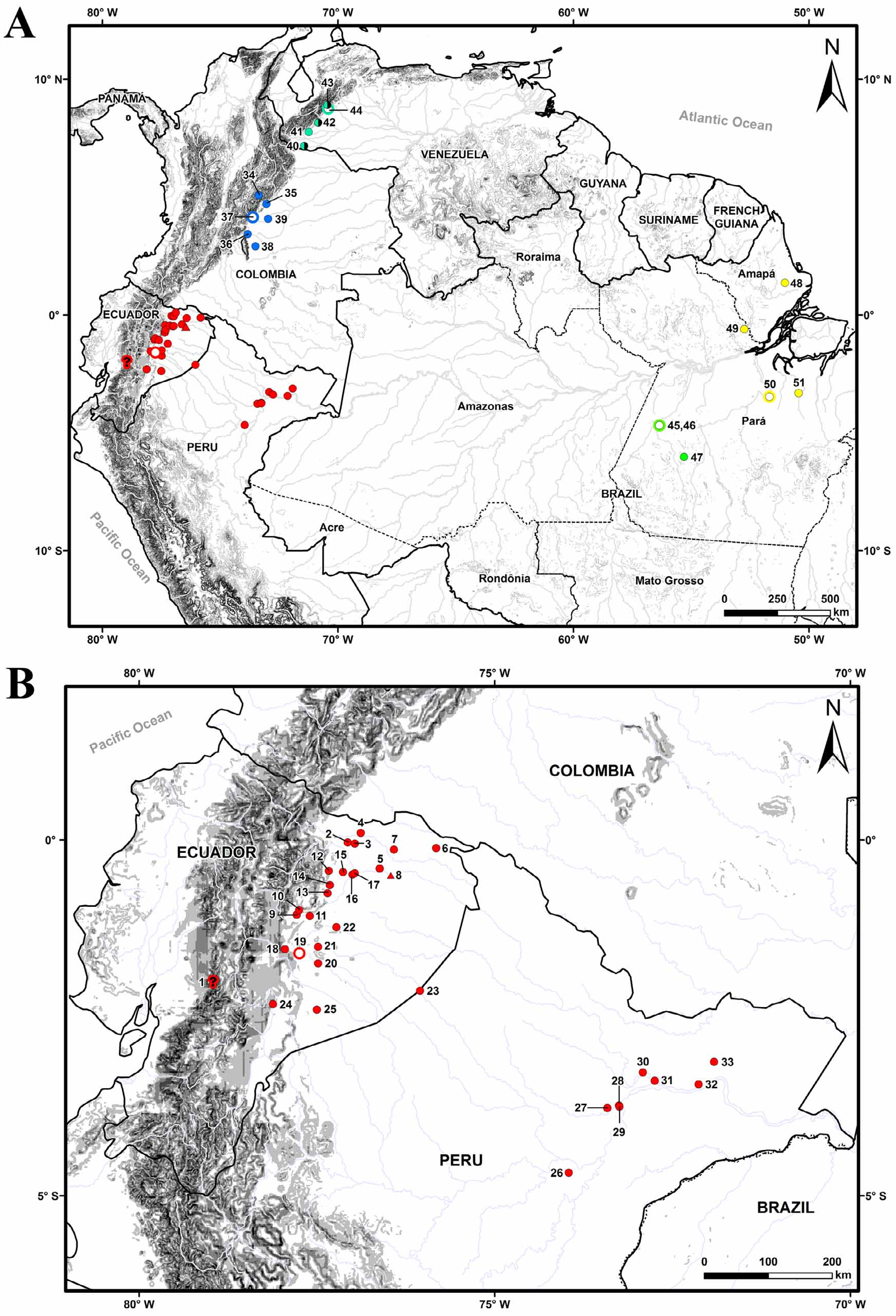
Distribution. Interfluvium Tapajós–Xingu, state of Pará, Brazil ( Fig. 3 View FIGURE 3 A).
Remarks. Rodrigues (1980) pointed out that the type-series was collected in an area with predominance of the palm Euterpe oleracea (“açaizal”), along a trail known as ‘Boa Fé’, in front of Cachoeira do Limão, Tapajós river, at approximately 0 4o 41´S 45o21´W. It is possible that it is the same locality where MPEG 27708–27718 have been collected, community ‘Boa Fé’ being the old name of this locality. Similar to the type-series, these specimens were in an “açaizal” in a flooded depression within the forest; they were found mostly on the palm Euterpe oleracea , one specimen on a tree trunk, between 30 and 200 cm above the ground. Contrary to what was observed by Rodrigues (1980), we also found G. humeralis in the “açaizal”, in syntopy with G. tapajonicus . The Euterpe oleracea forest was one of the few in the area still well-preserved, due to the exploitation in other similar environments of the palm heart for the food market. The species was not found in the more disturbed ‘açaizais’, nor outside this habitat.
Gonatodes nascimentoi sp. nov. ( Fig. 1 View FIGURE 1 D, 11, 12)
Holotype (number in bold between parentheses refer to localities in Fig. 3 View FIGURE 3 ): MPEG 25596 (field number BML 446), an adult male from Fazenda Caracol, right margin of Rio Xingu, Município Anapu, Pará, Brazil ( 3o27’30”S and 51o40’33”W) ( 50), collected in pitfall trap inside primary forest, by A. A. Lima, M. J. Sturaro and R. A. T. Rocha, 12 January 2008.
Paratypes (numbers in bold between parentheses refer to localities in Fig. 3 View FIGURE 3 ): BRASIL. AMAPÁ. Município Ferreira Gomes. Floresta Nacional do Amapá ( 01º06’37’’N and 51º53’37’’W) ( 48): IEPA /FL 361, male, leg. J. Lima, June 2005. PARÁ. Município Almeirim. Monte Dourado, forest near Estação Ecológica do Jari ( 0o35’27’’S and 52º44’09’’W) ( 49): MPEG 23822–27, 5 males and a female, leg. T. A. Gardner and M. A. Ribeiro-Jr, May– June 2005; MPEG 27719, female, leg. M. J. Sturaro and J. O. Gomes, January 2009. Município Anapu. Typelocality ( 50): MPEG 25164, male, leg. A. A. Lima and R. A. T. Rocha, November 2007; MPEG 25595, 25597, a juvenile, a female and a male, leg. A. A. Lima, M. J. Sturaro and R. A. T. Rocha, January 2008; MPEG 25598– 25601, a male and 3 females, leg. A. A. Lima, M. J. Sturaro, R. A. T. Rocha, P. L. V. Peloso and F. S. Rodrigues, March 2008. Município Portel. Fazenda Riacho Monte Verde (~ 3o15’S 50o19’W) ( 51): MPEG 24643, female, leg. T.C.S. Avila Pires and J. O. Gomes, February 2007; MPEG 24644–50, 6 males and a female, leg. J. O. Gomes, March 2007.
Diagnosis. A relatively large Gonatodes , with maximum SLV of 55.7 mm. Scales around midbody 118–143. Ventral scales in a longitudinal row 49–60. Proximal subdigital lamellae as wide as digits, in total 18–23 under fourth finger, 23–28 under fourth toe. Three, occasionally four, lateral rows of scales on distal portion of fingers and toes. Tail with midventral scales distinctly wider than long, forming a repetitive sequence of two single midventrals (one after the other), each in contact laterodistally with one scale per side, followed by a divided (only on proximal portion of tail, when present) or single midventral in contact laterodistally with two scales per side (respectively 1’1’2” and 1’1’1”). A white suprahumeral bar, bordered by black, both in males and females (but thinner in the latter); from very short (hardly higher than long) to almost reaching the middorsal region. Males with a vermiculated pattern of light and dark (in life respectively drab or orange-yellow, and reddish-brown) spots on head and body dorsally; gular region light with dark oblique streaks or redddish with white oblique streaks (orange with orange-yellow streaks in life).
Description. Maximum SVL in males of 55.7 mm (MPEG 23822), in females of 54.0 mm (MJS 035). Tail round in cross section, tapering toward tip, 1.1–1.3 (1.15 ± 0.09, N =8) times SLV. Head length 0.21–0.28 (0.24 ± 0.02, N =24) times SLV, 1.2–1.6 (1.40 ± 0.08, N =24) times as long as wide, 1.2–1.5 (1.40 ± 0.10, N =24) times as wide as high. Snout round, moderately elongate ( Fig 11 View FIGURE 11 A), gently sloping toward top of head. Neck slightly narrower than head and anterior portion of body. Body cylindrical. Limbs well developed, lower arm 0.14–0.17 (0.15 ± 0.01, N =24) times SVL, lower leg 0.13–0.17 (0.15 ± 0.01, N =24) times SVL.
Rostral convex, 1.8–3.0 (2.01 ± 0.28, N =24) times as wide as high; posterior margin with a shallow depression medially and slightly indented by 0–2 medial postrostrals, with or without a medial cleft extending anteriorly. Postrostrals 2 (12.5%), 3 (83.3%), or 4 (4.2%), laterals ones (supranasals) distinctly larger than medial one(s), when these are present ( N =24) ( Fig 11 View FIGURE 11 A). Nasal bordered by rostral, first supralabial (only a narrow contact in some specimens), 3 postnasals (N=24), and supranasal; internostril distance 0.14.–0.21 (0.17 ± 0.01, N =24) times head width. Supranasal roughly oval, circular or semicircular, 0.9–1.4 (1.13 ± 0.13; N =24) times as wide as long. Postnasals slightly larger than, or similar in size to, adjacent loreals. Scales on snout convex, hexagonal to round, juxtaposed, relatively uniform in size. Canthus rostralis rounded. Loreal region with scales slightly more elongate than those on snout, largest on row adjacent to supralabials, 10–13 (10.2 ± 0.8, N =24) loreals on a line between postnasals and anterior corner of eye. Top and posterior portion of head, as well as supraorbital region, with granular scales. A short supraciliary flap present, anteriorly with 7–12 (9.5 ± 1.4, N =24) enlarged and flattened scales, among which 0–3 (1.2 ± 1.3; N =24) small, conical spines. Pupil round, eye diameter 0.20–0.27 (0.22 ± 0.02, N =24) times head length. Scales on temporal region similar to those on top of head. Ear-opening much smaller than eye, oval, posterior to, and at same level of, commissure of mouth. Supralabials 5–7 (6.2 ± 0.5; N =24), distinctly enlarged anteriorly and decreasing in size posteriorly, one or two of them posterior to centre of eye, followed to corner of mouth by small scales.
Mental large, distinctly wider anteriorly than posteriorly, with posterior margin forming a wide angle or a right angle, 1.2–1.4 (1.28 ± 0.7; N =24) times as wide as long; 2 (95.8%), or 3 (4.2%) postmentals ( Fig. 11 View FIGURE 11 B). Scales on chin flat, smooth, polygonal, juxtaposted, larger anteriorly, decreasing in size posteriorly. Infralabials 5–8 (6.4 ± 0.9; N =24), distinctly enlarged anteriorly and decreasing in size posteriorly; 1–3 of them posterior to centre of eye, followed to corner of mouth by small scales.
Scales on nape small and granular, becoming slightly larger on sides of neck. Scales on throat anteriorly granular, posteriorly flat, smooth, hexagonal or round, increasing in size toward the posterior region, with a short transitional zone between the anterior and posterior parts.
Dorsals granular, increasing in size toward the flanks. Ventrals larger than dorsals, roughly hexagonal, flat, smooth, imbricate, in oblique rows; 52–60 (56.3 ± 2.4; N =22) scales along the midventral line between anterior margin of forelimbs and vent; 18–24 (21.2 ± 1.3; N =22) scales in a transverse line at midbody, with a short transitional zone between ventrals and scales on flanks. Scales around midbody 118–143 (129.9 ± 6.4; N =22). Scales on preanal plate similar to ventrals, except for those bordering vent, which are very small. Escutcheon present in males on posterior portion of belly and on 4–5 rows (body-knee direction) on ventral surface of thighs.
Scales on anterodorsal surface of forelimbs flat, smooth, roundish or rhomboidal, imbricate, largest close to the wrist; on posterodorsal and ventral surface of forelimbs convex, smooth, rhomboidal, juxtaposed, relatively small. Scales on anterodorsal surface of thighs and ventral surface of hind limbs flat, smooth, roundish or rhomboidal, imbricate and relatively larger; on posterodorsal surface of thighs and dorsal surface of lower legs smaller, convex, smooth, rhomboidal, juxtaposed.
Lamellae under second ( II) through fourth ( IV) fingers (proximal lamellae in parentheses): II: 15–19 (4–6), III: 18–22 (5–6), and IV: 18–23 (6–8) ( Fig 11 View FIGURE 11 C). Lamellae under second through fourth toes (proximal lamellae in parentheses): II: 15–19 (5–6), III: 18–23 (5–7), and IV: 23–28 (9–12) ( Fig. 11 View FIGURE 11 D). Claws exposed, non-retractile, between two basal scales. Fingers and toes with three, occasionally four, lateral scales distally, between the fourth (counted from the claw towards the hand) subdigital lamella, and the dorsal scale.
Scales on tail dorsally and laterally relatively small, rhomboidal, flat, smooth, imbricate. On ventral surface of tail scales smooth, flat, imbricate, increasing in size toward midventral line; midventral scales, except close to the base of the tail, distinctly wider than long, forming a repetitive sequence of two single midventrals (one after the other), each in contact laterodistally with one scale per side, followed by a divided (only on proximal portion of tail, when present) or single midventral scale in contact laterodistally with two scales per side – respectively 1’1’2” and 1’1’1” in the codification of Avila-Pires (1995: Figure 2 View FIGURE 2 ) ( Fig. 11 View FIGURE 11 E).
Measurements of holotype (in millimeters): SVL 48.7, TL 54.0, HL 11.7, HW 7.9, HD 6.8, ED 2.6, IND 2.0, SSL 0.8, SSW 1.12, RSL 1.3, RSW 2.5, MSL 2.6, MSW 3.2, LAL 8.1, LLL 7.8, HSH 5.34, HSL 2.6.
Scale counts of holotype: SAM 127 scales, VLR 55, VTR 21, SL 6, IL 5, PR 3, SP 1, PN 3, LS 12, PM 2, SCS 8, SSC 3, PL2F 5, DL2F 12, LS2F 3, PL3F 4, DL3F 14, LS3F 4, PL4F 6, DL4F 13, LS4F 4, PL2T 5, DL2T 12, LS2T 4, PL3T 5, DL3T 15, LS3T 3, PL4T 10, DL4T 13, LS4T 3, RSE 4.
Color in preservative. In males ( Fig. 1 View FIGURE 1 D), dorsal surface of head with reddish-brown and beige vermiculations. Sides of head, in some specimens, with oblique and/or longitudinal, beige and brown bands. Back and flanks, base of tail and hind limbs with relatively large, beige color and dark-brown vermiculations. A conspicuous, white suprahumeral vertical bar with black margin, which varies from only slightly to distinctly higher than long (see under “Remarks” below). Ventral surface of head and gular region beige with black streaks, in some specimens dark-brown with beige streaks, in contact or not at midvental line; chest beige; belly darkbrown; underside of limbs and base of tail brown. Dorsal surface of tail brown, underside dark-brown or black; a white band may be present distally, forming a complete ring around the tail. Escutcheon area (belly and thighs) light gray.
In females, dorsal surface of head and limbs with brown and beige irregular spots. Back gray with dorsolateral pairs of black and white spots; flanks gray with black and brown irregular spots. Suprahumeral bar conspicuous, white with black margins, thinner than that of males; in some cases they almost reach the middorsal region. Ventral surface of head and gular region white with black oblique streaks, in contact or not at midventral line; belly and underside of limbs light-gray. Tail gray dorsally and white ventrally; original tail distally with white and black bands that form complete rings around the tail.
Color in life (Photos of MPEG 23822, 23824–25, 25596–99; field notes of MPEG 24643): In males ( Fig. 12 View FIGURE 12 A–B), head dorsally and laterally with orange and orange-yellow, or orange and drab, vermiculations. On back vermiculations may be smaller than, or similar in size to, those on head and the lighter spots are frequently bordered by black (occasionally also on head). Suprahumeral bar or ocellus white with black margins. Flanks with smaller, bluish-white and black vermiculations. Head ventrally and gular region orange with orange-yellow streaks, chest orange. Remaining ventral region plumbeous, lighter on escutcheon areas (belly and thighs). Tail dark gray to black all around, except near the base, where it is similar to the back; distal portion with white spots. Color in life of MPEG 24643, a male, was described based on the color guide of Smithe (1975): Head dorsally “buff” (24) and “mahogany red” (132B); body “brick red” (132A) with “chamois” (123D) vermiculation; a white bar with black margins; lower flanks “blackish neutral gray” (82) with “pearl gray” (81) spots. Ventrally, head and chest (until forearm) “buff” (24) with convergent “scarlet” (14) streaks; otherwise ventral region and tail mainly “blackish neutral gray” (82), midventrally medium (84) to light (85) neutral gray; tail dorsally with a darker and lighter pattern (TCSAP, field notes).
Females ( Fig. 12 View FIGURE 12 C–D) dorsally brown or grayish-brown, with a paired series of dark brown to black irregular, transversely elongate, spots. Suprahumeral bar white with black margins, thinner than those in males. Head ventrally and gular region white with black streaks, chest light-gray. Tail gray with black spots all around; posterior portion with white spots.
Distribution. Eastern Brazilian Amazonia, in the states of Pará and Amapá. Until now it is only known south of the Amazon river in the interfluvium Xingu–Tocantins, Pará state, and north of the Amazon river east of Paru river, in the states of Pará and Amapá ( Fig. 3 View FIGURE 3 A).
Etymology. The species is named after Francisco Paiva do Nascimento, who worked for many years in Museu Paraense Emílio Goeldi and contributed to our knowledge of Amazonian reptiles, most of all about the snakes from eastern Amazonia.
Remarks. The suprahumeral spot varies among populations and, to a smaller degree, within populations. In the only specimen from Amapá examined, the spot has the shape of a short bar, almost an ocellus. Specimens from Pará, north of the Amazon River, show a vertical bar reaching dorsally at least the dorsolateral region, in some cases almost the middorsal region. South of the Amazon, all specimens from Monte Verde present a short vertical bar (not reaching the dorsolateral region, but always with dorso-ventral axis longer than antero-posterior axis), while in the specimens from Anapu the vertical bar is similar to those from northern Pará (although one specimen presents a vertical bar and a small ocellus above the bar, on both sides).
Specimens were collected by active search or in pitfall traps, in primary or little-disturbed forest. Six specimens were found on the buttresses or trunks of live trees, between 15–50 cm above ground; one was inside the loose bark of a dead tree, 120 cm above ground; a male and a female were on a fallen trunk; and at the Xingu river a pair was found on a rock, 50 cm above ground. At this locality specimens hid inside rock crevices or tree trunk holes when disturbed. Weights vary between 0.2–5.1 g.
Gonatodes riveroi sp. nov. ( Figs. 1 View FIGURE 1 E, 13, 14)
Gonatodes concinnatus: Vanzolini, 1955: 126 (part); 1968: 26 (part); Mechler, 1968: 331 (part); Peters & Donoso-Barros, 1970 (part); Rivero-Blanco, 1979: 94 (part); Sanchez, Castaño & Cardenas, 1995: 317; Bartlett & Bartlett, 2003: 158 (part).
Holotype (number in bold between parentheses refer to localities in Fig. 3 View FIGURE 3 ): MZUSP 49153 (field number FMEDEM 1465), adult male, from Villavicencio, State of Meta, Colombia (~ 4o09’S and 73o37’W) ( 37), collected by W. W. Lamar and F. Medem, 22 October 1977.
Paratypes (numbers in bold between parentheses refer to localities in Fig. 3 View FIGURE 3 ): COLOMBIA. BOYACÁ. Garagoa ( 34): CAS 71238, USNM 84968, USNM 92490–92, a male and 4 females, leg. Nicéforo Maria, 1931. BOYACÁ–CUNDINAMARCA– META border. Guaicáramo (according to Paynter Jr., 1997 in the hills near confluence of Rio Guavio with Rio Upía, at eastern base of the eastern Andes) ( 35): USNM 84972, female, leg. Nicéforo Maria, 1931. META. San Juan de Arama ( 36): KU 192635–36, a male and a female, leg. M. Chin, August 1967. Type-locality ( 37): MCZ 132796–97, a juvenile and a female, July 1972; AMNH 35280–87, AMNH 35290– 91, 5 males and 5 females, leg. Nicéforo Maria, January 1927; KU 110579 –80, a juvenile and a male, collector not specified, July 1967; KU 151933–35, 3 males, collector not specified, May 1967; MCZ 19213, male, January 1924; MCZ 77388–89, a male and a female, collector and date not specified; MCZ 110009–14, a juvenile, 3 males and 2 females, June 1968; MCZ 110741 –42, 2 males, leg. G. Gorman, June 1968; MZUSP 2145–46 (former AMNH 35292–93), 2 females, leg. Nicéforo Maria, no date; MZUSP 49152, female, leg. M. Lugor and F. Medem, October 1977; MZUSP 49154, female, leg. W. W. Lamar and F. Medem, October 1977; USNM 84969, male, leg. Nicéforo Maria, 1931. Villavicencio, Finca “El Buque” ( 37): MZUSP 49155–62, 3 males and 5 females, leg. E.Thierry and F. Medem, October 1977. Villavicencio, Instituto 'Roberto Franco' ( 37): MCZ 77391–97, 2 juveniles, 2 males and 3 females, collector not specified, July 1963; MCZ 146137, male, collector not specified, October 1974. 5 km NE of Villavicencio ( 37): UMMZ 127206, male, leg. W. Mobely and K. Adler, July 1965. Rio Negro, c. 12 km from Villavicencio, Finca 'Las Orquideas' (“below” [south of?] road Bogotá–Villavicencio) ( 37): MCZ 150022, male, leg. F. Medem, 1975. Güejar River, Finca Guadualito ( 38): MZUSP 44777, female, leg. L. Klein and F. Medem, September 1967. Puerto Lopez ( 39): AMNH 97350, juvenile, leg. A.V. Hutchison and Mahoney, January 1966; AMNH 98406, female, leg. V.H. Hutchison, April 1966.
Diagnosis. A moderately large Gonatodes , with maximum SLV of 47.6 mm. Scales around midbody 96–123. Ventral scales in a longitudinal row 44–55. Proximal subdigital lamellae as wide as digits, in total 14–21 under fourth finger, 17–25 under fourth toe. Two or three lateral rows of scales on distal portion of fingers and toes. Tail with midventral scales distinctly wider than long, forming a repetitive sequence of two single midventrals (one after the other), each in contact with one laterodistal scale per side, followed by a divided (only on proximal portion of tail, when present) or single midventral in contact with two laterodistal scales per side (respectively 1’1’2” and 1’1’1”). A white suprahumeral bar bordered by black, present both in males and females, although more conspicuous in males; it reaches dorsally at least up to the dorsolateral region, in some females it almost reaches the middorsal region; never in the form of an ocellus. Males with head dorsally without vermiculation; back and limbs finely vermiculated; gular region without dark streaks.
Description. Maximum SVL in males of 47.6 mm (USNM 84968), in females of 47.1 mm (USNM 92490). Tail round in cross section, tapering toward tip, 1.0–1.3 (1.17 ± 0.11, N =11) times SLV. Head length 0.22–0.28 (0.24 ± 0.01, N =61) times SLV, 1.3–1.6 (1.46 ± 0.05, N =57) times as long as wide, 1.2–1.8 (1.38 ± 0.10, N =56) times as wide as high. Snout round, moderately elongate ( Fig. 13 View FIGURE 13 A), gently sloping toward top of head. Neck slightly narrower than head and anterior portion of body. Body cylindrical. Limbs well developed, lower arm 0.11– 0.16 (0.13 ± 0.01, N =62) times SVL, lower leg 0.12–0.16 (0.14 ± 0.01, N =62) times SVL.
Rostral convex, 1.6–2.3 (1.96 ± 0.13, N =61) times as wide as high; posterior margin with a shallow depression medially and slightly indented by 0–3 medial postrostrals, with or without a median cleft extending anteriorly. Postrostrals 2 (4.9%), 3 (82%), 4 (4.9%) or 5 (8.2%), lateral ones (supranasals) distinctly larger than medial one(s), when these are present ( N =61) ( Fig 13 View FIGURE 13 A). Nasal bordered by rostral, first supralabial (only a narrow contact in some specimens), 2 (96.8%) or 3 (3.2%) postnasals (N=63), and supranasal; internostril distance 0.20.–0.27 (0.24 ± 0.02, N =55) times head width. Supranasal roughly oval or circular, 0.9–1.6 (1.23 ± 0.15; N =62) times as wide as long. Postnasals slightly larger than, or similar in size to, adjacent loreals. Scales on snout convex, hexagonal to round, juxtaposed, relatively uniform in size. Canthus rostralis rounded. Loreal region with scales slightly more elongate than those on snout, largest on row adjacent to supralabials, 9–11 (9.8 ± 0.7, N =61) loreals on a line between postnasals and anterior corner of eye. Top and posterior portion of head, as well as supraorbital region, with granular scales. A short supraciliary flap present, anteriorly with 6–12 (8.3 ± 1.0, N =60) enlarged and flattened scales, among which 0–5 (1.7 ± 1.4; N =60) small, conical spines. Pupil round, eye diameter 0.19–0.26 (0.22 ± 0.01, N =56) times head length. Scales on temporal region similar to those on top of head. Ear-opening much smaller than eye, oval, posterior to, and at same level of, commissure of mouth. Supralabials 5–8 (6.1 ± 0.6; N =62), distinctly enlarged anteriorly and decreasing in size posteriorly, one or two of them posterior to centre of eye, followed to corner of mouth by small scales.
Mental large, distinctly wider anteriorly than posteriorly, with posterior margin forming a moderate or wide angle, 1.1–1.5 (1.27 ± 0.08; N =59) times as wide as long; 1 (1.6%), 2 (76.2%), 3 (19.0%), or 4 (3.2%) postmentals ( Fig. 13 View FIGURE 13 B). Scales on chin flat, smooth, polygonal, juxtaposted, larger anteriorly, decreasing in size posteriorly. Infralabials 4–7 (5.4 ± 0.7; N =63), distinctly enlarged anteriorly and decreasing in size posteriorly; one or two of them posterior to centre of eye, followed to corner of mouth by small scales.
Scales on nape small and granular, becoming slightly larger on sides of neck. Scales on throat anteriorly granular, posteriorly flat, smooth, hexagonal or round, increasing in size toward the posterior region, with a short transitional zone between the anterior and posterior parts.
Dorsals granular, increasing in size toward the flanks. Ventrals larger than dorsals, roughly hexagonal, flat, smooth, imbricate, in oblique rows; 44–56 (50.8 ± 2.3; N =58) scales along the midventral line between anterior margin of forelimbs and vent; 15–21 (18.0 ± 1.2; N =62) scales in a transverse line at midbody, with a short transitional zone between ventrals and scales on flanks. Scales around midbody 96–123 (110.1 ± 5.6; N =60). Scales on preanal plate similar to ventrals, except for those bordering vent, which are very small. Escutcheon present in males on posterior portion of belly and on 3–5 rows (body-knee direction) on ventral surface of thighs.
Scales on anterodorsal surface of forelimbs flat, smooth, roundish or rhomboidal, imbricate, largest close to the wrist; on posterodorsal and ventral surface of forelimbs convex, smooth, rhomboidal, juxtaposed, relatively small. Scales on anterodorsal surface of thighs and ventral surface of hindlimbs flat, smooth, roundish or rhomboidal, imbricate and relatively larger; on posterodorsal surface of thighs and dorsal surface of lower legs smaller, convex, smooth, rhomboidal, juxtaposed.
Lamellae under second ( II) through fourth ( IV) fingers (proximal lamellae in parentheses): II: 13–17 (5–6), III: 15–19 (4–6), and IV: 14–21 (5–8) ( Fig 13 View FIGURE 13 C). Lamellae under second through fourth toes (proximal lamellae in parentheses): II: 12–18 (5–7), III: 16–21 (5–8), and IV: 17–25 (8–12) ( Fig. 13 View FIGURE 13 D). Claws exposed, non-retractile, between two basal scales. Fingers and toes with three, occasionally two, lateral scales distally, between the fourth (counted from the claw towards the hand) subdigital lamella, and the dorsal scale.
Scales on tail dorsally and laterally relatively small, rhomboidal, flat, smooth, imbricate. On ventral surface of tail scales smooth, flat, imbricate, increasing in size toward midventral line; midventral scales, except close to the base of the tail, distinctly wider than long, forming a repetitive sequence of two single midventrals (one after the other), each in contact laterodistally with one scale per side, followed by a divided (on proximal portion of tail, when present) or single midventral scale in contact laterodistally with two scales per side – respectively 1’1’2” and 1’1’1” in the codification of Avila-Pires (1995: Figure 2 View FIGURE 2 ) ( Fig. 13 View FIGURE 13 E).
Measurements of holotype (in millimeters): SVL 42.5, TL 46.0 (distally regenerated), HL 9.8, HW 6.7, HD 4.7, ED 2.0, IND 1.7, SSL 0.7, SSW 0.8, RSL 1.1, RSW 2.1, MSL 2.2, MSW 2.6, LAL 5.3, LLL 6.2, HSH 2.8, HSL 1.57.
Scale counts of holotype: SAM 116 scales, VLR 51, VTR 17, SL 6, IL 6, PR 3, SP 1, PN 3, LS 9, PM 2, SCS 8, SSC 2, PL2F 6, DL2F 11, LS2F 3, PL3F 6, DL3F 13, LS3F 3, PL4F 7, DL4F 11, LS4F 3, PL2T 6, DL2T 11, LS2T 3, PL3T 6, DL3T 13, LS3T 3, PL4T 11, DL4T 12, LS4T 3, RSE 4.
Color in preservative. In males ( Fig. 1 View FIGURE 1 E), dorsal surface of head reddish-brown, spotless. Back and flanks, base of tail and hind limbs with relatively small, beige and brown vermiculations. A large, conspicuous, white suprahumeral bar with black margins, extending dorsally at least up to the dorsolateral region. Ventral surface of head, gular region and chest brown, without oblique streaks; belly dark-brown or dark-gray; underside of limbs beige. Tail dark brown dorsally and brown ventrally. Escutcheon area (belly and thighs) light gray.
In females, dorsal surface of head and limbs light-brown or gray, with black irregular spots; brown and black oblique and/or longitudinal bands may be present on sides of head. Back gray with dorsolateral pairs of black spots; flanks gray with black and brown irregular spots. A moderately large, conspicuous, white suprahumeral bar, with black margins, but less defined than that on males; in some specimens it almost reaches the middorsal region. Ventral surface of head and gular region white with dark oblique streaks, in contact or not at midventral line; belly and underside of limbs white or beige. Tail gray dorsally and white ventrally; original tail distally with white and black bands that form complete rings around the tail.
Color in life (photo of UMMZ 127206, male): Head dorsally and laterally brown. Back with brown and yellow vermiculations. Suprahumeral bar white with black margins. Tail dark-brown ( Fig. 14 View FIGURE 14 ).
Distribution. Central Colombia east of the Andes ( Fig. 3 View FIGURE 3 A). Sanchez et al. (1995) reported G. concinnatus in Colombia from “Amazonas, Casanare, Boyacá, Cundinamarca, Meta, Pasto, Orinoco, Guaviare.” It is necessary, however, to verify whether all these records refer to the species here described.
Etymology. The species is named after Dr. Carlos Rivero-Blanco, who made an extensive study on Gonatodes , as well as contributions to environmental conservation in Venezuela.
Remarks. Rivero-Blanco (1979) considered the specimens from Villavincencio and vicinity, in Meta, Colombia, as intergrades between Gonatodes concinnatus concinnatus , from Peru and Ecuador, and G. c. ligiae , from Venezuela.


| MPEG |
Museu Paraense Emilio Goeldi |
| MZUSP |
Museu de Zoologia da Universidade de Sao Paulo |
| IEPA |
Istituto di Entomologia Agraria dell'Universita |
| CAS |
California Academy of Sciences |
| USNM |
Smithsonian Institution, National Museum of Natural History |
| MCZ |
Museum of Comparative Zoology |
| AMNH |
American Museum of Natural History |
| UMMZ |
University of Michigan, Museum of Zoology |
No known copyright restrictions apply. See Agosti, D., Egloff, W., 2009. Taxonomic information exchange and copyright: the Plazi approach. BMC Research Notes 2009, 2:53 for further explanation.
|
Kingdom |
|
|
Phylum |
|
|
Class |
|
|
Order |
|
|
Family |
|
|
Genus |
Gonatodes tapajonicus Rodrigues, 1980
| Sturaro, Marcelo José & Avila-Pires, Teresa C. S. 2011 |
Gonatodes tapajonicus
| Avila-Pires 1995: 283 |
| Vanzolini 1986: 10 |
| Rodrigues 1980: 309 |
Gonatodes concinnatus :
| Bartlett 2003: 158 |
| Sanchez 1995: 317 |
| Rivero-Blanco 1979: 94 |
| Mechler 1968: 331 |