
Montesauria indonesiana Constantinescu, 2019
|
publication ID |
https://doi.org/10.24349/acarologia/20194324 |
|
DOI |
https://doi.org/10.5281/zenodo.4525207 |
|
persistent identifier |
https://treatment.plazi.org/id/C84C4327-7767-FF9E-FE28-FE0FFB6487FC |
|
treatment provided by |
Felipe |
|
scientific name |
Montesauria indonesiana Constantinescu |
| status |
sp. nov. |
Montesauria indonesiana Constantinescu sp. n.
Figures 1 – 4 View Figure 1 View Figure 2 View Figure 3 View Figure 4
Zoobank: 6F70104D-14FE-4968-AE23-C08E8B391B45
Type material — Holotype male and 16 paratypes ( 7 male and 9 female), from Aplonis panayensis (Scopoli) ( Passeriformes , Sturnidae ), INDONESIA, Kalimantan Island, Lhok Tuan, Kutai National Park , 22 May 1991, bird inventory number 15717, no other data.
Type deposition — Holotype male (ANA 801), 6 male (ANA 802–807) and 8 female (ANA 808–815) paratypes in MGAB collection, 1 male and 1 female paratypes in DZUnesp-RC collection.
Etymology — The specific name indonesiana refers to the name of the country ( Indonesia) where the mite was collected.
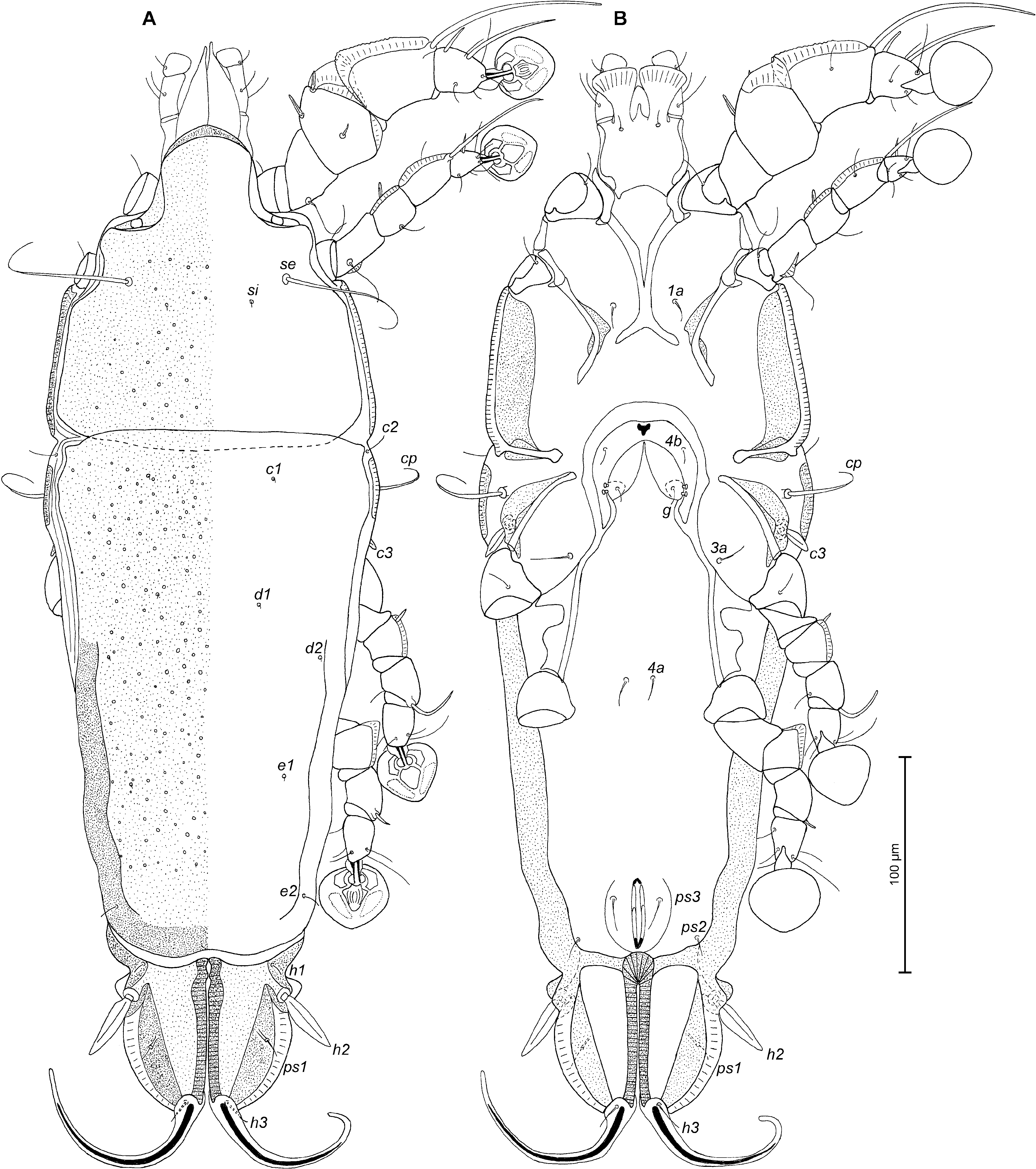
Description — Male. Length of idiosoma 322 (308–312), width 140 (120–140), length of hysterosoma 212 (204–208). Prodorsal shield: entire, anterior margin with lateral margins slightly concave at level of scapular setae, posterior margin with acute median extension, length along midline 110 (104–108), width at posterior margin 118 (100–118), surface with numerous pit-like lacunae. Scapular setae se separated by 65 (58–64). Humeral shields small, separated from epimerites III, not encompassing setae cp. Setae c2 situated dorsally on soft tegument. Subhumeral setae c3 lanceolate, 18 (16–18) × 8 (6–8). Hysteronotal shield: greatest length from anterior margin to lobar apices 200 (196–204), width at anterior margin 122 (102–120), anterior margin slightly convex, surface of this shield with numerous pit-like lacunae. Prodorsal and hysteronotal shields separated by narrow band of soft tegument. Opisthosomal lobes short, roughly trapezoidal, with pair of small extensions at base of setae h2 and with extensions at base of setae h3, terminal cleft V-shaped, 24 (22–24) long. Setae f2 absent. Setae h3 lanceolate, with acute tip, 38 (36–37) long, 12 (10–11) wide, setae h2 150 (140–160) long, 5 (4–5) wide. Supranal concavity circular, well outlined. Setae ps2 66 (54–60) long, thickened in basal part and with filiform apical part, setae ps1 filiform, about 10 (6–8) long, situated on margins of terminal cleft slightly anterior to level of setae h3 ( Fig. 1A View Figure 1 ). Distance between bases of dorsal setae: c2–d2 104 (84–100), d2–e2 96 (84–100), e2–h3 52 (48–52), d1–d2 46 (40–50), e1–e2 22 (22–26), ps2–h1 10 (6–10), h2–h2 40 (42–44), h3–h3 22 (20–22), ps2–ps2 70 (64–66).
Epimerites I fused into a Y, posterior end of sternum connected to medial part of epimerites II ( Fig. 1B View Figure 1 ). Coxal fields I, II without wide sclerotized areas. Rudimentary sclerites rEpIIa present. Coxal fields II and III open. Epimerites IVa present. Genital arch small, 16 (17–20) in length, 26 (24–28) in width, basal sclerite of genital apparatus shaped semicircular, aedeagus straight, sword-shaped, 70 (62–65) long, extending to or beyond anterior margin of anal suckers. Genital shield absent, adanal shields present, small, circular, surrounding base of setae g. Anal suckers 14 (13–14) in diameter, corolla without indentations, surrounded by radially striated area. Opisthoventral shields small, circular, surrounding base of setae ps3 ( Fig. 1B View Figure 1 ). Distance between ventral setae: 3a–4b 30 (28–32), 4b–4a 28 (28–32), 4a–g 44 (42–46), g–ps3 40 (40–41), ps3–ps3 66 (62–64), ps3–h3 38 (34–37).
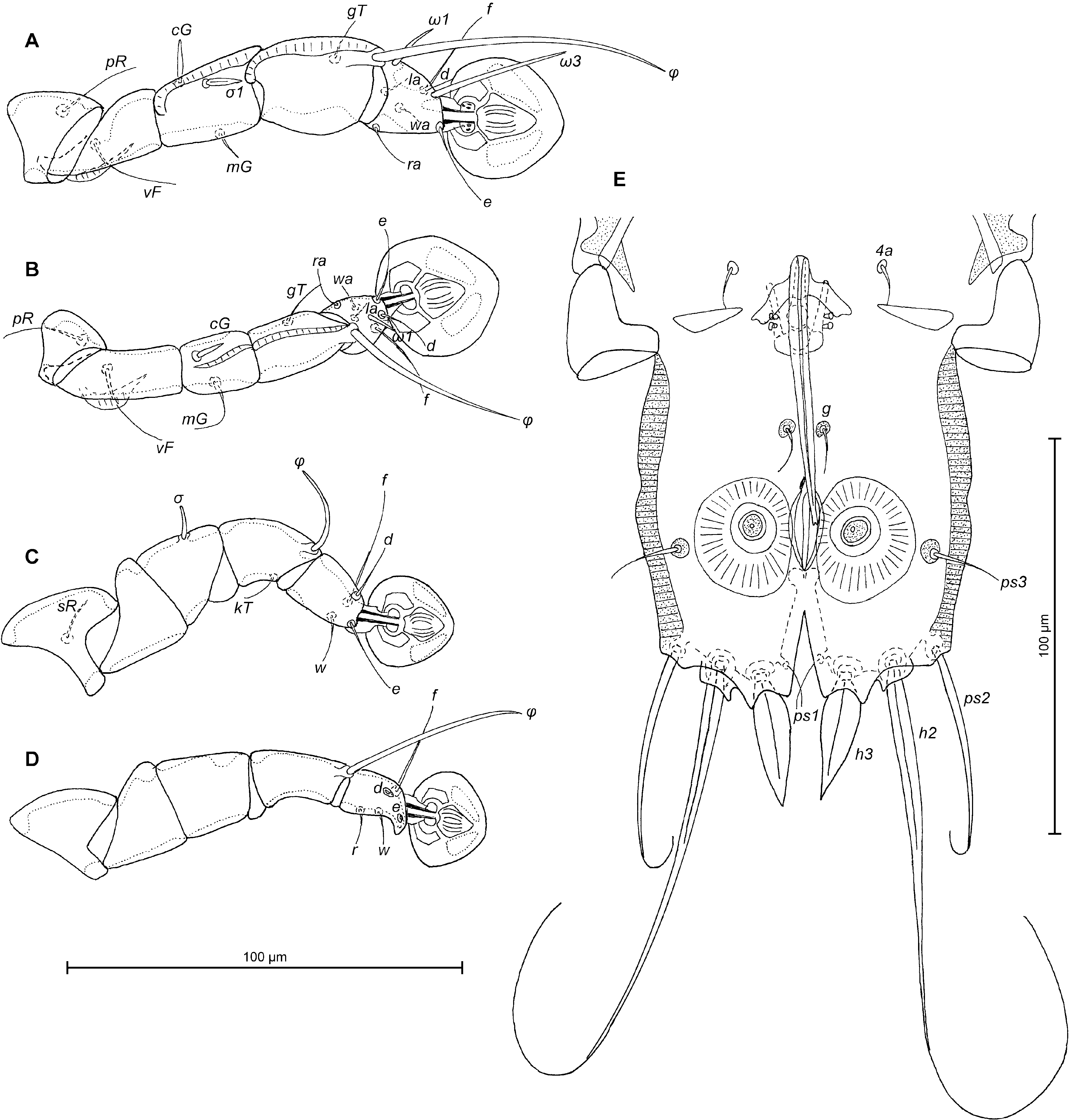
Legs I noticeably longer and thicker than legs II. Genua and tibiae I, II with narrow dorsal crest, femora I, II with ventro-basal crest. Solenidion σ1 of genu I lanceolate, setae cG I and cG II spine-like ( Fig. 2A, B View Figure 2 ). Tarsus IV 20 (20–22) long, with small apical claw-like process, modified setae d, e button-like ( Fig. 2D View Figure 2 ). Length of solenidia: ω1 I 11 (12–14), ω1 II 6 (6–9), φ I 68 (60–68), φ II 46 (40–44), φ III 24 (20–24), φ IV 25 (20–26).
Female (range for 5 paratypes). Length of idiosoma excluding terminal appendages 452–460, width 139–160, length of hysterosoma 300–312. Prodorsal shield: shape as in male, except for convex posterior margin, 144–152 long, 122–142 wide, posterior part with pit-like lacunae ( Fig. 3A View Figure 3 ). Scapular setae se separated by 68–76. Humeral shields small, not encompassing setae cp. Setae c2 situated on soft tegument. Setae c3 lanceolate, 17–18 × 6–7. Prodorsal and hysteronotal shields touching each other. Anterior and lobar parts of hysteronotal shield well delimited from each other by a transverse groove. Hysteronotal shield: anterior margin slightly convex, posterior margin concave in median part, length 236–248, width at anterior margin 130–148, with pit-like lacunae. Length of lobar region excluding terminal appendages 70–78, width at level of setae h2 83–84, median part of anterior margin slightly convex. Terminal cleft as narrow V, 56–60 long, extended beyond level of setae h2. Lateral margins of opisthosomal lobes posterior to setae h2 membranous. Setae f2 absent, setae h1 on lobar shield, near its anterior margin. Setae h2 spindle-like, 36–38 long, 8–10 wide. Setae h3 filiform, 16–21 in length, about 1/6 of the length of terminal appendages. Setae ps1 situated close to lateral margins of opisthosomal lobes ( Fig. 3A View Figure 3 ). Distance between bases of dorsal setae: c2–d2 82–104, d2–e2 100–116, e2–h2 44–48, h2–h3 54–60, d1–d2 34–47, e1– e2 56–60, h1–h2 14–16, ps1–h2 22–29, h1–h1 60–61, h2–h2 68–72.
Epimerites I fused into a Y, posterior end of sternum with acute extensions directed laterally but not connected with epimerites II ( Fig. 3B View Figure 3 ). Lateral parts of coxal fields I without heavily sclerotized areas, coxal fields II with heavy sclerotized areas. Epimerites II do not reach the level of anterior margin of epigynum. Epimerites IVa absent. Translobar apodemes of opisthosomal lobes present, fused with ventral sclerotization of lobar region and posterolateral margins of anterior hysteronotal shield. Epigynum horseshoe-shaped, greatest width 52–65, apodemes of ovipore connected with coxal apodemes IIIa. Head of spermatheca weakly sclerotized, primary spermaduct with ball-shaped enlargement, secondary spermaducts 2 times longer than distance between enlargement of primary spermaduct and spermatheca ( Fig. 4E View Figure 4 ). Distance between pseudanal setae: ps2–ps2 56–58, ps3–ps3 20–24, ps2–ps3 24–28.
Legs I hypertrophied, tibia and genu I strongly inflated on dorsal side, heavily sclerotized and 2.5–3 times thicker than respective segments of legs II. Tibia I with well-developed dorsal crest, its proximal part rounded with indentations, distal part of genu I with large angular paraxial crest and with narrow antaxial crests. Tibia and genu II with narrow dorsal crest and femur II with ventro-basal crest. Genu IV with rounded dorsal crest. Solenidion σ1 of genu I lanceolate, setae cG I and cG II spine-like ( Fig. 4A, B View Figure 4 ). Length of solenidia: ω1 I 16 (14–16), ω1 II 8 (6–10), φ I 72 (66–72), φ II 44 (40–48), φ III 24 (22–24), φ IV 7 (7–8).
Differential diagnosis. The new species, Montesauria indonesiana sp. n., belongs to the pachypa species group. This new species is most similar to M. mainati ( Trouessart, 1885) , which was originally described from Common hill myna Gracula religiosa Linnaeus, 1758 ( Passeriformes , Sturnidae ) in Indonesia ( Trouessart 1885) and later redescribed ( Mironov 2006). Both species have similar ornamentation of the dorsal shields and setae cG I and cG II are spiniform in male and female. In males of both species, setae h3 are lanceolate, setae ps2 are thickened in the basal part and filiform in apical part, and epimerites IVa are present. In females, the terminal cleft is extended beyond the level of setae h2. M. indonesiana sp. n. clearly differs from M. mainati by the following character states in males (corresponding character states of M. mainati are in parentheses): the prodorsal and hysteronotal shields are separated by a narrow band of soft tegument ( vs. both shields almost touching), the terminal cleft has a triangular anterior end ( vs. round), the rudimentary sclerites rEpIIa are present ( vs. absent), the adanal shields are present ( vs. absent), the opisthoventral shields are small, circular, surrounding the base of setae ps3 ( vs. narrow, with short extension bearing setae ps3). In females of the new species, the anterior and lobar parts of hysteronotal shields are well delimited from each other by a transverse groove ( vs. poorly delimited by a pair of thin striae on the sides of the opisthosoma), the dorsal crest of genu IV has a round angle vs. (acute angle), the dorsal crest of tibia I has its proximal margin rounded, with indentations vs (. sharp and without indentations), the dorsal crest of genu IV has a round angle vs (. acute angle).


| MGAB |
Muzeul de Istorie Naturala "Grigore Antipa" |
No known copyright restrictions apply. See Agosti, D., Egloff, W., 2009. Taxonomic information exchange and copyright: the Plazi approach. BMC Research Notes 2009, 2:53 for further explanation.
|
Kingdom |
|
|
Phylum |
|
|
Class |
|
|
Order |
|
|
SuperFamily |
Analgoidea |
|
Family |
|
|
SubFamily |
Pterodectinae |
|
Genus |