
Kalasiris martini Hodgson & Richmond
|
publication ID |
https://doi.org/10.11646/zootaxa.4092.1.7 |
|
publication LSID |
lsid:zoobank.org:pub:5F6A20FB-CE78-4F9D-9777-02BD773B3924 |
|
DOI |
https://doi.org/10.5281/zenodo.6081482 |
|
persistent identifier |
https://treatment.plazi.org/id/41D7CF0C-1BB9-46E6-908E-4E95C111EABB |
|
taxon LSID |
lsid:zoobank.org:act:41D7CF0C-1BB9-46E6-908E-4E95C111EABB |
|
treatment provided by |
Plazi |
|
scientific name |
Kalasiris martini Hodgson & Richmond |
| status |
sp. nov. |
Kalasiris martini Hodgson & Richmond , spec. n.
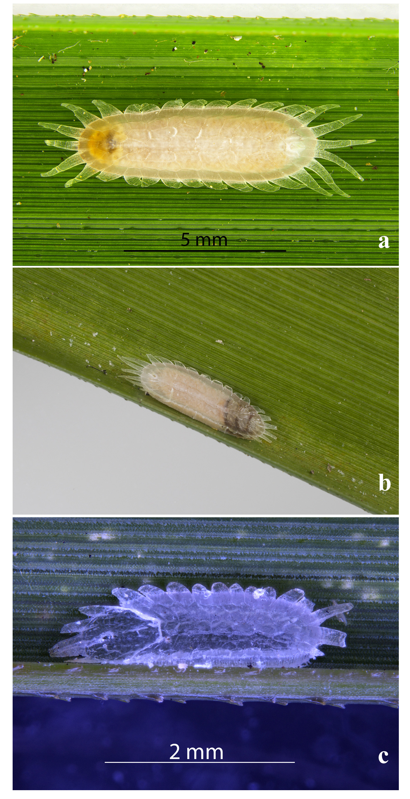
( Figs 1 View FIGURE 1 & 2 View FIGURE 2 ).
Material examined. Holotype and paratype adult females: NEW ZEALAND (AK), Avondale South Domain; Blockhouse Bay, on Gahnia setifolia (Cyperaceae) , 15.ix.2014, N.A. Martin ( New Zealand Arthropod Collection (NZAC)): one slide with two specimens in good condition, holotype specimen clearly marked near center of slide. Also paratype adult females: as previous but dated 16.ix.2014 (NZAC): two slides each with a single specimen (fair-good) and one slide with a pharate pupa. [Note that Avondale South Domain has recently been renamed Gittos Domain and is also known as Sandy Bay Reserve].
Note. Described from all four adult female specimens.
Adult female. Unmounted material. Fresh specimens greenish yellow, covered in a transparent glassy test, with 29 triangular waxy extensions around margin ( Fig. 1 View FIGURE 1 ), forming a ledge above a short vertical side. Older specimens with post-reproductive adult crumpled up at anterior end, appearing dark through test ( Fig. 1 View FIGURE 1 b); remainder of test full of eggs. Young specimens in alcohol pale to dark brown.
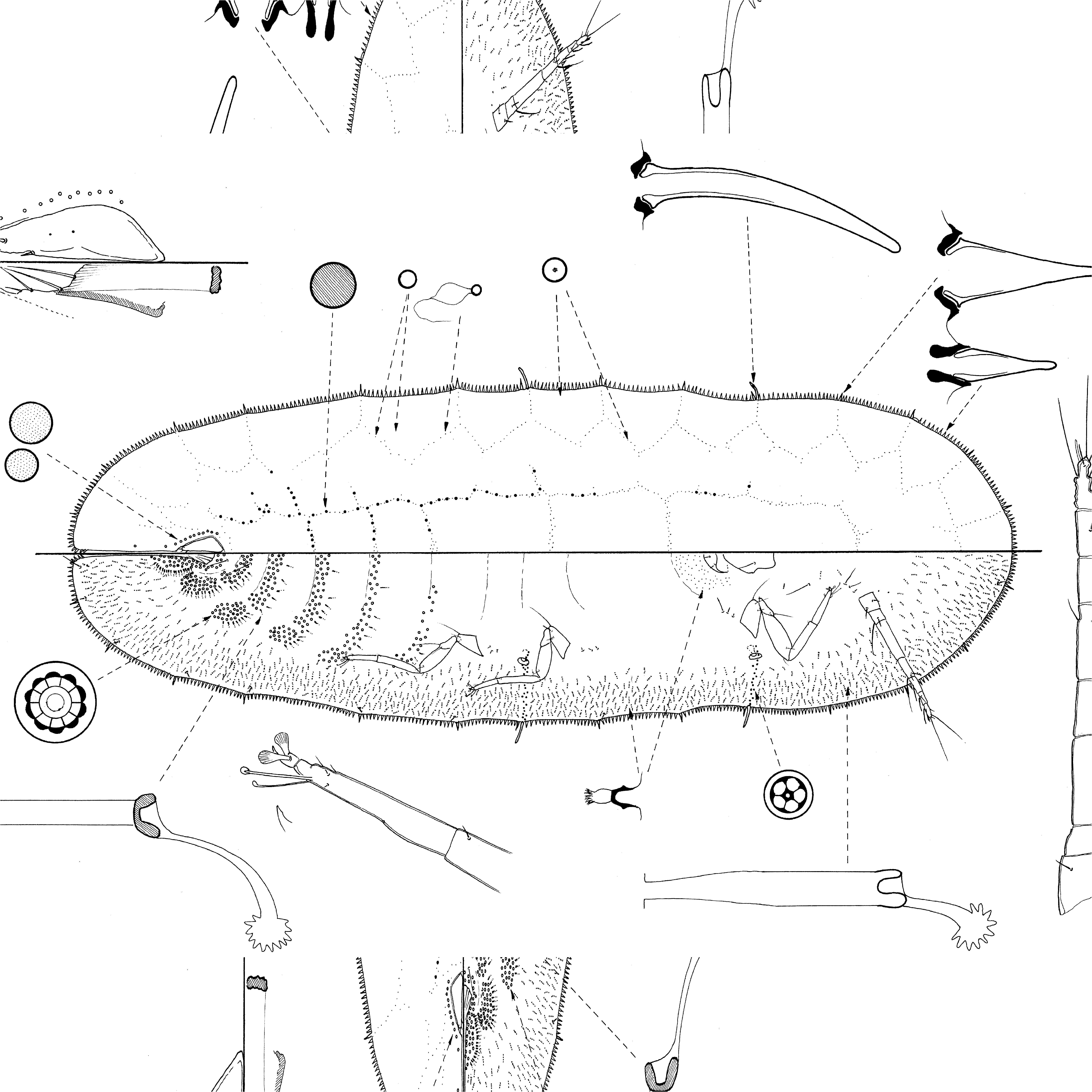
Mounted material. Body elongate oval; length 3.7–7.9 mm; breadth 1.1–2.25 mm; anal cleft about 1/9th body length; stigmatic clefts absent.
Dorsum. Derm membranous. Dorsal setae absent. Dorsal pores in a reticulate pattern, with reticulation areas in probably 9 longitudinal rows, with 8 (9?) reticulation areas medially between anal plates and anterior margin and with 29 areas around margin (but see under Discussion below). Dorsal pores of possibly five types: (i) microductules, each about 0.5 µm wide, with a long balloon-like inner ductule, main pore marking position of reticulation lines, possibly absent elsewhere; (ii) simple pores, each small and probably flat, about 1.0 µm wide, sparse throughout dorsum, including between reticulation lines; (iii) a slightly larger simple pore, each about 2.0– 2.5 µm wide with a dark centre, sparse within reticulation lines and possibly forming a sparse line along margin; (iv) slightly larger, sclerotised macropores, each probably convex and 3.0–3.5 µm wide, delimiting submedial reticulation lines, at least on abdomen, with a few extending onto thorax and also in more medial radial lines, and (v) a similar-sized simple pore, each rather variable in size, 2.0–4.0 µm wide, probably flat, in a sparse band or line on each side of anal plates. Preopercular pores, dorsal tubercles and dorsal tubular ducts absent. Anal plates together widest at anterior 1/5th to 1/4th, tapering to a narrow apex, each 180–200 µm long, combined widths 130– 165 µm; with 0–2 minute pores on dorsal surface of each plate; length of setae: anterior inner margin setae 13–25 µm; posterior inner margin 16–20 µm; apical 35–40 um and outer margin setae 16–20 µm (actually on dorsal surface near apex); each anal plate with a narrow supporting bar. Anogenital fold with 2 pairs of setae along anterior margin and 1 or 2 setae on each lateral margin; longest 50–55 µm long. Anal tube quite long; anal ring with 6 setae.
Margin. Marginal setae in a single line, of two sizes, both lanceolate and spinose, with rather blunt apices and narrow basal sockets: (i) shorter 16–20 µm long, with 51–63 on each side between stigmatic clefts, and (ii) larger setae, similar in shape to (i) but each 30–45 µm long, located at ends of each reticulation line, with 8 between stigmatic areas anteriorly; three on each side between stigmatic areas and seven on each abdominal margin; marginal setae extending only a short distance up anal cleft margin. Stigmatic areas without stigmatic sclerotisations and without a cleft, each area with a single rather blunt spine, generally slightly curved, with broad basal sockets; each 50–60 µm long. Eyespot present on margin, each about 8–10 µm long.
Venter. Derm membranous. Pregenital disc-pores each about 8 µm wide with mainly 10 loculi; in bands across all abdominal segments but most abundant posteriorly; each band with a mediolateral group; with none laterad to metacoxae. Spiracular disc-pores each about 4–5 µm wide with mainly 5 loculi, in bands 1–4 pores wide; with 25– 42 in each band; with a few extending medially over each peritreme. Ventral microducts of one size, each about 1.5 µm wide, in a sparse submarginal band and in a dense group posterior to mouthparts. With 0–2 preantennal pores just anterior to each antenna. Ventral tubular ducts of two types as for genus: (i) shorter duct with outer ductule 15– 17 µm long and with a fine inner ductule 11–14 µm long, frequent on either side of anal cleft and across all abdominal segments in a line or band anterior to multilocular disc-pores, but very sparse or absent medially, and (ii) longer ducts, each with outer ductule about 25 µm long and inner ductule 8 µm long, in a broad submarginal band, extending medially to spiracles and also a few present between clypeolabral shield and antennae. Ventral setae: ventral anal lobe setae 15–25 µm long; with one pair of anterior anal cleft setae; with long pregenital setae restricted to pairs medially on abdominal segments VI and VII; short setae medially on each abdominal segment few; with 2 or 3 minute setae near each coxa but with one longer seta near each procoxa; with 3 pairs of interantennal setae, posteriormost setae unusually long; with 1–3 submarginal setae on each side between stigmatic areas. Antennae 8-segmented, with distinct segmentation between segments III and IV, and between IV and V; total length 500–550 µm; length of apical setae 80–90 µm; fleshy seta on each antenna particularly long. Length of clypeolabral shield 165–170 µm. Width of spiracular peritremes: anterior 50–60 µm, posterior 58–70 µm. Legs elongate, with a separate tibia and tarsus, but no tibio-tarsal articulatory sclerosis; distribution of leg setae as for family; claws with a minute denticle; claw digitules similar and broad; tarsal digitules dissimilar, one shorter and narrower than other; tarsal campaniform pores absent; lengths (metathoracic): coxa 160–175 µm, trochanter + femur 225–240 µm, tibia 165–175 µm, tarsus 100–110 µm, claw 20–23 µm.
Notes. Although the exact layout of the reticulation lines on the mounted specimens was hard to determine, the presence of 14 larger marginal setae on each side clearly suggests that there are a total of 29 marginal waxy plates. The small size of the pores and their sparseness made delineation of the other rows of reticulation plates difficult but the photographs strongly suggest that there are three pairs in addition to the central line. These are not shown in the figure.
Hodgson & Henderson (2000) suggested that the following adult female characteristics diagnosed Kalasiris : (i) marginal setae particularly abundant, with some extending up margins of anal cleft; (ii) at least a few larger simple pores dorsally on either side of anal plates; (iii) two types of ventral tubular ducts, a longer type submarginally and a shorter type medially and mediolaterally on abdominal segments; (iv) ventral tubular ducts present in a submarginal band near to or on margin, the ducts often lying radially, and (v) ventral tubular ducts absent medially on thorax and restricted to between clypeolabral shield and antennae on head. Clearly K. martini agrees with this diagnosis.
Male test and pupa. Described from single specimen.
Mounted material. Male test ( Figs 1 View FIGURE 1 c & 3): Glassy with a reticulate pattern of polygonal plates, in seven longitudinal rows. Length of test at least 2 mm. Test with 8 medial plates anterior to anal plates, and with more or less 3 lateral lines of plates on each side, each plate of rather variable shape; with 29 plates around outer margin.
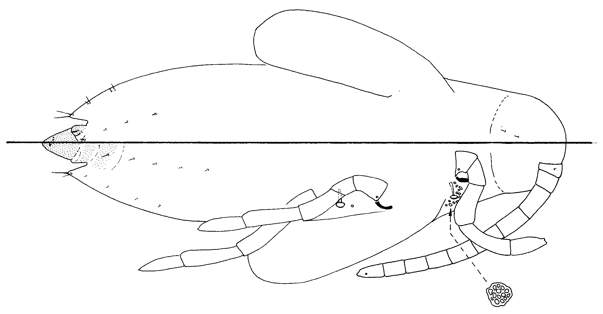
Pupa ( Fig. 4 View FIGURE 4 ): Division into head, thorax and abdomen clear, although segmentation obscure apart from on abdomen. Derm membranous, with small dermal spinules. All ducts and pores (bar spiracular disc-pores) absent and setae few. Of moderate size: length 1.8 mm.
Head. Without mouthparts or eyes. Setae: two pairs of setae present posteriorly on dorsum. Antennae ten segmented, total length 900 µm (ratio to total body length 1:2).
Thorax. No setae located. With 12–15 spiracular disc-pores associated with each anterior spiracle, distributed anteriorly and laterad to peritreme, extending medially full length of spiracular apodeme; with 1 disc-pore just anterior to each posterior spiracle; shape and number of loculi in each disc-pore highly variable. Spiracles: width of peritremes 25 µm. Legs each with a small triangular finger on apex, probably an incipient claw; length of metathoracic legs 950 µm. With a pair of long wing-buds on each side, extending to about abdominal segment III, mildly sclerotised: length 725 µm, width 225 µm (ratio length to width 1:0.32).
Abdomen. Segmentation fairly distinct, anteriormost segment considered to represent segment II (see Hodgson & Henderson, 2000, p. 54), so that there are seven visible segments (segments II to VIII) anterior to penial sheath. With 2 ante-anal setae dorsally on segment VIII; with 1 pair of small ventral setae on segments II–VII; dorsopleural setae: 1 pair on segments V–VIII; 0 or 1 small ventropleural seta on segments V–VII. Lateral lobe of VII rather pointed, each about half length of penial sheath, each with a long apical seta (45 µm long) and two shorter setae laterally. Segment VIII with a pair of prominent, mildly sclerotised lobes dorsally on either side of penial sheath, each with 2 stout setae. Penial sheath longer than lateral lobes of segment VII and a little longer than wide (165 µm long, 125 µm wide at base; ratio length to width 1:0.75); with a pair of minute setae on dorsal surface and numerous small pores on apex.
Notes: The pupae of Kalasiris species are rather similar to those of Aphenochiton , Crystallotesta ( fagi -group), Ctenochiton and Umbonichiton from which it is difficult to separate them. The pupae of Kalasiris share the following characters: (i) body moderate to quite large in size; (ii) spiracular disc-pores associated with each anterior spiracle mainly in a group dorsad and laterad to peritreme, none extending mesad to inner margin of spiracular peritreme; (iii) with or without disc-pores associated with each posterior spiracle but, when present, few; (iv) lobes on abdominal segment VII well developed; (v) lobes of abdominal segment VIII moderately large and obvious, with 1 or more setae; (vi) ante-anal setae present, and (vii) the segmental arrangement of the dorsopleural setae.
Generic characters. Kalasiris is closest to Ctenochiton Maskell and Crystallotesta Henderson & Hodgson in having abundant multilocular disc-pores medially across all abdominal segments. It also resembles species of Crystallotesta in having 2 types of ventral tubular duct, small spiracles, abundant marginal setae and large simple pores laterad to the anal plates. However, Kalasiris differs from Crystallotesta in lacking tubular ducts medially on the thorax (present in Crystallotesta ) and in having the submarginal tubular ducts tending to lie radially (more random in Crystallotesta ). Adult female Crystallotesta leptospermi (Maskell) is superficially rather similar to K. martini in having: (i) a rather elongate body; (ii) eight-segmented antennae, (iii) macropores more or less restricted to more medial reticulation lines; (iv) quite large reticulation setae on margin, and (v) long stigmatic spines. However C. leptospermi has a few tubular ducts medially on the thorax, and the distribution of the reticulation plates is different, with only approximately 5 plates between anal plates and anterior margin ( K. martini has eight). It is clear that these two genera ( Crystallotesta and Kalasiris ) are very close and may need to be synonymised when more species are discovered.
Name derivation. The species name martini refers to Dr. Nicholas Martin, Research Associate, Landcare Research, Auckland, New Zealand, who collected these insects and also many other scale insects in New Zealand, several of them new to science.
No known copyright restrictions apply. See Agosti, D., Egloff, W., 2009. Taxonomic information exchange and copyright: the Plazi approach. BMC Research Notes 2009, 2:53 for further explanation.