
Trichomycterus poikilos, Ferrer & Malabarba, 2013
|
publication ID |
https://doi.org/10.1590/S1679-62252013000200001 |
|
persistent identifier |
https://treatment.plazi.org/id/CA2E0317-9238-8870-EAE7-F156FCE4FCA1 |
|
treatment provided by |
Carolina |
|
scientific name |
Trichomycterus poikilos |
| status |
sp. nov. |
Trichomycterus poikilos View in CoL , new species
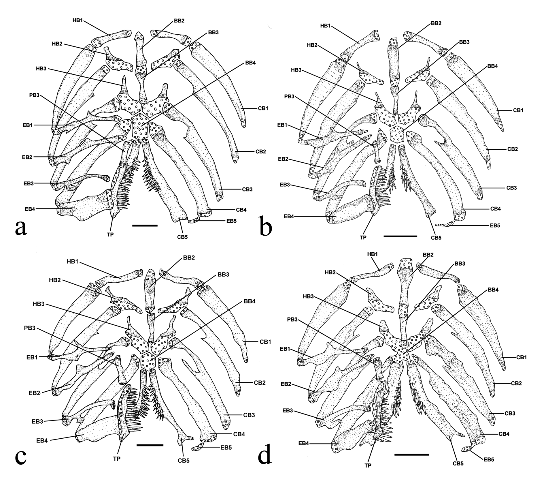
Figs. 2c View Fig , 3c View Fig , 4c View Fig , 5c View Fig 6c View Fig , 11b View Fig , 13 View Fig , 14 View Fig
Trichomycterus sp. 3. Becker et al. (2013: Table 1, listed, Taquari-Antas river basin).
Holotype. UFRGS 16239 View Materials , 63.3 mm SL, Brazil, Rio Grande do Sul State, municipality of Júlio de Castilhos, arroio Passo dos Buracos or Tipiaia on road BR-158, alto rio Jacuí basin, 29º06’50”S 53º39’04”W, 20 May 2011, A. T. Thomaz, F. R. Carvalho, J. Ferrer & L. R. Malabarba. GoogleMaps
Paratypes. All from Brazil, Rio Grande do Sul State. Alto Jacuí basin. UFRGS 16240 View Materials , 10 View Materials (3 c&s), 33.1-66.8 mm SL collected with holotype. UFRGS 11856 View Materials , 2 View Materials , 41.4-45.2 mm SL, municipality of Júlio de Castilhos , unnamed stream tributary to rio Iauí , 29º07’S 53º22’W, 5 Aug 2009, A. R. Cardoso & V. A. Bertaco. UFRGS 14992 View Materials , 6 View Materials (1 c&s), 40.5-92.5 mm SL, municipality of Cruz Alta , rio Passo Novo , 28º38’43”S 53º33’36”W, 20 May 2011,A. T. Thomaz, F. R. Carvalho, J. Ferrer & L. R. Malabarba. MCN 16089, 5 (1 c&s), 52.0- 60.9 mm SL, municipality of Estrela Velha, unnamed stream tributary to rio Jacuí , 29º10’20”S 53º09’27”W, 19 Jan 1999, W. R. Koch & C. J. Mansan. MCP 18253, 1 View Materials , 65.6 mm SL, municipality of Faxinal do Soturno , unnamed stream tributary to rio Soturno , 29º25’29”S 53º30’59”W, 28 Jul 1995, G. F. Rey, M. P. Barros & N. F. Fontoura. MCP 21231, 2 View Materials , 46.8-54.1 mm SL, municipality of Agudo, arroio Linha Louca on road to Dona Francisca dam, 29º29’09”S 53º17’09”W, 22 Aug 1998, J. F. P. Silva, R. E. Reis & V. A. Bertaco. MCP 21237, 1 View Materials , 59.7 mm SL, municipality of Arroio do Tigre , arroio Tamanduá, 29º16’39”S 53º03’10”W, 24Aug 1998, J. F. P. Silva R. E. Reis & V. A. Bertaco. MCP 21252, 1 View Materials , 66.9 mm SL, municipality of Espumoso, unnamed stream on road from Espumoso to Campos Borges , 28º45’51”S 52º55’10”W, 24Aug 1998, J. F. P. Silva R. E. Reis & V. A. Bertaco. MCP 21464, 1 View Materials , 64.7 mm SL, municipality of Fortaleza dos Valos, arroio Lajeado Fortaleza, 28º46’45”S 53º11’15”W, 11 Oct 1998, E. H. Pereira, J. F. P. Silva & R. E. Reis. MCP 21508, 3 View Materials , 37.4-59.7 mm SL, municipality of Fortaleza dos Valos, unnamed stream tributary to rio Lajeado Fortaleza , 28º47’39”S 53º12’17”W, 11 Oct 1998, E. H. Pereira, J. F. P. Silva & R. E. Reis. MCP 21522, 1 View Materials , 64.5 mm SL, municipality of Fortaleza dos Valos, unnamed stream tributary to rio Ingai , 28º54’29”S 53º17’08”W, 11 Oct 1998, E. H. Pereira, J. F. P. Silva & R. E. Reis. MCP 22127, 1 View Materials , 43.6 mm SL, municipality of Santo Antônio do Planalto , rio da Glória, 28º21’02”S 52º43’16”W, 18 Jan 1999, E. H. Pereira, J. F. P. Silva & R. E. Reis. MCP 22193, 1 View Materials , 64.0 mm SL, municipality of Passo Fundo, arroio Pinheiro Torto, 28º22’23”S 52º26’38”W, 19 Jan 1999, E. H. Pereira, J. F. P. Silva & R. E. Reis. MCP 22219, 3 View Materials , 24.9-43.3 mm SL, municipality of Nicolau Vergueiro, arroio Quebra Dentes, 28º36’09”S 52º27’23”W, 19 Jan 1999, E. H. Pereira, J. F. P. Silva & R. E. Reis. MCP 22699, 6 View Materials (1 c&s), 26.1-79.0 mm SL, municipality of Cruz Alta, rio Passo Novo , 28º38’43”S 53º33’35”W, 2 Apr 1999, E. H. Pereira, R. E. Reis & V. A. Bertaco. MCP 22778, 1 View Materials , 54.6 mm SL, municipality of Santa Bárbara do Sul , arroio das Figueiras on road from Cruz Alta to Saldanha Marinho, 28º26’39”S 53º12’37”W, 2 Apr 1999, E. H. Pereira, R. E. Reis & V. A. Bertaco. MCP 26540, 2 View Materials , 43.9-56.6 mm SL, municipality of Agudo, unnamed stream tributary to rio Jacuí , 29º31’38”S 53º16’01”W, 10 Nov 2000, A. R. Cardoso, C. Kaefer, J. F. P. Silva & V. A. Bertaco. MCP 26974, 1 View Materials , 51.2 mm SL, municipality of Ibarama, unnamed stream tributary to reservoir of Dona Francisca dam, 2 Feb 2001, R. E. Reis & V. A. Bertaco. MCP 46948, 7 View Materials (1c&s), 34.9-61.0 mm SL, municipality of Nova Jacuí, unnamed stream on road from Estado Velho to Nova Jacuí, 29º06’20”S 53º12’32”W, 11 Oct 1998, E. H. Pereira, J. F. P. Silva & R. E. Reis. Rio Taquari- Antas basin. CIUFSC 2117, 4, 13.8-64.1 mm SL, municipality of Passo Fundo, unnamed stream at Floresta Nacional of Passo Fundo , 28º18’46”S 52º11’30”W, 24 Nov 2008, B. Marterer. MCN 15679, 1, 61.0 mm SL, municipality of Guabiju, arroio Erval on the road between Nova Araçá and São Jorge, rio Carreiro basin, 28º33’34”S 51º43’12”W, 16 Apr 1998, W. R. Koch. MCP 21203, 1 View Materials , 51.2 mm SL, municipality of Soledade, unnamed stream on road from Soledade to Arvorezinha, 28º48’17”S 52º22’20”W, 25 Aug 1998, J. F. P. Silva R. E. Reis & V. A. Bertaco. MCP 22237, 2 View Materials , 73.4-75.4 mm SL, municipality of Vila Maria, arroio Porongas, 28º31’34”S 52º08’22”W, 20 Jan 1999, E. H. Pereira, J. F. P. Silva & R. E. Reis. MPEG 24031 View Materials , 2 View Materials , 39.8-48.3 mm SL, municipality of Nova Bassano, arroio Caçador, tributary to rio Carreiro , 28º42’16”S 51º50’38”W, 10 Mar 2010, J. Ferrer & J. M. Wingert. MZUSP 110993 View Materials , 5 View Materials , 38.0- 74.5 mm SL, municipality of Nova Alvorada, rio Lajeado Engenho Velho , rio Guaporé basin, 28º39’23”S 52º09’40”W, 14 May 2010, C. Vogel, G. Rosa, L. de Fries & L. Santos. UFRGS 13892 View Materials , 2 View Materials , 50.6-56.9 mm SL, municipality of Nova Bassano, arroio Caçador, tributary to rio Carreiro , 28º42’16”S 51º50’38”W, 7 Dec 2010, J. Ferrer & J. M. Wingert. UFRGS 16241 View Materials , 6 View Materials (1 c&s), 32.0- 71.2 mm SL, municipality of Nova Alvorada, rio Lajeado Engenho Velho , rio Guaporé basin, 28º39’23”S 52º09’40”W, 14 May 2010, C. Vogel, G. Rosa, L. de Fries & L. Santos. UFRGS 16342 View Materials , 2 View Materials (1 c&s), 59.8-64.2 mm SL, municipality of Vila Maria, rio Jordão , rio Guaporé basin, 28º32’48”S 52º06’38”W, 13 May 2010, C. Vogel, G. Rosa, L. de Fries & L. Santos. Rio Pardo basin. MCP 26725, 2 View Materials , 34.6-39.6 mm SL, municipality of Barros Cassal, arroio Pessegueiro on reservoir of Corsan, rio Pardo basin, 29º05’04”S 52º36’02”W, 08 Dec 2000, J. F. P. Silva & J. R. Marinho GoogleMaps .
Non-type material. All from Brazil, Rio Grande do Sul State. Rio do Sinos basin. MCP 26148, 7 View Materials (1 c&s), 31.3-57.0 mm SL, municipality of Igrejinha , arroio Solitária , 29º35’S 50º46’W, 10 Jun 2000, M. Moreira, R. Costa & U. Schulz GoogleMaps .
Diagnosis. Trichomycterus poikilos is distinguishable from all congeners except T. brachykechenos ; T. mboycy Wosiacki & Garavello, 2004 ; and T. naipi Wosiacki & Garavello, 2004 by the modal possession of I+5 pectoral-fin rays (one among 82 specimens with I+6; vs. I+6 or more pectoral-fin rays) and the first ray of the pectoral fin not prolonged as a filament ( Figs. 13-14 View Fig View Fig ; vs. the first ray of the pectoral fin prolonged as a filament). Trichomycterus poikilos is distinguished from T. brachykechenos by having the posterior fontanel extending from the parieto-supraoccipital to the frontals ( Fig. 2c View Fig ; vs. the posterior cranial fontanel restricted to the parietosupraoccipital), the origin of dorsal fin located at the vertical falling between the tip of the pelvic fin and the anterior insertion of the anal fin ( Figs. 13-14 View Fig View Fig ; vs. the origin of dorsal fin located at the vertical through the last third of the pelvic fin), the maxillary barbel length (33.2-60.4% vs. 68.2-87.7% HL), and the number of odontodes of the opercular patch (13-17 vs. 8-11). Trichomycterus poikilos is distinguished from T. mboycy by the pectoral-fin length (7.7-9.1% vs. 9.9- 13.8% SL) and skin color ( Figs. 13-14 View Fig View Fig ; yellow background vs. gray background); and from T. naipi in the number of odontodes of the interopercular patch (20-32 vs. 12-15), number of dorsal-fin rays (I-II+6-7 vs. I+8), and absence of a thin unpaired dark dorsosagittal body stripe in larger specimens ( Fig. 13 View Fig ; vs. presence of a thin unpaired dark dorsosagittal stripe in larger specimens). Additionally, T. poikilos is distinguished from remaining congeners in the laguna dos Patos system by having the origin of the dorsal fin located at the vertical falling between the tip of the pelvic fin and the anterior insertion of the anal fin ( Figs. 13-14 View Fig View Fig ; vs. at the vertical through the last third of the pelvic fin in T. balios and T. diatropoporos ); 20-23 teeth on ceratobranchial 5 ( Fig. 3c View Fig ; vs. 36-39 in T. balios and 12-13 in T. diatropoporos ); 23-25 teeth on plate connected to pharyngobranchial 4 ( Fig. 3c View Fig ; vs. 35-40 in T. balios and 18-20 in T. diatropoporos ); color pattern of the dorsal and lateral surfaces of body mottled dark brown over a light yellow background or with three stripes with notched borders (dorsosagittal, midlateral, and ventrolateral) ( Figs. 13-14 View Fig View Fig vs. the dorsal and lateral surfaces of body with circular black blotches variable in size in T. balios and T. tropeiro , the lateral surface of body with dark blotches variable in size and shape progressively larger and coalescent dorsally thus forming extensive black pigmented regions with light yellow gaps in T. diatropoporos ). Trichomycterus poikilos is further distinguishable from T. diatropoporos by the insertion of the first dorsal-fin basal radial anterior to the neural spine of the 20 th- 22 th vertebrae ( vs. anterior to the neural spine of the 17 th or 18 th vertebrae). Trichomycterus poikilos is further distinguishable from T. tropeiro by the presence of the pelvic girdle and pelvic fins ( Figs. 13-14 View Fig View Fig ; vs. absence); and the absence of the pores i1 and i3 of the infraorbital sensory canal ( Fig. 2c View Fig ; vs. presence).
Description. Morphometric data for holotype and paratypes in Table 3. Body elongate, trunk roughly cylindrical and gradually compressed towards caudal fin. Dorsal profile of trunk convex along anterior half, then straight to insertion of dorsal fin. Ventral profile of trunk straight to slightly convex. Dorsal and ventral profile of caudal peduncle straight.
Head depressed, trapezoidal from dorsal view, wider posteriorly. Dorsal profile straight and ventral profile straight to slightly convex. Snout rounded from dorsal view. Eyes readily visible, anteroposteriorly elliptical and dorsally oriented; orbital rim not free, eyes covered with skin thin and transparent. Each eye located over posterior termination of shallow small longitudinal crest beginning at posterior nostril and making eyes visible from lateral view.
Nostrils of same size and smaller than diameter of eye. Anterior nostril surrounded by fleshy flap of integument posterolaterally continuous with nasal barbel. Posterior nostril surrounded anterolaterally by thin flap of integument. Gill openings not constricted but united with isthmus anteriorly forming a free fold. Mouth subterminal with corners posteriorly oriented. Lower lip with conspicuous fleshy lobes along lateral limits internal to origin of rictal barbels. Lips with small papillae; papillae largest on inner surface of upper lip.
Barbels with large bases and tapering gradually towards tips. Tip of nasal barbel usually reaching to between pore i11 and pore po1 or at most to middle of distance between them. Origin of nasal barbels on posterolateral portion of integument flap around anterior nostril. Maxillary barbel usually extending between to anterior and posterior margins of interopercular patch of odontodes. Rictal barbel slightly shorter than maxillary barbel.
Mesethmoid with anterior margin slightly concave, cornua short and thick, width of their bases similar to their length ( Fig. 2c View Fig ).Anterior cranial fontanel restricted to small rounded opening situated between frontals and epiphyseal bar. Posterior cranial fontanel long and narrow extending from posterior portion of frontals to parieto-supraoccipital. Epiphyseal bar longer than wide. Antorbital short and anteriorly expanded. Tendon-bone supraorbital elongate, anteriorly expanded and approximately three times larger than antorbital.Anterior portion of sphenotic anterolaterally directed from dorsal view.Sphenotic, prootic and pterosphenoid totally fused. Vomer arrow-shaped with long posterior process extending to parasphenoid. Parasphenoid with long and pointed process extending to basioccipital. Anterior portion of Weberian complex fused to basioccipital. Weberian capsule with lateral opening smaller than lateral profile of capsule.
Premaxilla rectangular with 74-76 curved, flat vertically to slightly conical and blunt teeth (n = 3). Teeth of variable size, irregularly distributed in three rows. Maxilla large, boomerangshaped and shorter than premaxilla. Lower jaw with 79-91 curved, flat vertically and blunt teeth of variable size (n =3). Few teeth at base of coronoid process and three discernible rows near dentary symphysis ( Fig. 11b View Fig ). Autopalatine with anterior and mesial margins straight, distal margin straight to slightly concave and small posterior process extending slightly over metapterygoid.
Metapterygoid large and laminar and connecting with quadrate through cartilage. Hyomandibula well-developed. Preopercle long and narrow and in contact with ventral margins of quadrate and hyomandibula. Opercular patch of odontodes rounded with 13-17 conical odontodes (n = 9). Interopercular patch of odontodes elongate with 20-32 conical odontodes (n = 9) more concentrated posteriorly. Odontodes of both opercular and interopercular patches gradually curving medially and increasing in size posteriorly.
Ventral hypohyal triangular ( Fig. 4c View Fig ). Anterior ceratohyal elongate and widening at anterior and posterior limits. Posterior ceratohyal short and triangular. Nine branchiostegal rays (eight in one side of 1 specimen) (n = 9); five on anterior ceratohyal, one on posterior ceratohyal and three on interceratohyal cartilage. Last four branchiostegal rays widest. Dorsal hypohyal and interhyal absent. Urohyal with expanded anterior head; two elongate lateral processes with wide bases narrowing distally and bearing rounded tips; and laminar, elongate, narrow posterior process ( Fig. 5c View Fig ).
Basibranchial 1 absent. Basibranchials 2 and 3 connected to each other of approximately equal lengths with cartilage at tips ( Fig. 3c View Fig ). Ossified portion of basibranchial 2 distinctly wider than basibranchial 3. Hexagonal basibranchial 4 completely cartilaginous. Hypobranchial 1 of similar size as but narrower than basibranchial 2 with cartilage at tips. Hypobranchials 2 and 3 with narrow ossified portion slightly more elongate on hypobranchial 3, and large area of cartilage; cartilage slightly larger in hypobranchial 3. Hypobranchial 4 absent. Five elongate narrow ceratobranchials with cartilage at tips. Ceratobranchials 2 and 3 with concavity along posterior margins; concavity larger in ceratobranchial 3. Ceratobranchial 5 expanded posteromedially with 20-23 conical, elongate pointed teeth arranged in 3 rows (n = 2). Five epibranchials; first three elongate and narrow with cartilage at tips. Epibranchials 1 and 2 with triangular process along anterior margins; process larger in epibranchial 1. Epibranchial 3 with robust uncinate process along posterior margin. Epibranchial 4 rectangular. Epibranchial 5 small, narrow, curved and completely cartilaginous. Pharyngobranchials 1 and 2 absent. Pharyngobranchial 3 similar in form to hypobranchial 1 but shorter with cartilage at tips. Pharyngobranchial 4 curved and well ossified connected to plate with 23-25 conical, elongate pointed teeth arranged in 3 rows; teeth growing in length posteriorly (n = 2).
Sensory canals on head with simple (non-dendritic) tubes ending in single pores. Supraorbital sensory canal complete with pores s1, s3 and s6 ( Fig. 2c View Fig ). Pore s1 located between anterior nostrils, pore s3 located in same longitudinal row of pore s1 after posterior nostrils and pore s 6 in interorbital space. Infraorbital sensory canal incomplete; pores i1 and i3 absent, pore i10 located behind eye and pore i11 located lateral to posterior margin of eye. Postotic pore po1 located lateral to anterior margin of opercular patch of odontodes. Postotic pore po2 located lateral to middle of length of opercular patch of odontodes. Lateral-line very short with 2 pores located above insertion of pectoral fin and just posterior of gill openings.
Pectoral fin with distal margin rounded, I+5* rays (1 of 82 specimens with I+6), first ray short and not prolonged as filament. Pelvic fin with distal margin rounded reaching at most anterior margin of urogenital papilla, I+4* rays (1 of 82 specimens with I+3). Inner margin of pelvic fins very close basally. Pelvic girdle with two basipterygia united medially by cartilage with two elongate bifid processes (external process and internal process) and medial process short (in two of eight specimens medial process united to internal process). Pelvic splint thin, commashaped and parallel to first pelvic-fin ray. Urogenital papilla nearer tip of pelvic fin than origin of anal fin.
Dorsal fin with distal margin rounded, semicircular when fin expanded with one to three unsegmented rays (n = 9), I-II+6-7 rays (n = 82), usually II+7*. Dorsal-fin origin located at vertical between tip of pelvic fin and anterior insertion of anal fin, usually in the papillae urogenital. Dorsal-fin basal radials 7-8 (n = 9); first inserting anterior to neural spine of 20 th to 22 th vertebrae.
Anal fin slightly smaller than dorsal fin with distal margin rounded; one to three unsegmented rays (n = 9), II+4-5-6* rays (n = 82), usually II+5. Anal-fin origin located at vertical between anterior insertion and middle of dorsal fin base.Analfin basal radials 5-6 (n = 9); first inserting anterior to haemal spine of 22 th to 24 th vertebrae.
Caudal fin with distal margin rounded to straight with dorsal and ventral lobes rounded at limits. Procurrent caudalfin rays 15-19 dorsally (n = 9) and 11-15 ventrally (n = 9). Principal rays I+5+5-6+I-II (n = 82), usually I+5+6+I*, branched rays splitting up to three times. Lower caudal plate with parhypural and hypurals 1 and 2 co-ossified and fused to compound caudal centrum ( Fig. 6c View Fig ). Upper caudal plate with uroneural and fused hypurals 3, 4, and 5.
Vertebrae 38-40 (n = 9); ribs 12-15 (n = 9) with first rib straight and thickest and last rib rudimentary.
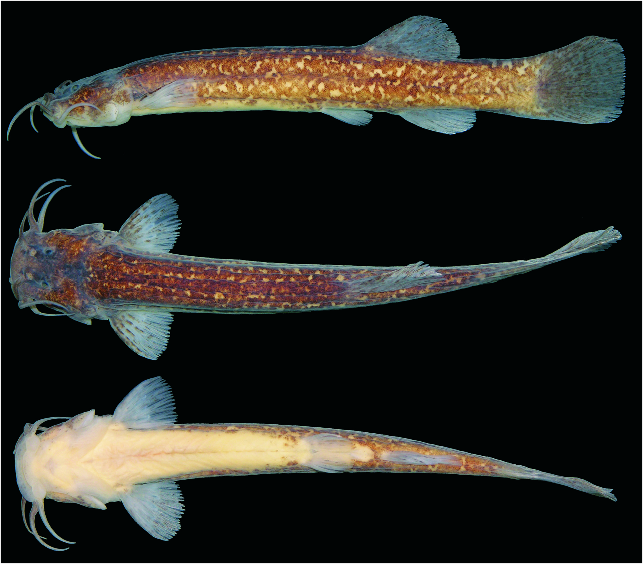
Coloration in alcohol. Trichomycterus poikilos demonstrates a variable color pattern. Most of types present the dorsum and lateral surface of body mottled dark brown over a light yellow background ( Fig. 13 View Fig and 14a View Fig ). Some specimens have a wide unpaired dorsosagittal stripe and midlateral and ventrolateral bilaterally paired stripes with notched borders ( Fig. 14c View Fig ). Smallest specimens with midlateral stripe more evident and ventrolateral stripe diffuse ( Fig. 14d and 14e View Fig ). Ventral surface of body light yellow with diffuse and small black spots between pectoral-fin insertion and near pelvicfin insertion. Area between pelvic and anal fins and caudal peduncle mottled dark brown over light yellow background. Head dark brown mottled over light yellow background dorsally and yellow ventrally. Pectoral, dorsal, anal and caudal fins spotted basally with dark pigmentation becoming inconspicuous towards tips, and lighter along margins. Pelvic fin unpigmented. Barbels with dark pigmentation dorsally and lighter ventrally. Large specimens (greater than 46.6 mm SL) with pigmentation arranged in two distinct layers; mottled dark brown or with three stripes (dorsosagittal, midlateral, and ventrolateral) with notched borders on inner skin layer and small black spots on outer skin layer ( Fig. 13 View Fig , 14 View Fig a-c).
Distribution and ecological notes. Trichomycterus poikilos is distributed in the rio Jacuí, rio Pardo and rio Taquari-Antas basins, in the laguna dos Patos system ( Fig. 8 View Fig ). The species is widely distributed in the upper portion of rio Jacuí basin, occurring at elevations of 124 to 700 m a.s.l. One lot is from the headwaters of rio Pardo basin, at 625 m a.s.l. In the Taquari- Antas basin the species occurs in the upper portions of the Carreiro, Guaporé, and Forqueta rivers, between 417 and 688 m a.s.l. The type locality is a rapid flow river with clear water, a rocky bottom and submerged vegetation ( Fig. 9c View Fig ), located at approximately 405 m a.s.l. At this site the specimens were associated with submerged vegetation and collected with Ancistrus brevipinnis (Regan, 1904) , Eurycheilichthys limulus Reis & Schaefer, 1998 , and Rineloricaria cadeae (Hensel, 1868) . The stomach of six c&s specimens contained aquatic larvae of Diptera (Simuliidae) and Lepidoptera; and nymphs of the Ephemeroptera and Plecoptera.
Remarks. Trichomycterus poikilos demonstrates intraspecific ontogenetic color pattern variation. Although Bockmann & Sazima (2004) considered the body pigmentation pattern highly conserved in known species of Trichomycterus , a intraspecific variation in coloration is present in T. caipora Lima, Lazzarotto & Costa, 2008 ( Lima et al., 2008), in T. iheringi ( Eigenmann, 1917) associated to body size and microhabitat preference ( da Silva et al., 2010), and in T. santanderensis Castellanos- Morales, 2007. The ontogenetic variation in T. poikilos is clear in lot UFRGS 14992 (six specimens with 40.5-92.5 mm SL), in which the smallest specimens have one obvious midlateral stripe on the body that gradually become indistinguishable in the largest specimens ( Fig. 14 View Fig ). This ontogenetic variation is not observed in all samples. In other lots all specimens have a mottled pattern (UFRGS 14500, 11 specimens with 32.0- 74.5 mm SL) regardless of body size. Besides coloration no additional differences were found between these populations that allow us to separate them and ontogenetic variation in the color pattern seems to appear only in some populations.
The color pattern of some specimens of T. poikilos – lateral surface of body with widened stripes – is very similar to T. itatiayae Miranda Ribeiro, 1906 ; T. naipi ; T. pauciradiatus Alencar & Costa, 2006; T. perkos , and T. reinhardti ( Eigenmann, 1917) (Barbosa & Costa, 2008; Wosiacki & Garavello, 2004; Alencar & Costa, 2006; Datovo et al., 2012; Eigenmann, 1917), but differs from these by other characters (see diagnosis). The midlateral stripe is present in juveniles of other species of Trichomycteridae , but absent in adults (see Datovo et al., 2012).
Seven specimens from the rio do Sinos basin are poorly preserved and were not measured or counted (MCP 26148), however a mottled color pattern over a light yellow background is present in all specimens, and one c&s specimen possesses the lower jaw with blunt teeth which leads us to believe it to be T. poikilos .
Etymology. From the Greek poikilos meaning variegated, varicolored, in reference to the intraspecific color pattern variation of the new species. An adjective.
Trichomycterus brachykechenos , new species Figs. 2d View Fig , 3d View Fig , 4d View Fig , 5d View Fig 6d View Fig , 15 View Fig , 16 View Fig
Holotype. MCN 18929, 61.1 mm SL, Brazil Rio Grande do Sul, State, municipality of Caraá, rio do Sinos , 29º45’44"S 50º19’39"W, 30 Jun 2006, B. B. Calegari, M. A. Azevedo & R. Hirano. GoogleMaps
Paratypes. All from Brazil, Rio Grande do Sul State, rio do Sinos basin. MCP 46949, 3 View Materials , 26.3-70.9 mm SL, Brazil, Rio Grande do Sul State, municipality of Caraá , rio do Sinos , 29º44’58”S 50º16’59”W, 30 Jun 2006, B. B. Calegari, M.A.Azevedo & R. Hirano.All the following lots from Brazil, Rio Grande do Sul State, municipality of Caraá, rio do Sinos, 29º43’32”S 50º16’56”W. MCN 18930, 2, 36.3-53.7 mm SL, 28 Nov 2007, M. A. Azevedo & T. Aguzzoli. MCN 18931, 2, 39.4-47.9 mm SL, 26 Jun 2007, M. A. Azevedo, T. Aguzzoli & M. Pairet. MCP 46950, 1 View Materials , 51.6 mm SL, 14 Mar 2007, F. Becker, M. A. Azevedo & T. Aguzzoli. UFRGS 16244 View Materials , 1 View Materials (c&s). 51.6 mm SL, 30 Jul 2007, M. A. Azevedo, T. Aguzzoli & M. Pairet. UFRGS 16245 View Materials , 5 View Materials (1 c&s), 35.0- 61.9 mm SL, 2 Feb 2007, M. A. Azevedo & T. Aguzzoli GoogleMaps .
Diagnosis. Trichomycterus brachykechenos is distinguishable from all congeners except T. megantoni Fernández & Chuquihuamaní, 2007 by its elongate posterior cranial fontanel which is restricted to the parietosupraoccipital ( Fig. 2d View Fig vs. the posterior fontanel extending from the parieto-supraoccipital to the frontals or posterior fontanel separated in two openings not restricted to the parieto-supraoccipital). Trichomycterus brachykechenos is distinguished from T. megantoni by the modal possession of I+5 pectoral-fin rays (one among 15 specimens with I+6 vs. I+7-8 pectoral-fin rays), first ray of the pectoral fin not prolonged as a filament ( Fig. 15 View Fig ; vs. the first ray of the pectoral fin prolonged as a filament), absence of the interopercle thickened ventrally ( vs. presence). Trichomycterus brachykechenos is further distinguishable from all its congeners except T. mboycy , T. naipi , and T. poikilos by the modal possession of I+5 pectoral-fin rays (one among 15 specimens with I+6; vs. I+6 or more pectoralfin rays) and the first ray of the pectoral fin not prolonged as a filament ( Fig. 15 View Fig ; vs. first ray of the pectoral fin prolonged as a filament). Trichomycterus brachykechenos is further distinguishable from T. mboycy and T. naipi by the color pattern of the dorsal and lateral surface of body of a densely mottled dark brown over a light yellow background ( Fig. 15 View Fig ; vs. the lateral surface of body with small spots in T. mboycy ; the lateral surface of body with three dark longitudinal stripes in T. naipi ); the maxillary barbel length (68.2-87.7% vs. 50.3-63% HL in T. mboycy ; 34.1-68.2% HL in T. naipi ). Additionally, T. brachykechenos is distinguished from the remaining congeners in the laguna dos Patos system by the number of odontodes on the opercular patch (8-11 vs. 13 or more in T. balios , T. diatropoporos , T. poikilos , and T. tropeiro ) and the maxillary barbel length (68.2-87.7% vs. 37.8- 66.6% HL in T. balios ; 38.3-53.9% HL in T. diatropoporos ; 33.2-60.4% HL in T. poikilos ).
Description. Morphometric data for holotype and paratypes in Table 4. Body elongate, trunk roughly cylindrical and gradually compressed towards caudal fin. Dorsal profile of trunk convex along anterior half then straight to insertion of dorsal fin. Ventral profile of trunk straight. Dorsal and ventral profiles of caudal peduncle slightly concave to straight.
Head depressed, trapezoidal to square in larger specimens from dorsal view, wider posteriorly. Dorsal profile straight and ventral profile straight to slightly convex. Snout rounded from dorsal view. Eyes readily visible, rounded and dorsally oriented, orbital rim not free, eyes covered with skin thin and transparent. Each eye located over posterior termination of shallow and small longitudinal crest beginning at posterior nostril and making eyes visible from lateral view.
Nostrils of same size and smaller than diameter of eye. Anterior nostril surrounded by fleshy flap of integument posterolaterally continuous with nasal barbel. Posterior nostril surrounded anterolaterally by thin flap of integument. Gill opening not constricted but united with isthmus anteriorly forming a free fold. Mouth subterminal with corners posteriorly oriented. Lower lip with conspicuous fleshy lobes along lateral limits internal to origin of rictal barbels. Lips with small papillae; papillae largest on inner surface of upper lip.
Barbels with large bases and tapering gradually towards tips. Tip of nasal barbel usually reaching opercular patch of odontodes, always to middle of distance between pore i11 and pore po1. Origin of nasal barbels on posterolateral portion of integument flap around anterior nostril. Maxillary barbel usually reaching or extending beyond pectoral-fin insertion, always extending beyond posterior margin of interopercular patch of odontodes. Rictal barbel slightly shorter than maxillary barbel.
Mesethmoid with anterior margin slightly concave to straight, cornua short and thick, width of their bases similar to their length. Anterior cranial fontanel absent or restricted to very small opening situated between frontals ( Fig. 2d View Fig ). Epiphyseal bar absent. Posterior cranial fontanel elongate and restricted to parieto-supraoccipital. Antorbital short and anteriorly expanded. Tendon-bone supraorbital elongate and rod-like shaped. Anterior portion of sphenotic directed anteriorly from dorsal view. Sphenotic, prootic and pterosphenoid totally fused. Vomer arrow-shaped with long posterior process extending to parasphenoid. Parasphenoid with long and pointed process extending to basioccipital. Anterior portion of Weberian complex fused to basioccipital. Weberian capsule with lateral opening smaller than lateral profile of capsule.
Premaxilla rectangular with 78 conical, curved and pointed teeth (n = 1). Teeth of variable size and irregularly distributed in up to three rows. Maxilla large, boomerang-shaped and shorter than premaxilla. Lower jaw with 75 conical, curved and pointed teeth of variable size (n = 1). Few teeth at base of coronoid process and three discernible rows near dentary symphysis.Autopalatine with anterior margins straight, mesial and distal margins concave and small posterior process ( Fig. 16 View Fig ) extending slightly over metapterygoid.
Metapterygoid large and laminar and connecting with quadrate through cartilage. Hyomandibula well-developed. Preopercle long and narrow and in contact with ventral margins of quadrate and hyomandibula. Opercular patch of odontodes rounded with 8-11 conical odontodes (n = 2). Interopercular patch of odontodes elongate with 22-30 conical odontodes (n = 2) more concentrated posteriorly. Odontodes of both opercular and interopercular patches gradually curved medially and increasing in size posteriorly.
Ventral hypohyal triangular ( Fig. 4d View Fig ). Anterior ceratohyal elongate widening at anterior and posterior limits. Posterior ceratohyal short and triangular. Eight branchiostegal rays (n = 2); five on anterior ceratohyal, one on posterior ceratohyal and two on interceratohyal cartilage. Last three branchiostegal rays widest. Dorsal hypohyal and interhyal absent. Urohyal with expanded anterior head, two elongate processes with wide bases narrowing distally and bearing pointed tips; and laminar, elongate, narrow posterior process ( Fig. 5d View Fig ).
Basibranchial 1 absent. Basibranchials 2 and 3 connected to each other, of approximately equal lengths with cartilage at tips ( Fig. 3d View Fig ). Ossified portion of basibranchial 2 distinctly wider than basibranchial 3. Basibranchial 4 completely cartilaginous. Hypobranchial 1 of similar size as but narrower than basibranchial 2 with cartilage at tips. Hypobranchials 2 and 3 with narrow ossified portion anterolaterally, larger on hypobranchial 2, and large area of cartilage distally; cartilage larger in hypobranchial 3. Hypobranchial 4 absent. Five elongate and narrow ceratobranchials with cartilage at tips. Ceratobranchials 2 and 3 with concavity along posterior margins; concavity larger in ceratobranchial 3. Ceratobranchial 5 expanded posteromedially with 21-22 conical, elongate and pointed teeth arranged in 3 rows (n = 1). Five epibranchials; first three elongate and narrow with cartilage at tips. Epibranchials 1 and 2 with triangular process along anterior margins; process slightly larger in epibranchials 1. Epibranchial 3 with robust uncinate process along posterior margin. Epibranchial 4 rectangular. Epibranchial 5 small and completely cartilaginous. Pharyngobranchials 1 and 2 absent. Pharyngobranchial 3 similar in form but shorter than hypobranchial 1 with cartilage at tips. Pharyngobranchial 4 curved and well ossified connected to plate with 22-25 conical, elongate and pointed teeth arranged in 3 rows; teeth growing in length posteriorly (n = 1).
Sensory canals on head with simple (non-dendritic) tubes ending in single pores. Supraorbital sensory canal complete with pores s1, s3 and s6. Pore s1 located between anterior nostrils, pore s3 located in same longitudinal row of pore s1 after posterior nostrils and pore s 6 in interorbital space. Infraorbital sensory canal incomplete; pores i1 and i3 absent, pore i10 located behind the eye and pore i11 located lateral to posterior margin of eye. Postotic pore po1 located lateral to anterior margin of opercular patch of odontodes. Postotic pore po2 located lateral to middle of length of opercular patch of odontodes. Lateral-line very short with 2 pores and located above insertion of pectoral fins and just posterior of gill openings.
Pectoral fin with distal margin rounded to truncate, I+5* (1 of 15 specimens with I+6), first ray short and not prolonged as filament. Pelvic fin with distal margin rounded reaching at most anterior margin of urogenital papillae, I+4* (one specimen with I+3) (n = 15). Inner margin of pelvic fin very close basally. Pelvic girdle with two basipterygia united medially by cartilage with two elongate bifid processes (external process and internal process) and medial process short. Pelvic splint thin, comma-shaped and parallel to first pelvic-fin ray. Urogenital papillae nearer tip of pelvic fin than origin of anal fin.
Dorsal fin with distal margin rounded, semicircular when fin expanded with two unsegmented rays (n = 2), II-III+6-8 rays (n = 15), usually II+7* (n = 15). Dorsal-fin origin located at vertical through last third of pelvic fin. Dorsal-fin basal radials 7-8 (n = 2); first inserting anterior to neural spine of 19 th vertebrae.
Anal fin slightly smaller than dorsal fin with distal margin rounded, two unsegmented rays (n = 2), II+4*-5 rays (n = 15), usually II+5. Anal-fin origin located at vertical through middle of dorsal-fin base.Anal-fin basal radials 6 (n = 2); first inserting anterior to haemal spine of 21 th or 22 th vertebrae.
Caudal fin with distal margin rounded. Procurrent caudalfin rays 15-16 (n = 2) and 10-13 ventrally (n = 2). Principal rays I+5+6+I (n = 15), branched rays splitting two times. Lower caudal plate with parhypural and hypurals 1 and 2 co-ossified and fused to compound caudal centrum ( Fig. 6d View Fig ). Upper caudal plate with uroneural and fused hypurals 3, 4, and 5.
Vertebrae 37-38 (n = 2); ribs 12 (n = 2) with first rib straight and thickest and last rib rudimentary.
Coloration in alcohol. Dorsal and lateral surfaces of head and body densely mottled dark brown over a light yellow background and progressively lighter ventrally ( Fig. 15 View Fig ). Ventral surface of head and trunk yellow. Area between pelvic and anal fins and ventral surface of caudal peduncle light brown. Pectoral, dorsal, anal, and caudal fins weakly pigmented with small light brown spots and lighter along margins. Pelvic fin unpigmented. Barbels with dark pigmentation dorsally and lighter ventrally.
Distribution and ecological notes. Trichomycterus brachykechenos is apparently endemic to the rio do Sinos, in the laguna dos Patos system ( Fig. 8 View Fig ). At the locality where most types ( 11 specimens) were collected, the rio do Sinos is narrow (ranging from 5 to 9 m a.s.l.) and shallow (depth less to 1 m) with fast current and rapids, a distinct slope, clear waters, a substrate consisting mainly of large rocks, and conserved riparian vegetation ( Fig. 9d View Fig ). The type locality is located at elevation of approximately 109 m a.s.l. and the two other type lots were from 169 m and 266 m a.s.l. The stomach contents of one c&s specimen contained larvae of the Chironomidae (Diptera) and pupa of the Diptera .
Remarks. The cranial fontanel demonstrates several modifications in the Trichomycteridae . The basal subfamilies Copionodontinae and Trichogeninae possess a completely open anterior cranial fontanel extended as a long slit from the mesethmoid to the epiphyseal bar ( de Pinna, 1992a). Ituglanis has only the posterior cranial fontanel which is reduced to a small round opening restricted to the parieto-supraoccipital (Costa & Bockmann, 1993) or is even absent in I. macunaima Datovo & Landim, 2005 ; I. mambai Bichuette & Trajano, 2008 ; and some I. epikarsticus Bichuette & Trajano, 2004 (Datovo & Landim, 2005; Bichuette & Trajano, 2008, 2004, respectively). A completely closure of cranial fontanels also occurs in the subfamilies Glanapteryginae , Stegophilinae , and Vandelliinae ( Baskin, 1973) . Bockmann et al. (2004) identified the anterior cranial fontanel partially or completely closed as a derived feature that corroborates the monophyly of clade 2 ( sensu de Pinna 1992a, 1998).
In the Trichomycterus species the cranial fontanel is normally divided in two openings separated by epiphyseal bar: the anterior fontanel which is a small rounded opening situated between frontals, and the posterior fontanel which is long and narrow extending from posterior portion of frontals to parieto-supraoccipital.A reduced cranial fontanel is present in at least other three species of the genus: T. cachiraensis Ardila Rodríguez, 2008 , T. sketi Castellanos-Morales, 2011 from Colombia, and T. megantoni from Peru.
Trichomycterus cachiraensis possess the posterior cranial fontanel reduced to a small round opening restricted to the parieto-supraoccipital – condition identical to that reported for Ituglanis (Costa & Bockmann, 1993) – and the anterior cranial fontanel variable ( Ardila Rodríguez, 2008: fig. 4). Trichomycterus sketi has three openings with triangle shape: the anterior cranial fontanel between frontals, and the posterior cranial fontanel divided in one orifice between frontals and the other in the parieto-supraoccipital ( Castellanos-Morales, 2010: fig. 3).
The cranial fontanel of Trichomycterus megantoni is illustrated and described as “reduced between frontals and supraoccipital” (Fernández & Chuquihuamaní, 2007: fig. 3), a shape very similar to T. brachykechenos , which is absent or restricted to a very small opening anteriorly and elongate and restricted to the parieto-supraoccipital posteriorly ( Fig. 2d View Fig ). Fernández & Chuquihuamaní (2007) stated that the peculiar cranial fontanel of Trichomycterus megantoni is probably an autopomorphy, but cannot be ascertained as unique in Trichomycterinae because some species are rare or do not have their anatomy examined. Indeed, the discovery of T. brachykechenos confirms that the condition is not unique in the subfamily Trichomycterinae .
Nevertheless, the several differences between the two species (see diagnosis) lead us to believe that they are not closely related and that this reduction is homoplastic among the two species. It reinforces the assumption of Fernández & Chuquihuamaní (2007) that cranial fontanel reduction must be interpreted as homoplastic among Trichomycterus species, Ituglanis , glanapterygines and stegophilines, according to the current understanding of the trichomycterid phylogeny.
Etymology. From the Greek brachys meaning short, and kechenos meaning gap, opening, in reference to the short posterior cranial fontanel of the new species. A name in apposition.
Key to the species of Trichomycterus View in CoL from laguna dos Patos system
1. Pelvic fins absent.………............ Trichomycterus tropeiro View in CoL
1’. Pelvic fins present.………………………………………... 2
2. Lateral surface of body with circular black blotches ..............….............................................. Trichomycterus balios View in CoL
2’. Lateral surface of body mottled, with stripes or blotches variable in shape and never circular ……………………. 3
3. Maxillary barbel extending beyond posterior margin of interopercular patch of odontodes, usually reaching or extending beyond posterior margin of the pectoral-fin insertion ...…………….… Trichomycterus brachykechenos View in CoL
3’. Maxillary barbel not extending beyond posterior margin of interopercular patch of odontodes ……………..….….. 4
4. Lateral surface of body with blotches, I+6 pectoral-fin rays ................................................ Trichomycterus diatropoporos View in CoL
4’. Lateral surface of body mottled or with stripes, I+5 pectoralfin rays ..................…………...….... Trichomycterus poikilos View in CoL


| T |
Tavera, Department of Geology and Geophysics |
| R |
Departamento de Geologia, Universidad de Chile |
| V |
Royal British Columbia Museum - Herbarium |
| MCP |
Pontificia Universidade Catolica do Rio Grande do Sul |
No known copyright restrictions apply. See Agosti, D., Egloff, W., 2009. Taxonomic information exchange and copyright: the Plazi approach. BMC Research Notes 2009, 2:53 for further explanation.
|
Kingdom |
|
|
Phylum |
|
|
Class |
|
|
Order |
|
|
Family |
|
|
Genus |