
Echinoderes dujardinii Claparède, 1863
|
publication ID |
https://doi.org/10.37828/em.2016.6.1 |
|
DOI |
https://doi.org/10.5281/zenodo.8033228 |
|
persistent identifier |
https://treatment.plazi.org/id/CC5787B4-FFB7-D93E-DDB5-1757FD8FFD25 |
|
treatment provided by |
Felipe |
|
scientific name |
Echinoderes dujardinii Claparède, 1863 |
| status |
|
Echinoderes dujardinii Claparède, 1863 View in CoL View at ENA
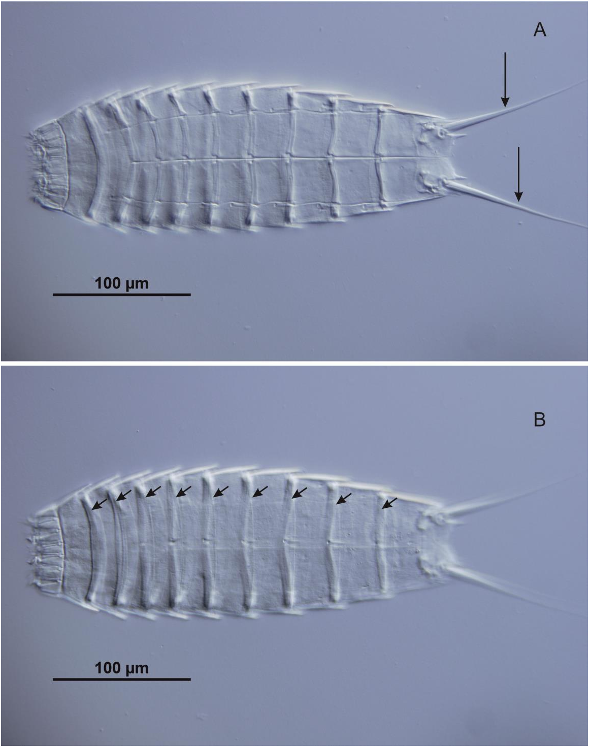
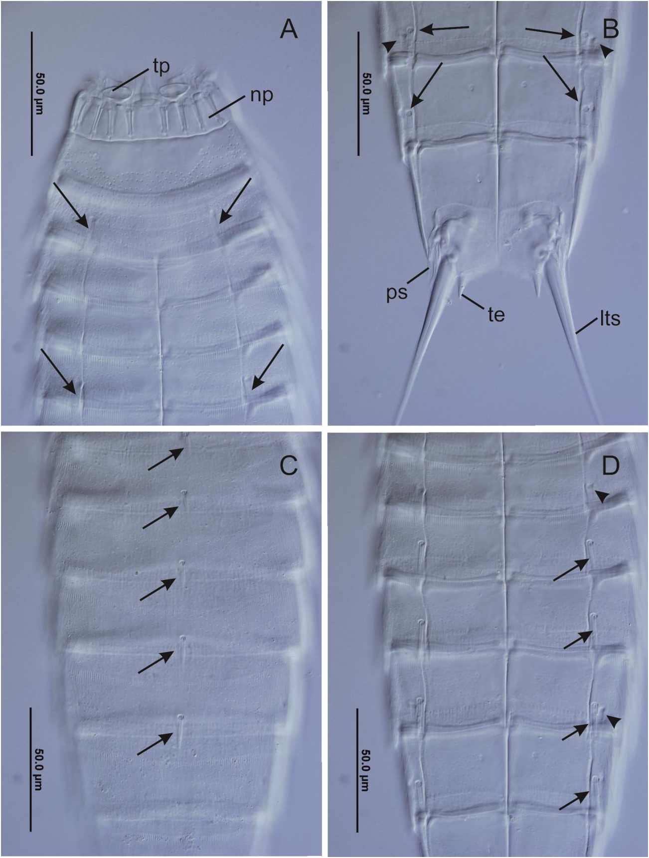
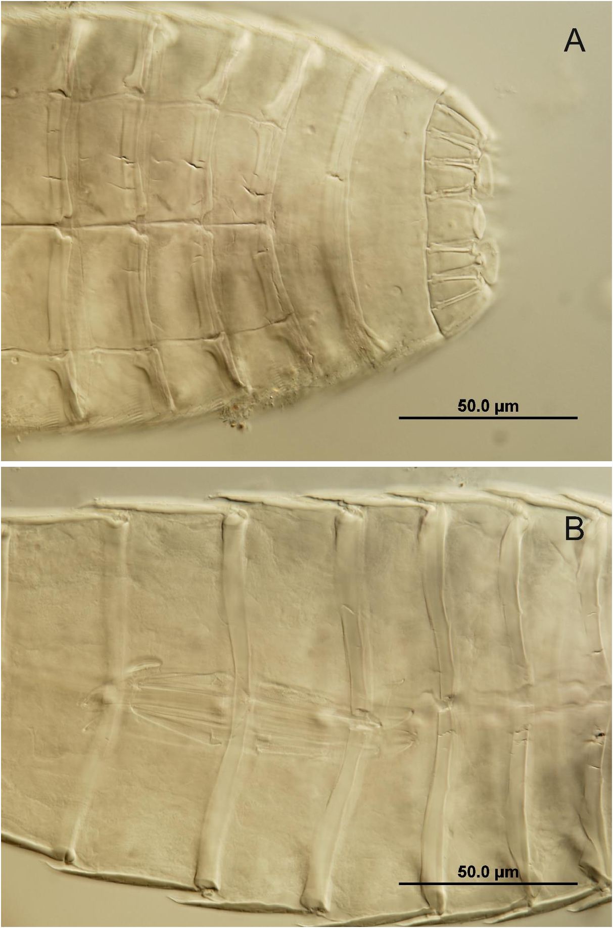
( Figures 2-4 View Figure 2 View Figure 3 View Figure 4 )
The specimens of the phylum Kinorhyncha obtained at Sinop Bay have been identified beyond doubt as Echinoderes dujardinii Claparède, 1863 . The body of the adult is divided into introvert, neck and trunk.
Introvert. None of the specimens obtained had the introvert extended, so no information can be given on the mouth cone, oral styles and the structure and arrangement of scalids.
Neck. The neck is formed by 16 cuticular plates named placids, radially arranged and articulated directly with the first segment of the trunk. All placids have a trapezoid shape and are approximately equal in size, except for the midventral one, wider and slightly extended distally ( Figs. 2A View Figure 2 , 3A View Figure 3 , 4A View Figure 4 ). Some of the placids bear additional plates, the trichoscalid plates, on which the trichoscalids of the last scalid ring articulate ( Fig. 3A View Figure 3 ). The cuticle between adjacent placids is soft and appears folded, sometimes mistakenly named as interstitial placids.
Trunk. The trunk is divided into 11 segments. As a diagnostic character of the genus, the two first segments are closed rings of cuticle ( Fig. 4A View Figure 4 ). Segments 3 to 11 are formed by a dorsal tergal plate articulated with two ventral sternal plates ( Fig. 4A View Figure 4 ). Hence a ventral view of the animal shows the sternal plates plus a narrow lateral strip of the tergal plate, the so-called lateroventral area. Every segment has an anterior cuticular thickening, the pachycyclus ( Figs. 2B View Figure 2 , 4B View Figure 4 ) and a posterior edge specialized as a fringe of cuticular tips, the pectinate fringe that overlaps the anterior edge of the next segment. The sizes of the whole trunk and the individual segments agree with the ones reported in the descriptions of the species. The cuticular surface is covered by minute hairs, arising from perforations arranged following characteristic patterns. Other cuticular specializations include sensory spots, glandular cell outlets and cuticular scars marking attachment sites for the underlying musculature. The most important taxonomic feature is the presence, nature and distribution of cuticular appendages, namely spines and tubes ( Figs. 3 View Figure 3 A-D).
Tubes occur in lateroventral positions on segment 2; in lateroventral position on segment 5 ( Fig. 3A View Figure 3 ) and in lateral accessory position on segment 8, very close to the lateroventral spine on that segment ( Figs. 3B, D View Figure 3 ). Additionally a laterodorsal tube occur on segment 10. Short middorsal spines occur on segments 4 to 8, slightly increasing in length posteriorly. Spines are located in lateroventral positions on segments 6-9 ( Fig. 3D View Figure 3 ).
Segment 11 ( Fig. 3B View Figure 3 ) bears a pair of big lateral terminal spines, about 150 µm long ( Fig. 2A View Figure 2 ). Additionally, females present one pair of lateral terminal accessory spines nearly 50 µm long. Males have three pairs of flexible penile spines instead. Tergal plate of segment 11 projected posteriorly as one pair of short and pointed tergal extensions.
No known copyright restrictions apply. See Agosti, D., Egloff, W., 2009. Taxonomic information exchange and copyright: the Plazi approach. BMC Research Notes 2009, 2:53 for further explanation.
|
Kingdom |
|
|
Phylum |
|
|
Class |
|
|
Order |
|
|
Family |
|
|
Genus |