
Ptilocera Wiedemann, 1820
|
publication ID |
https://doi.org/10.5281/zenodo.278481 |
|
DOI |
https://doi.org/10.5281/zenodo.6189540 |
|
persistent identifier |
https://treatment.plazi.org/id/D10887FC-DF39-FFA7-8BA5-FC5EBB80F368 |
|
treatment provided by |
Plazi |
|
scientific name |
Ptilocera Wiedemann, 1820 |
| status |
|
Ptilocera Wiedemann, 1820 View in CoL View at ENA
Ptilocera Wiedemann, 1820: 7 View in CoL
Type species: Stratiomys quadridentata Fabricius , by monotypy.
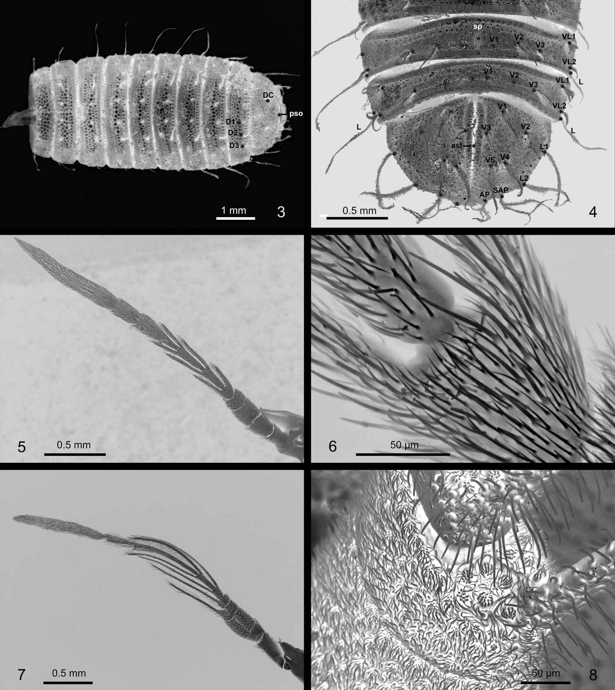
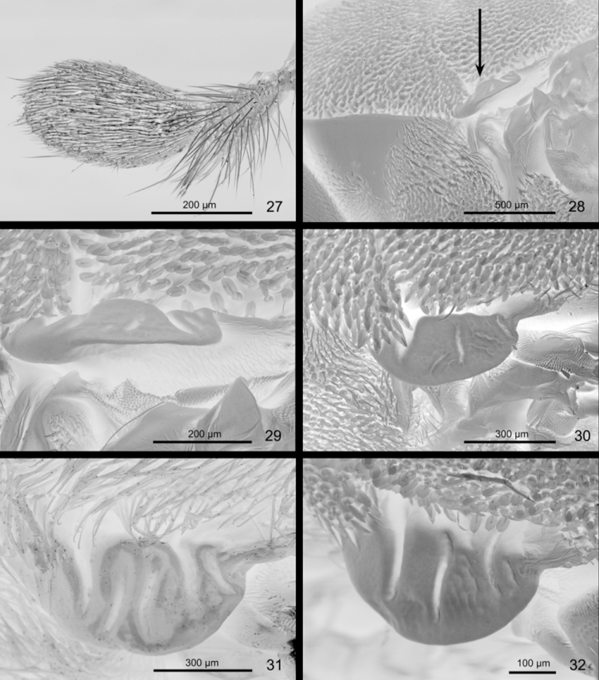
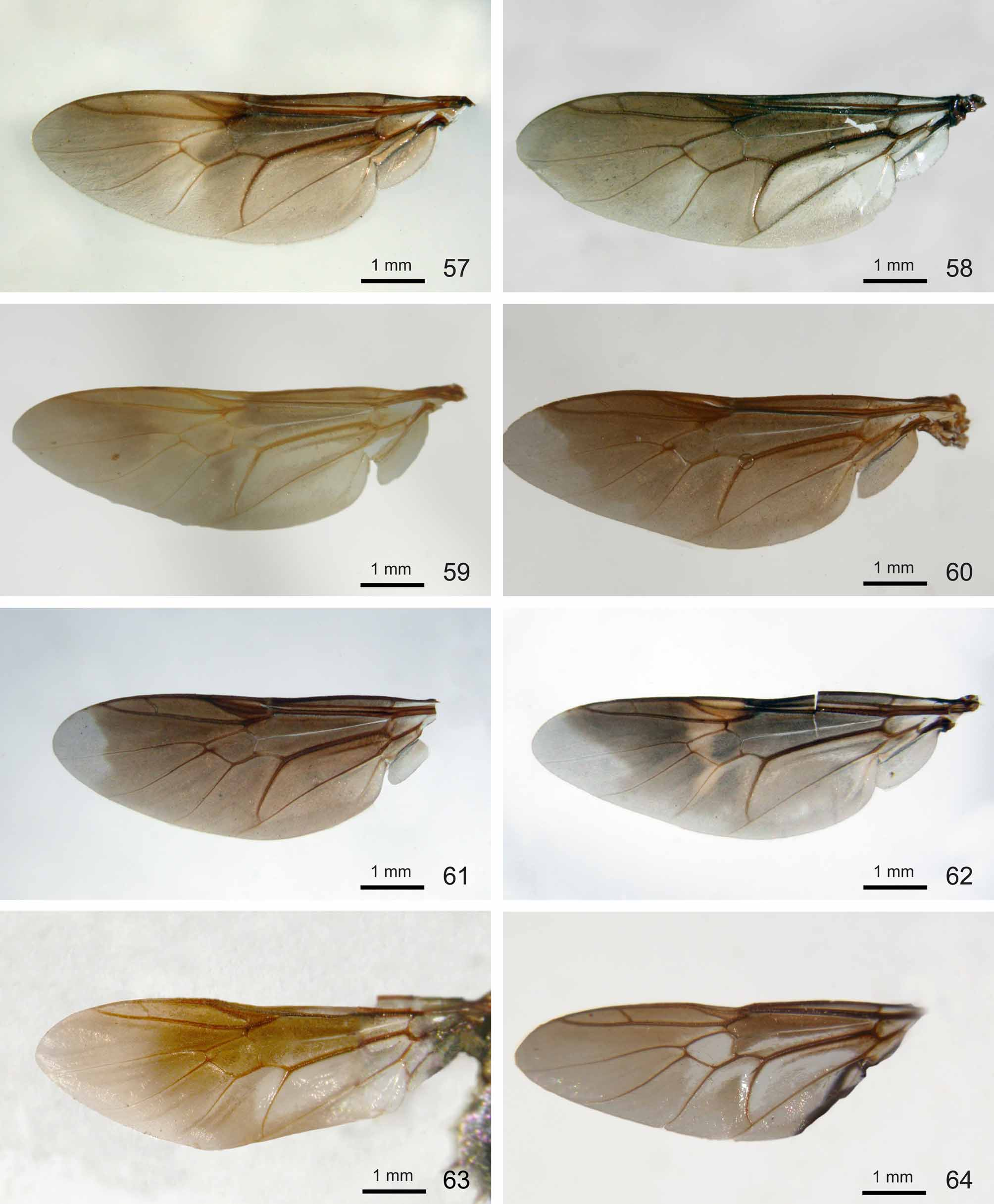
Diagnosis. The genus is characterized by the following set of characters: (1) flagellum consisting of 8 flagellomeres, medial 3‒4 of them provided with slender lateral projections ( Figs 5, 7 View FIGURES 3 – 8 ), (2) thorax with leathery, rounded and flat prealar prominence in front of wing ( Figs 28–34 View FIGURES 27 – 32 View FIGURES 33 – 38 ), (3) scutellum with four moderately long spines (Fig. 43), (4) vein R2+3 starting close to or at crossvein R-M (Fig. 44), (5) vein R4 present ( Figs 57–73 View FIGURES 57 – 64 View FIGURES 65 – 73 ), (6) abdomen short, rounded to subquadrate, distinctly convex dorsally (e.g. Fig. 64 View FIGURES 57 – 64 ), and (7) aedeagal complex with short cylindrical posterolateral papillae ( Figs 56 View FIGURES 51 – 56 , 99 View FIGURES 95 – 100 ). From these characters probably only characters (1) and (7) are autapomorphic for this genus although partly shared with Isomerocera in a modified form (see Introduction). Character (1) is probably secondarily reduced in the male of P. s i m p l e x sp. nov. and character (2) was also found in some other Pachygastrinae (e.g. in Camptopteromyia de Meijere, Gabaza Walker , Gnorismomyia Kertész , Lophoteles Loew , Pegadomyia Kertész , Pseudopegadomyia Rozkošný & Kovac and Saldubella Kertész ) as well as in taxa outside of Pachygastrinae (e.g. in some genera of Clitellariinae , as a subtriangular prominence in Adoxomyia Kertész and as a strong spine in Clitellaria Meigen and Nigritomyia Bigot ).
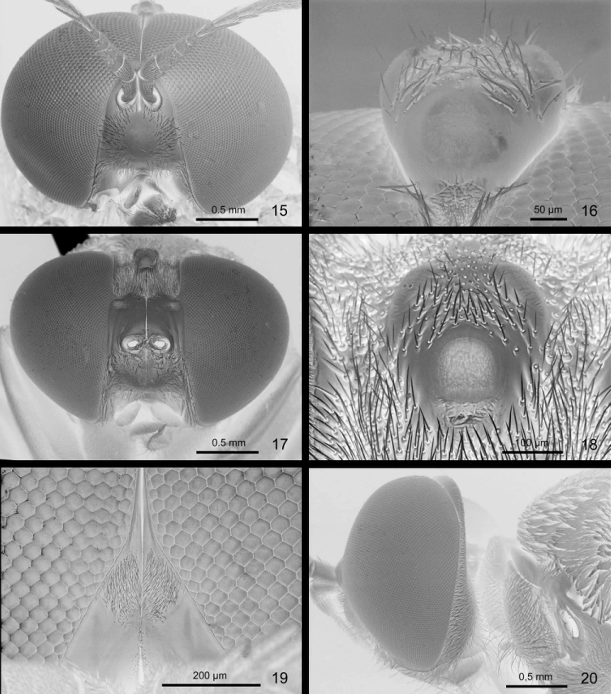
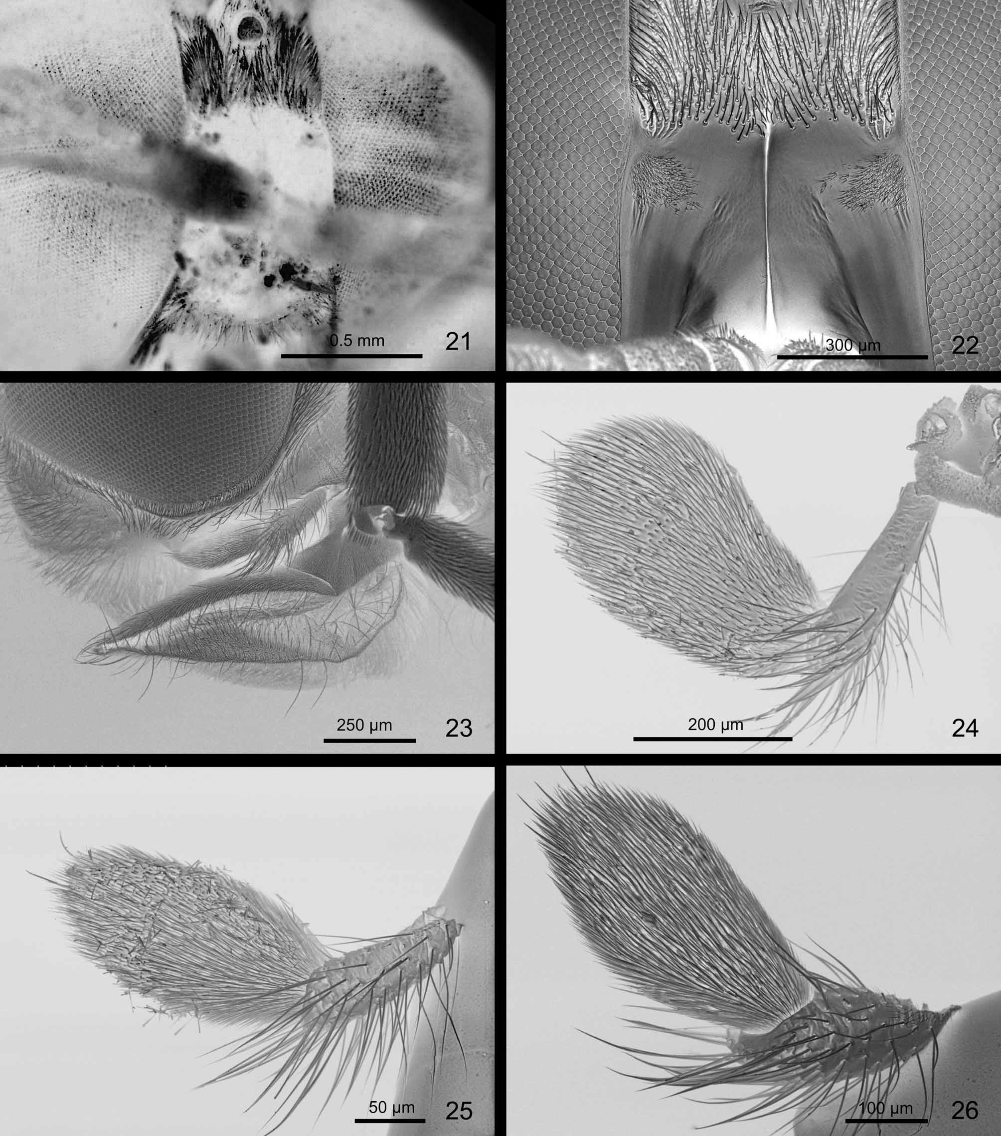
Description. Male. Head with strongly holoptic eyes contiguous for a relatively long distance, ommatidia in upper part of eye larger than in lower third ( Fig. 15 View FIGURES 15 – 20 ). Postocular area usually narrow, more distinct only in lower third of head in lateral view ( Fig. 20 View FIGURES 15 – 20 ). Upper frons barely broader than diameter of anterior ocellus, tapered anteriorly and reaching to about middle of distance between anterior ocellus and frontal tomentose spot above antennae ( Fig. 15 View FIGURES 15 – 20 ). Ocellar tubercle ( Fig. 16 View FIGURES 15 – 20 ) distinctly prominent in lateral view, vertex beyond it transversely oblong, not longer than ocellar tubercle in dorsal view. Lower frons above antennae ( Fig. 19 View FIGURES 15 – 20 ) shining black, slightly protuberant, usually with distinct medial groove and rounded, medianly divided silverish tomentose spot in upper half. Antenna ( Fig. 5 View FIGURES 3 – 8 ) usually more than twice as long as head, scape about twice as long as pedicel, longer only in P. simplex sp. nov. Two basal flagellomeres cylindrical, with scattered sensory pits, first of them 1.5–2.0 times as long as broad and second usually shorter than broad. Each of flagellomeres 3–5 with ventral and dorsal slender projections, flagellomeres 6–8 short but densely haired. Last flagellomere usually 4–5 times longer than preceding one, relatively shorter only in P. kerteszi sp. nov. Face prominent, tubercle-like, rounded in profile. Proboscis with fleshy and stout labellum, palpus relatively long, two-segmented, with apical segment oval and flattened.
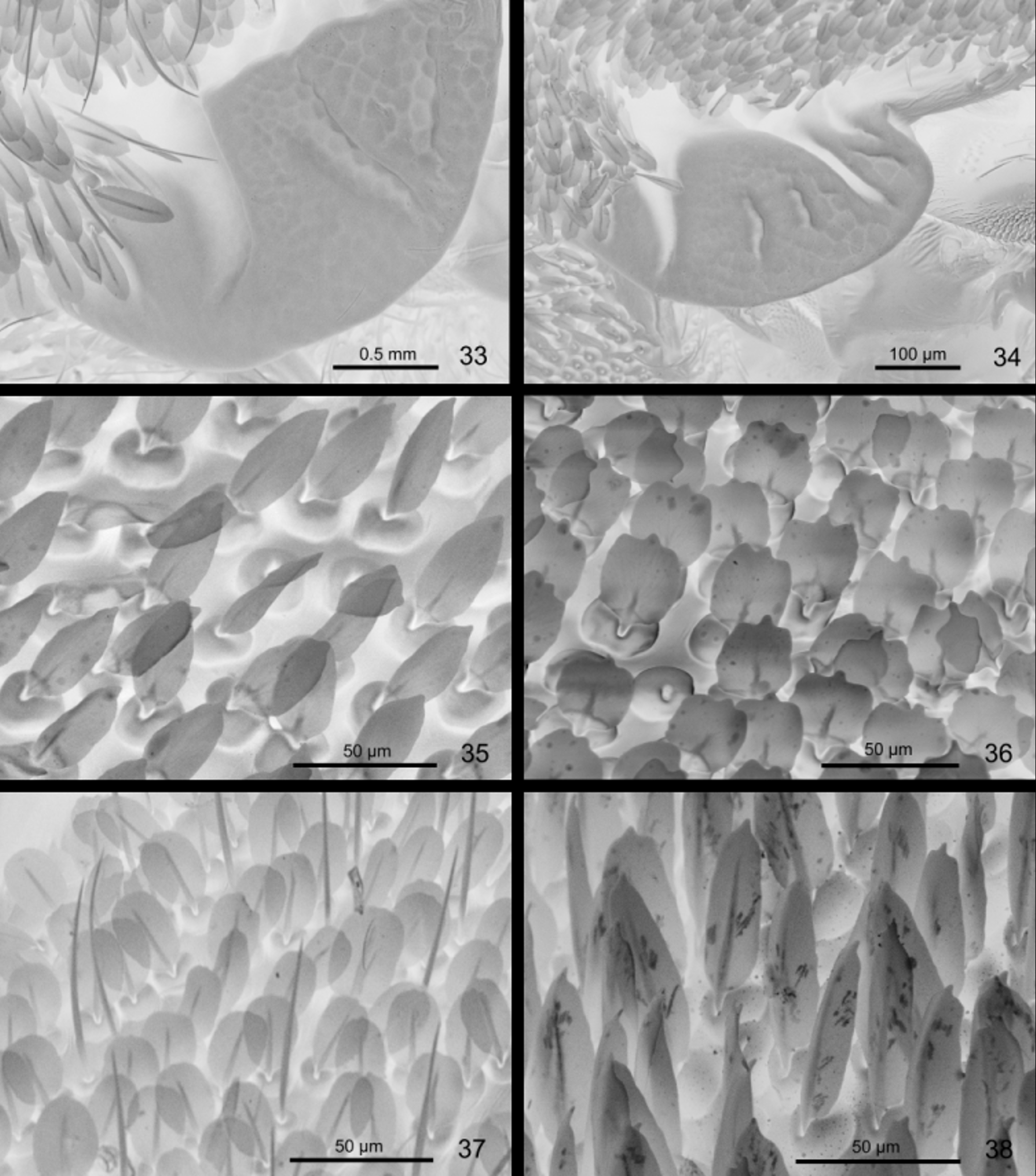
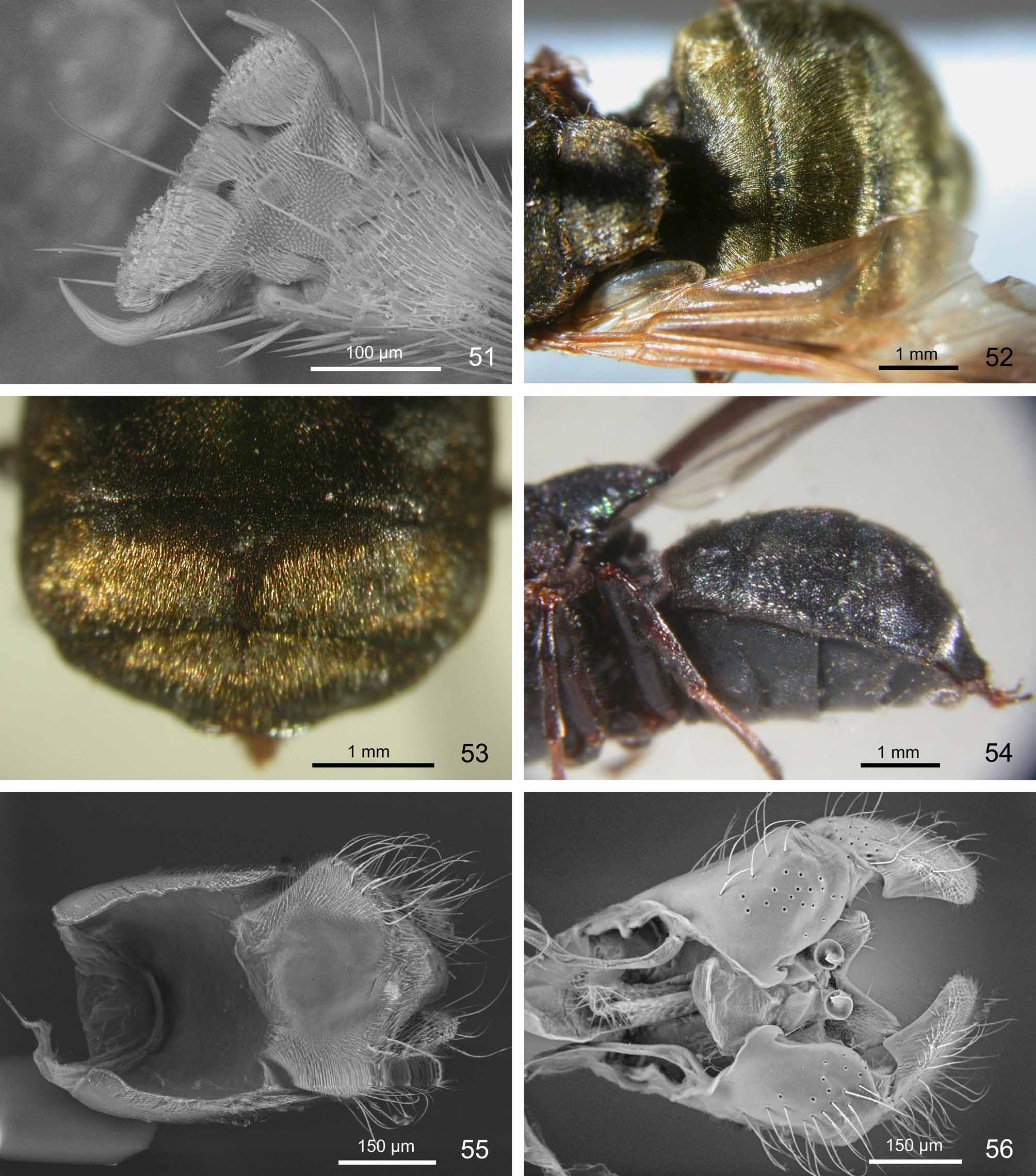
Thorax longer than broad, scutum with markedly prominent postpronotal callus and slightly dilated toward wing bases. Leathery, rounded and flat prealar prominence in front of wing base well developed as in females (cf. Figs 29–34 View FIGURES 27 – 32 View FIGURES 33 – 38 ). Scutellum about at same level as scutum, subquadrate or more rounded posteriorly, with two pairs of slightly upcurved and pointed spines, distance between middle pair of spines usually same as or longer than that between medial and lateral spines. Scutum and scutellum mostly covered with metallic iridescent scales in different arrangement and density. Scales usually more or less elliptical, subquadrate or tapered, often pointed apically, with semi-globular to globular bases, similar to those in females (cf. Figs 35 View FIGURES 33 – 38 –39). Wing membrane densely covered with microtrichia. In some species an irregular transverse yellowish streak from wing margin to lower margin of discal cell or even to anterior cubital cell less or more distinct ( Figs 57 View FIGURES 57 – 64 , 65 View FIGURES 65 – 73 ). Vein R2+3 arising slightly before, beyond or just at crossvein R-M (Fig. 44) and vein R4 always present, no veins reaching posterior wing margin, all faint before end. Calypter usually with dense and mostly wool-like, black marginal fringe (cf. Fig. 46 View FIGURES 45 – 50 ). Halter relatively long, usually with darkened knob ( Fig. 48 View FIGURES 45 – 50 ) and dense surface sensillae ( Fig. 49 View FIGURES 45 – 50 ). Legs dark, only mid and hind basitarsomeres sometimes paler. Apical tarsomeres and pretarsus as in Figs 50–51 View FIGURES 45 – 50 View FIGURES 51 – 56 .
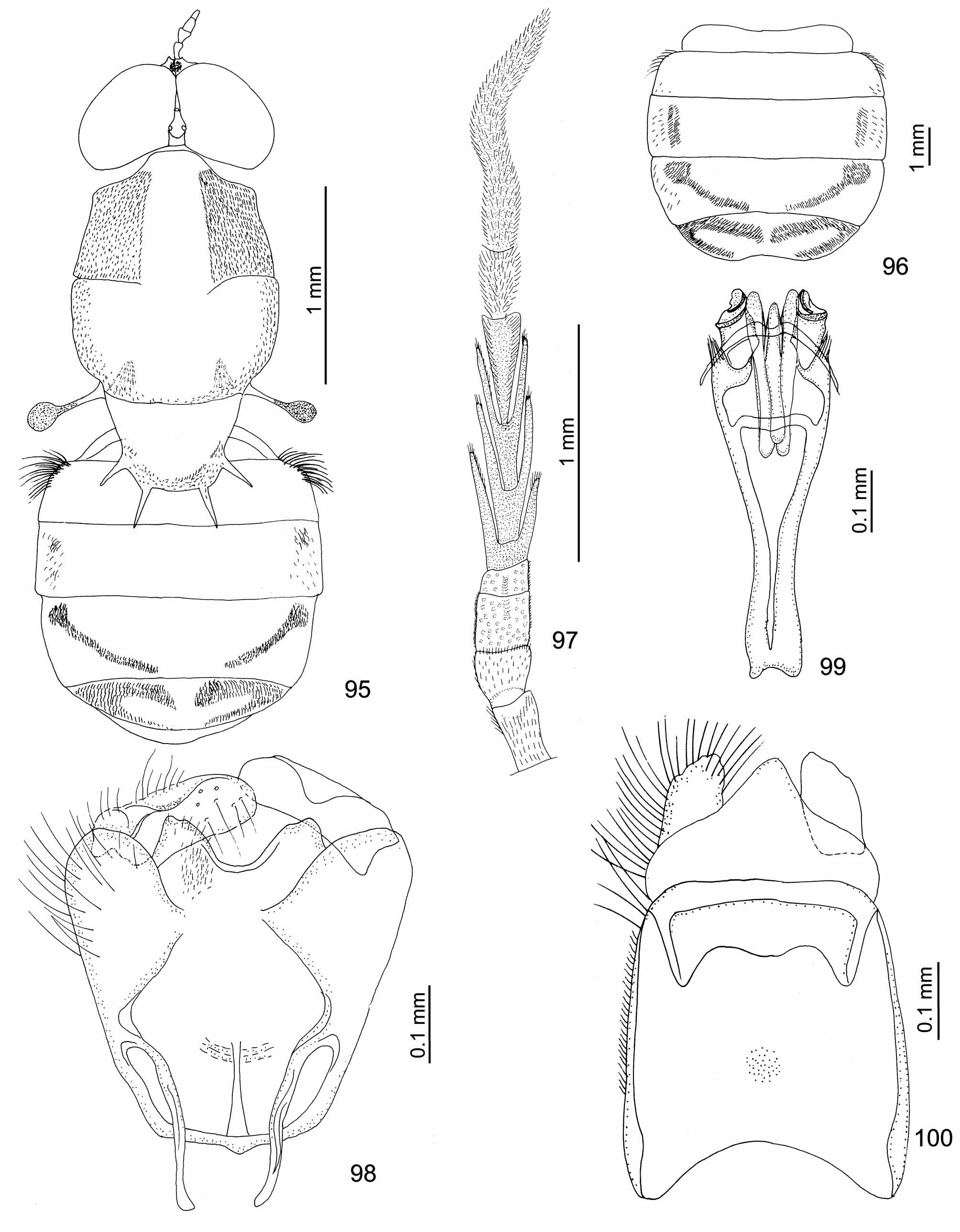
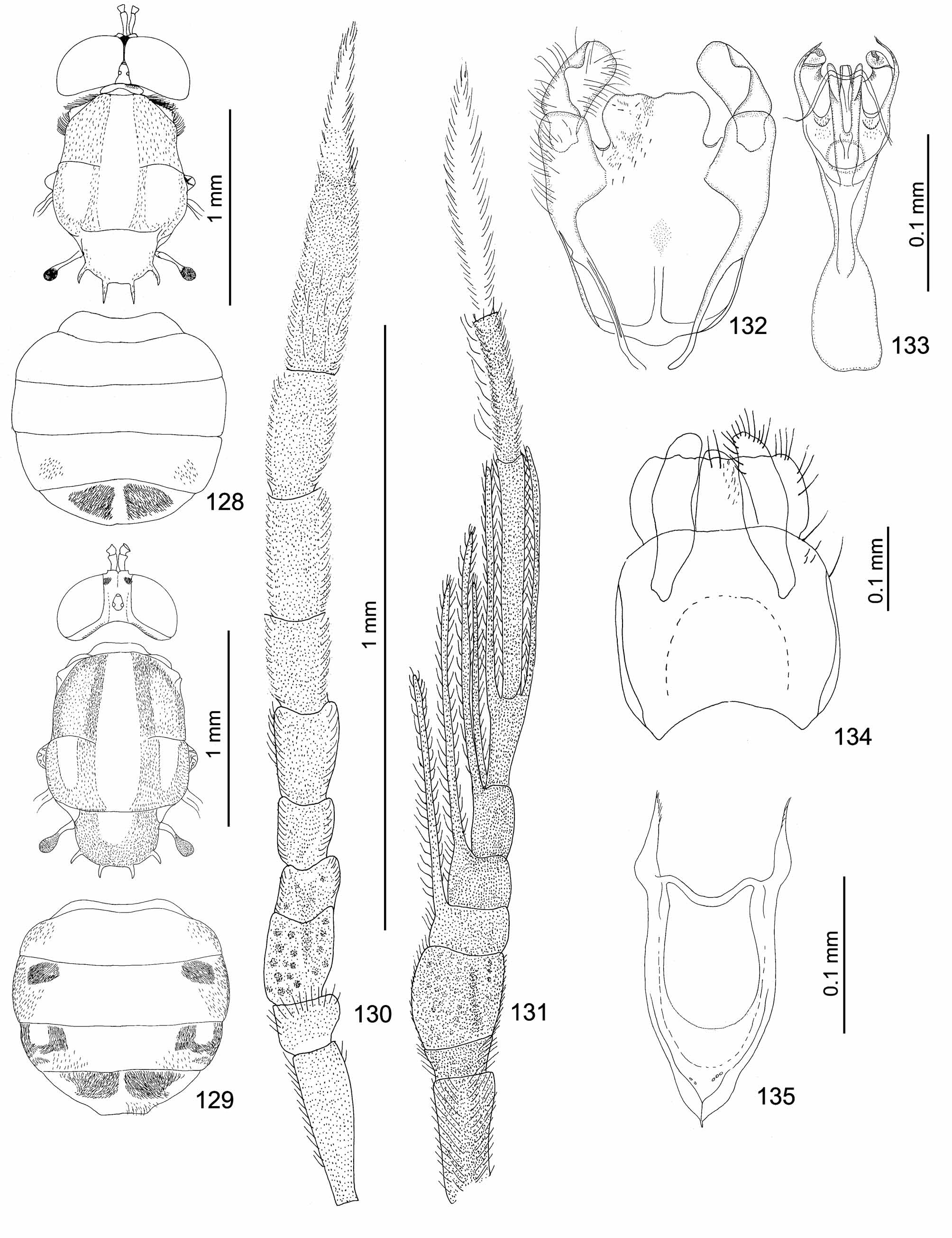
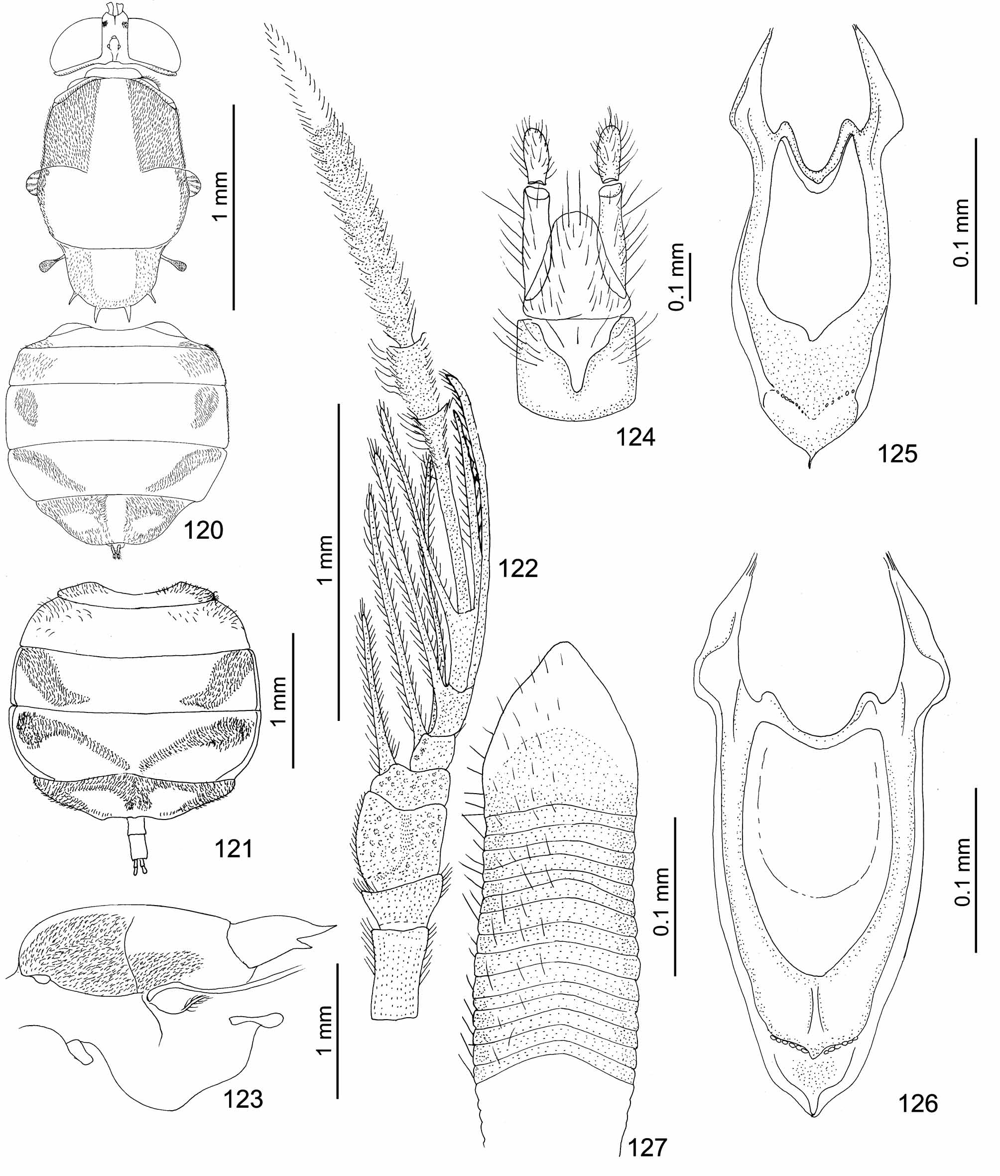
Abdomen rounded, barely longer than broad, with prominent but rounded anterolateral corners, slightly convex dorsally and ventrally. Silverish white (rarely golden yellow) hair patches may form species-specific ornamentation, abdomen only rarely entirely bare dorsally (male of P. b e rg i). Male terminalia ( Figs 55–56 View FIGURES 51 – 56 ) specialised, with autapomorphic aedeagal complex. Epandrium usually subquadrate or slightly dilated proximally, more or less indented at proximal margin, proctiger mostly pentagonal, cerci only slightly reaching beyond epiproct. Genital capsule subquadrate, usually somewhat tapered toward proximal margin, medial process well developed, bilobate as a rule, simple only in P. s i m p l e x. Gonostylus leaf-shaped. True trifid phallic organ ( Fig. 87 View FIGURES 82 – 88 , in middle of distal part) protected by an enclosure consisting of a membrane between both gonocoxites and distally ending as a short cylindrical posterolateral papilla on each side of phallus. Phallic organ continuing proximally as flat, transversely margined or rounded aedeagal apodeme. Posterolateral papillae provided with transverse ridges in some species and/or their bases with tranverse rows of flat, hyaline and pointed spines. Only male of P. s im pl e x with additional posterolateral, pointed projections at outer side of each posterolateral papilla ( Fig. 133 View FIGURES 128 – 135 ).
Female. Head with dichoptic eyes separated by frons occupying about 1/5 of head width, size of ommatidia small on entire eye surface ( Fig. 17 View FIGURES 15 – 20 ). Ocellar tubercle ( Fig. 18 View FIGURES 15 – 20 ) and especially anterior ocellus prominent in profile but less distinct than in male. Postocular area well developed, about as broad as pedicel is long and shining in dorsal view, narrower and pilose laterally ( Fig. 20 View FIGURES 15 – 20 ). Frons black, depressed in middle and slightly protuberant above antennae in profile, granulose, short and densely pilose in upper half and shining in lower half, with medial groove in lower half ( Fig. 17 View FIGURES 15 – 20 ). Two subtriangular, subquadrate or rounded, whitish tomentose spots at eye margin below middle of frons. Scape and two basal flagellomeres usually stouter than in conspecific male. One unpaired projection present on flagellomere 2 and paired projections on flagellomeres 3–5, all projections much longer than in males, with combs of conspicous erect setulae ( Figs 7–9 View FIGURES 3 – 8 View FIGURES 9 – 14 ). Facial tubercle below antennae ( Fig. 17 View FIGURES 15 – 20 ) as in male but usually broader. Iridescent scales on scutum and scutellum sometimes even denser than in males, usually arranged in longitudinal stripes on scutum, in some cases of diagnostic value (cf. Figs 35 View FIGURES 33 – 38 –40). Scutellar spines short, reaching only about 1/3 of scutellum length (e.g. Fig. 43), almost entirely darkened. Anterior spiracle as in Fig. 47 View FIGURES 45 – 50 . Yellow transverse streak in middle of wing usually indistinct or absent. Wing microtrichia are usually less developed than in males, partly or extensively reduced especially in basal cells, anterior and posterior cubital, and anal cells ( Figs 58, 63 View FIGURES 57 – 64 , 71 View FIGURES 65 – 73 ). Calypter with white or black marginal hair fringe. Female terminalia ( Fig. 124 View FIGURES 120 – 127 ) with two-segmented cercus, the basal segment up to 5 times longer than apical one, tergite 10 subtriangular, shorter than basal segment of cercus. Tergite 9 subquadrate and emarginated distally, sternite 9 weakly sclerotized, almost 3.0 times as long as broad, subgenital plate (= sternite 8) ( Fig. 127 View FIGURES 120 – 127 ) longer than tergite 8, distally projecting in middle. Genital furca fairly uniform, elongate oval and pointed proximally, deepened in middle of frame distally, posterolateral projections long and pointed (cf. Figs 81 View FIGURES 74 – 81 , 93–94 View FIGURES 89 – 94 , 106–107). Intersegmental membranous areas long although usually shorter than neighbouring segments. Completely extended oviscape usually longer than sclerotized abdominal segments 1–5. Spermathecae not examined.
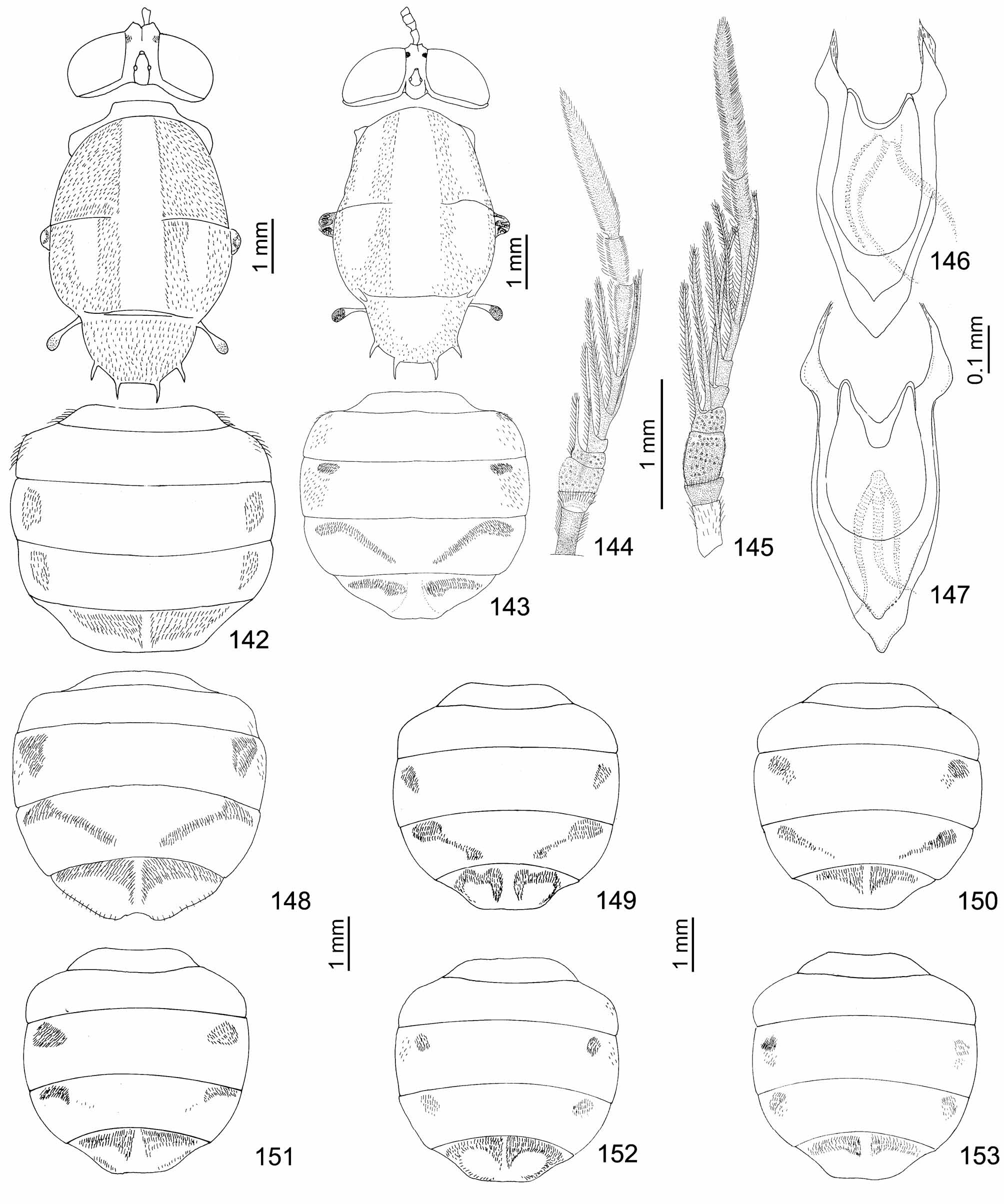
Diagnostic characters have been found especially on the antenna (the length and colour of basal segments, the shape of two basal flagellomeres, the length and colour of the ultimate and penultimate flagellomere), on the wing (presence or absence of the yellowish transverse streak, the extent of hyaline areas with reduced microtrichia) and the shape of hair patches on the abdomen. However, colour characters and the extent of abdominal hair patches are often considerably variable (cf. Figs 142–143, 148–153 View FIGURES 142 – 153 ). A complex of characters must be thus taken in consideration when making identifications. Our preliminary study of surface structures in some species indicates that the size, shape and density of scales, especially on the scutum and scutellum, might be a diagnostic character even in females (cf. Figs 37 View FIGURES 33 – 38 –39). Scales are often accompanied or replaced by slender or slightly dilated setulae inserted in globose or semiglobose basal structures (Figs 39, 40). Nevertheless, these surface structures need further study including a detailed comparison of scales and setulae among species, between both sexes and on different body parts. On the contrary, the shape of the prealar prominence seems to be fairly uniform within this genus, apart from some differences in the number and form of grooves on the dorsal surface (cf. Figs 29–34 View FIGURES 27 – 32 View FIGURES 33 – 38 ). The shape of the apical segment of the palpus ( Figs 24–27 View FIGURES 21 – 26 View FIGURES 27 – 32 ) shows only slight differences among the female specimens examined. The male terminalia do not provide a sufficiently diverse set of morphological structures in some related species and only those of P. s i m p l e x ( Figs 132–134 View FIGURES 128 – 135 ) display highly species-specific structures. Differences in the female terminalia are generally even less distinct.


No known copyright restrictions apply. See Agosti, D., Egloff, W., 2009. Taxonomic information exchange and copyright: the Plazi approach. BMC Research Notes 2009, 2:53 for further explanation.
|
Kingdom |
|
|
Phylum |
|
|
Class |
|
|
Order |
|
|
Family |
Ptilocera Wiedemann, 1820
| Mason, Franco & Rozkošný, Rudolf 2011 |
Ptilocera
| Wiedemann 1820: 7 |