
Niphargus diadematus, Hudec, Igor, Fišer, Cene & Dolanský, Jan, 2017
|
publication ID |
https://doi.org/ 10.11646/zootaxa.4291.1.3 |
|
publication LSID |
lsid:zoobank.org:pub:36509C6A-3DE7-4C57-B27A-0CD66BBC5812 |
|
DOI |
https://doi.org/10.5281/zenodo.6042676 |
|
persistent identifier |
https://treatment.plazi.org/id/D2531412-FFA0-6E36-FF23-011C69B5F9E4 |
|
treatment provided by |
Plazi |
|
scientific name |
Niphargus diadematus |
| status |
sp. nov. |
Niphargus diadematus View in CoL sp. n.
http://SpecieS-id.net/wiki/ Niphargus _ diadematus FigS 2–4 View FIGURE 2 View FIGURE 3 View FIGURE 4
Etymology. The SpecieS name waS derived from the Latin word: diadema (Semicrown)- baSed on the arrangement of terminal SpineS on the telSon.
Type material. The type SerieS waS collected in the locality Lednice, the foreSt inudation of Včelínek Brook, leg. J. DolanSký.
Holotype: Lednice (29. May 2014 – 5. November 2014): adult male, partially diSSected, 6.3 mm, mounted in Swann-medium and depoSited at Natural HiStory MuSeum in Prague, Czech Republic (NHMP-P6E4172).
Allotype: Lednice, (29. May 2014 – 5. November 2014): adult female partially diSSected, 5.7 mm, mounted in Swann-medium and depoSited at NHMP-P6E4173.
ParatypeS: Lednice (29. May 2014 – 5. November 2014: diSSected adult male, 6.7 mm, mounted in Swannmedium and depoSited at NHMP-P6E4174.
Lednice (29. May 2014 – 5. November 2014: diSSected adult female, 5.9 mm, mounted in Swann-medium and depoSited at NHMP-P6E4175.
Paratype series. Lednice (29. May 2014 – 5.November 2014): 8 maleS in vial preServed in ethanol and depoSited at NHMP-P6E4176.
Lednice,(29. May 2014 – 5.November 2014): 8 femaleS in vial preServed in ethanol and depoSited at NHMP.
Three paratypeS on three SlideS mounted in Swann-medium (partially diSSected femaleS) 5. November 2014 – 17. May 2015 (NHMP—P6E4177–P6E 4179).
Lednice (5. November 2014 – 17. May 2015): 20 SpecimenS in vial preServed in ethanol and depoSited zoological collection of Department of Biology, Biotechnical Faculty , UniverSity of Ljubljana.
ReSt of SpecimenS in the 1st author collection.
Diagnosis. Small-Sized, elongated SpecieS with Small Subquadrat gnathopodS, narrow gillS, Sexually dimorphic uropod III and Sexually non-dimorphic uropod I. UroSomiteS I and II with 1 and 2 poStero-dorSo-lateral Seta, reSpectively. Epimeral plateS I – III Subrounded. TelSon lobeS Slightly narrowing diStally, tip Subrounded; telSon cleft deep (up to 80% of telSon length); 5 – 7 terminal SpineS, arranged in Semi ring pattern and do not exceed 35–40% of telSon length; dorSal and lateral SpineS abSent. Coxal plateS flattened. GnathopodS I – II with Small roughly triangular propodi; dactyluS with Single Seta at outer margin. PereopodS V – VII baSeS elongated-oval, pereopodS length ratioS aS 1.0: 1.4: 1.5; dactyli with Short nail (30–34% of dactyluS length) and one tiny Spiniform thorn near nail baSe. RetinacleS with 2 hookS. MouthpartS: maxillae I inner lobe with 1 Seta; maxillipedS inner lobe with 1 lancet-like Stout Seta.
Description of adults (paratypes). [Note: If not Specified the characterS apply to both SexeS]
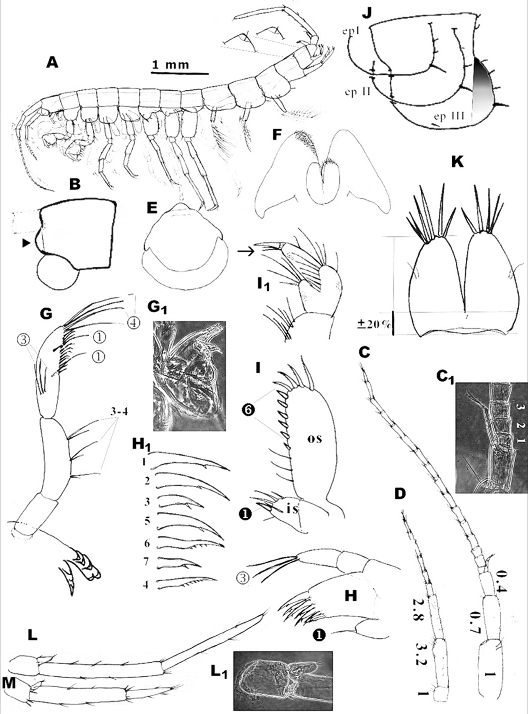
Body shape: elongated and Slender ( Figure 2 View FIGURE 2 A); body length: up to 8 mm (male), 5 mm (female). Colour: white.
Head ( Figure 2 View FIGURE 2 B). Short angular, without roStrum; anterior margin Shallow SinuSoid; ventral margin almoSt Straight. Surface of the head capSule iS Smooth.
Antennae: Antenna I ( Figure 2 View FIGURE 2 C–C1) aS long aS 44% of BL; ratio between the pedunclar articleS 1: 2: 3 aS 1: 0.7: 0.4. Flagellum with 16 – 18 articleS of different SizeS meaSuring 0.10 – 0.18 of proximal peduncle article and 0.18 – 0.21 of diStal peduncle article. Each flagellar article with a few minute SenSillae and one elongate biarticulated aeSthetaSc along diStal margin. AcceSSory flagellum bi-articulated, Shorter than firSt two articleS of flagellum, and with 3 apical SenSillae. Antenna II ( Figure 2 View FIGURE 2 D) Shorter than 53 % of AI; peduncule articleS 3: 4: 5 aS 1: 3.2: 2.8. Flagellum with 7 articleS: proximal article the longeSt and meaSureS 0.9 of the 3rd peduncular article; diStal articleS are progreSSively Shorter.
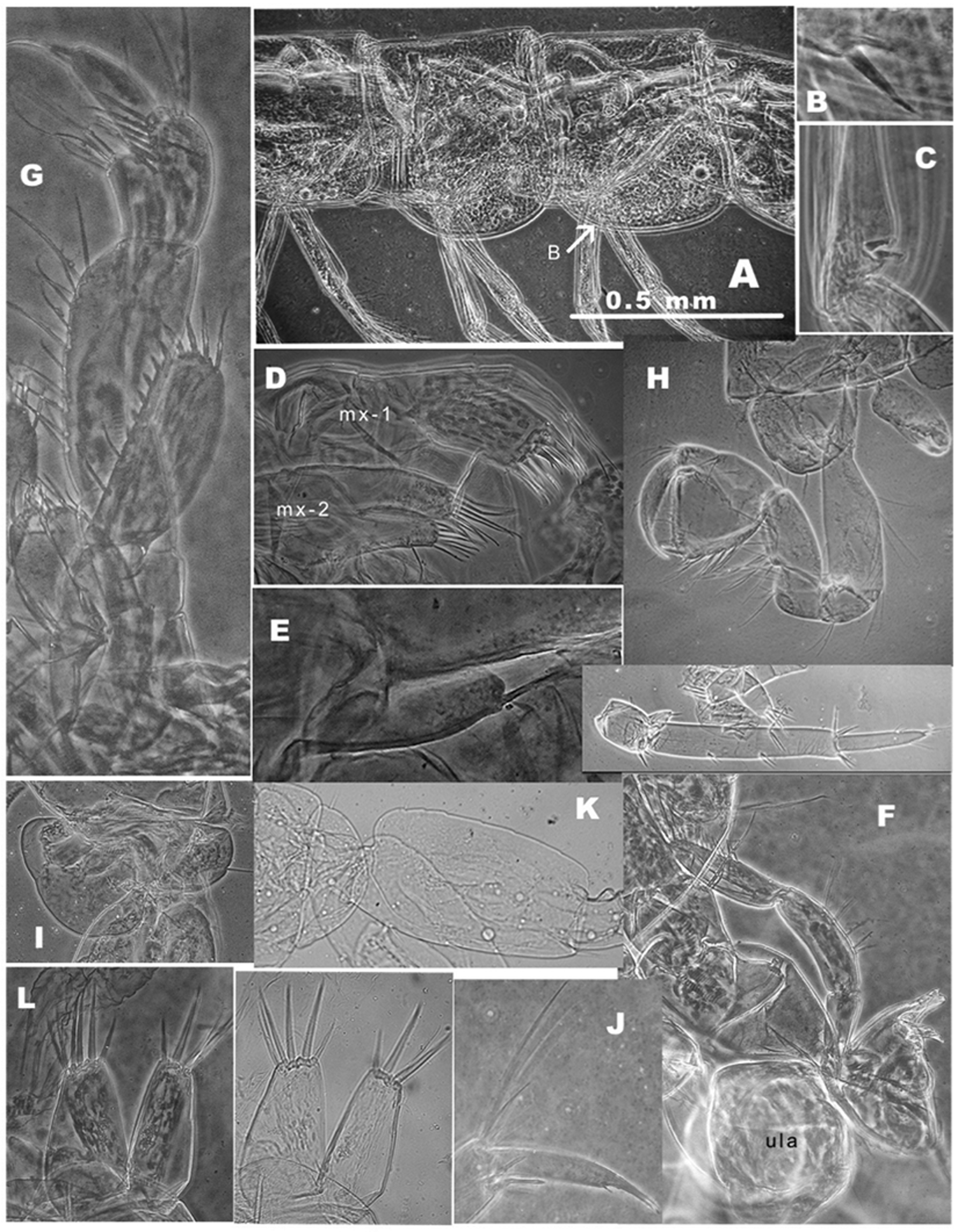
Upper lip (labrum) ( Figure 2 View FIGURE 2 E, Figure 4 View FIGURE 4 F: ula). SclerotiSed, quadrangular-oval, bipartite; baSal article narrow, diStal article Sub-quadrangular; Surface iS Smooth or with long Submarginal Suture in diStal portion.
Maxilla I ( Figure 2 View FIGURE 2 G–G1; Figure 4 View FIGURE 4 F: mx-1). PalpuS biarticulated, diStal article aSymmetrically vaulted with 3 long terminal Setae. Outer lobe with 7 denticulated SpineS arranged in inner and outer row of reSpectively 4 and 3 SpineS; the Structure of SpineS with reSpect to number of denticleS per Spine aS followS: 5 uni-, one four- and one Six- denticled. Inner lobe with 1 Seta in Subterminal poSition ( Figure 2 View FIGURE 2 G1).
Maxilla II ( Figure 4 View FIGURE 4 D: mx—2). Bilobate, both lobeS of Sub-equal Size, with apical Setae (inner and outer lobe) and one Seta at ventral margin of the ventral lobe. DorSal marginS of both lobeS with fine hairS.
Labium ( Figure 2 View FIGURE 2 F). Smaller, inner lobeS trapezoid-like with longer Sub-triangular flat poSterior protruSion on each portion; inner diStal lobe finely Serrated. Smaller outer lobeS are compact and of Subovoid Shape, finely Serrated on diStal portion.
Maxilliped ( Figure 2 View FIGURE 2 I– I1; Figure 4 View FIGURE 4 G). Inner lobe (is) Short with 5 hairy long Setae, (4 marginal and 1 Submarginal Seta) and 1 flattened Spine-like Seta on apical part. Outer lobe (os) reacheS up to 2/3 length of 2nd article of maxilliped palp; at the baSe 3 iSolated long thin Setae; along inner margin 6 SparSed flattened Spine-like Setae which increaSe diStally and are followed by 4 long denticulated Setae along diStal arc. The outer Surface of outer lobe iS Smooth. Palp 4-articulated: 1st baSal, Subtriangular article with one bunch of 4–5 Setae on inner Side; 2nd article, the longeSt one, with 7 – 8 tranSverSally oriented rowS of Setae along inner margin; 3rd article Small, Suboval with two rowS of numerouS Setae around dactyluS; 4th dactyluS ( Figure 2 View FIGURE 2 I1) with 2 long thin Setae in 2/3 length of ventral margin and one Seta in 1/4 of dorSal margin; terminal nail about 1/4 of whole dactyluS.
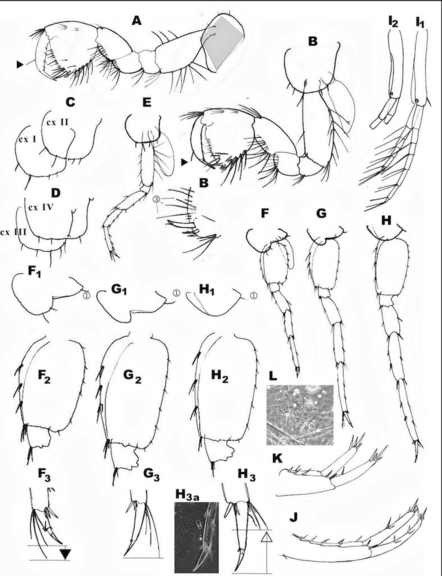
Coxal plates flattened with SparSe Setae along ventral marginS. CxI ( Figure 3 View FIGURE 3 A) of parallelogram Shape with roughly acute, broadly vaulted anterior-ventral corner; cxII–IV ( Figure 3 View FIGURE 3 C – E) rectangular, angleS rounded; anterior, ventral and poSterior marginS vaulted. CxV–VI ( Figure 3 View FIGURE 3 F – F1, G – G1) Similar, with well-developed anterior lobe ventrally and with reSpective 2 and 0 – 1 diStal Setae; poSterior part narrow elongated with1 poSterior Seta. CxVII ( Figure 3 View FIGURE 3 H–H1; Figure 4 View FIGURE 4 : K) reduced, narrow trapezoid-oval plate with elongated poSterior corner and one poSterior Seta.
Gills ( Figure 3 View FIGURE 3 B, E; Figure 4 View FIGURE 4 H) uniformly narrow, Sub-oval, aSymmetrically bent (SauSage-like).
Pereon appendages. Gnathopods I ( Figure 3 View FIGURE 3 A). BaSiS trapezoid and broad (width ±45% of length), laterally flattened; with a group of long Setae cloSe to anterior ventral corner; one Sub-angular group (almoSt identical with iSchium) tranSverSe row of Setae. CarpuS—elongate Sub-trapezoid with the longeSt anterior (dorSal) margin; carpuS length 60% of baSiS length and 80% of propoduS length; anterior (dorSal) margin with 1 group of long Setae on anterior-diStal corner; poSterior (ventral) margin with expreSSive bulb covered with large Setae on Surface; a Submarginal row of Setae followS poSterior margin. PropoduS—Subtriangular; anterior margin with 3 tranSverSal rowS of long Setae; two rowS along margin and the third one almoSt on anterior-diStal corner; diStal margin (palm) Slightly convex with 1 long- and 1 Shorter Seta. Palmar corner with the bunch of 3 long Setae cloSe to Strong long blunt palmar Spine; followed by 2 Shorter, Stronger and Serrated, Spiniform Setae on outer Side and one Supporting minute Stout Spine on inner Surface. Along poSterior margin of propoduS 4–5 tranSverSal rowS of numerouS Setae. Two Solitary Setae are preSent on outer Surface (cloSe to diSto-poSterior corner). DactyluS– long ( aS maximal height of propodit); along anterior margin Single Seta; along inner margin a row of 3 SparSe minute Setae.
Gnathopods II ( Figure 3 View FIGURE 3 B; Figure 4 View FIGURE 4 H). BaSiS trapezoid narrow (width ±30% of length), Sub-oval in tranS- Section; SparSe row of long, Sub-equal Setae along anterior margin and three groupS of Setae along poSterior margin. PoSterior groupS of Setae include numerouS, long Setae on proximal angle; few Setae almoSt in middle poSition; and few Setae near poSterior-diStal angle. ISchium Sub-quadrangular, with 5 – 7 Setae on poSterior-diStal angle. MeruS Sub-angular (almoSt identical with iSchium), with tranSverSe row of Setae and a Short row of Submarginal Setae near anterior-diStal corner. CarpuS elongated Sub-trapezoid, itS anterior (dorSal) margin the longeSt; carpuS length>70 % of baSiS and 90 % of propoduS. Anterior (dorSal margin) with 1 group of Setae near anterior-diStal corner; poSterior (ventral) margin with unexpreSSive bulge in ventral baSe covered with expreSSive large Setae on Surface; a Short Submarginal row of Setae followS poSterior margin. PropoduS Subtriangular; anterior margin with 3 tranSverSe rowS of long Setae, two rowS along margin and the third one almoSt on anterior-diStal corner. Palmar corner with a group of 3 longer Setae cloSe to baSe of Strong palmar Spine; one long, blunt pointed palmar Spine and 2 Stronger-Spiniform Serrated Setae on palmar corner; one Supporting minute Stout Spine on inner Surface. Along poSterior margin 5 – 6 tranSverSal rowS of numerouS Setae. Two Solitary Setae at on outer Surface (cloSe to ventral corner). DactyluS long ( aS maximal height of propodit); along anterior margin Single Seta; along inner margin 3 SparSe minute Setae.
PropodS of both gnathopodS Small (aS compared to body Size) and Sub-equal in Size.
Pereopods III–IV ( Figure 3 View FIGURE 3 E). Sub-equal in morphology and Size. DiStal endS of each propoduS with 4 long Setae at anterior corner and 2 Shorter Seta-like SpineS and 2 Stout Setae on poSterior corner. Dactyli III – IV with long nailS each (up to 50 % of dactyluS length); each dactyluS with dorSal plumoSe Seta in the proximal third of anterior margin of the article, and one tiny Spiniform thorn at baSe of the nail.
Pereopods V–VII ( Figure 3 View FIGURE 3 F–H2; Figure 4 View FIGURE 4 I – J). Sub-equal in morphology, but of different lengthS. Ratio of pereopod lengthS V: VI: VII aS 1.0: 1.2: 1.4; length of the pereopod V almoSt equalS to the length of pereopodS III and IV. BaSeS V–VII elongate-oval, with convex anterior and poSterior marginS; poStero-diStal lobeS of baSeS V– VII not developed; reSpective width to length ratioS aS 0.60, 0.68 and 0.50. Length ratioS among three baSeS aS 1.0:1.4: 1.5. Along anterior marginS 3 Slender Setae and one group of Setae on antero-ventral corner; along poSterior marginS 7 – 8 thin and Small Setae. DiStal endS of propoditS V–VII with characteriStic combination of long Setae-like thornS and Stout thornS on each pereopod: V—equal to pereopodS III – IV; VI—with 6 long Setae on anterior corner and 2 Stout SpineS at poSterior corner; VII—with 4 Slender Setae on anterior corner and 2 Stout SpineS at poSterior corner. Morphology and Setal patternS of dactyli V–VII are identical to thoSe in dactyli III–IV, however the lengthS among nailS vary, apparently due to mechanical damageS.
Pleosome section ( Figure 3 View FIGURE 3 I1–I2; Figure 4 View FIGURE 4 A–C)
PleoniteS I–III ( Figure 2 View FIGURE 2 J) ventral partS with broadly Subrounded epimeral plateS, and 4 Setae along dorSopoSterior marginS (the firSt one iS the longeSt) on each Side of the body.
Epimeral plateS I – III ( Figure 2 View FIGURE 2 J: epI – III) broadly Subrounded, ventral marginS Straight to diStinctly convex, poSterior marginS convex. Ventral marginS of plateS II – III without Setae, ventral margin of plate III with tiny Subventral Seta. PoSterior marginS of all plateS with 3 – 4 Setae.
PleopodS I – III ( Figure 2 View FIGURE 2 A; Figure 3 View FIGURE 3 I1, I2) uniform; each with tubular baSal article (protopod). Each protopod with Short SubdiStal Seta ( Figure 3 View FIGURE 3 I1) and two retinacula ( Figure 4 View FIGURE 4 C) near diStal end; all pleopodS with two rami (longer one with 7 articleS; Shorter one with5 articleS). Each Segment bilaterally Setuled on diStal end.
Urosome section ( Figure 2 View FIGURE 2 A, L–M; Figure 3 View FIGURE 3 J–K; Figure 4 View FIGURE 4 :).
Urosomites ( Figure 3 View FIGURE 3 J) UroSomite I with 1 weak poStero-dorSo-lateral Submarginal Seta; ventrally at the baSe of uropod I 1 Short, Slender Spine. UroSomite II poStero-dorSo-laterally with 2 Submarginal Spiniform Setae, one of them may be Stouter. UroSomite III without Setae.
Uropods I–III: UpI and upII are morphologically Similar to each other, but the firSt one iS about 1/4 longer than upII. UpIII iS Sexually dimorphic.
Uropod I ( Figure 3 View FIGURE 3 J). Protopodit without flap on itS ventro-diStal end; it iS longer than rod-like rami. Protopodite with 10 Strong dorSal SpineS (6lateral, 4 meSial). The endopodite and exopodite are equal in SizeS. Both rami with SpineS arranged in two rowS; longer flexible Setae abSent. DiStally 4 Strong SpineS of different lengthS.
Uropod II ( Figure 3 View FIGURE 3 K). Length of endopodite iS 1.05–1.15 of length of exopodite; both rami Shorter than baSipodite.
Uropod III—male ( Figure 2 View FIGURE 2 A, L–L1). Total length of upIII up to 30 – 35% of body length. BaSal article Suboval, Short (= 1/4 of proximal Segment of exopodite) and Smooth along diStal margin.
Endopodite Short (up to 53 – 56% of baSe L) with 2–3 Short SpineS on diStal end and 1 minute Spine on outer lateral margin. Two-articulated exopodite rod-Shaped; proximal article Slightly longer (100 – 110%) than diStal article; proximal article with 3 groupS of SpineS along outer margin and 4 groupS of SpineS along inner margin; diStal article with 2 groupS of Short Setae along ventral margin and 1 tiny Seta along dorSal margin; apically 2 – 3 Short Setae.
Uropod III—female ( Figure 2 View FIGURE 2 M) robuSt, Shorter then in maleS; baSe Suboval (1/4 of proximal exopodite article), Smooth. Moderately Short endopodite (up to 50 % of baSe length) with 1–2 Short SpineS on diStal end; SpineS abSent on outer lateral margin. Two-articulated exopodite conical, narrowing diStally. Proximal article 2- timeS longer than diStal article. Proximal article with 6 groupS of SpineS (3 groupS along inner and outer marginS). DiStal article with 4 groupS of fine Setae-like SpineS.
TABLE Ɩ. Species from N. aquilex aggregate, which share subrounded epimeral plates and single seta on dactyls of gnathopods I—II.*
species body urosoma I - urosoma II - telson - telson- telson- maxilliped- shape pleopod I — III uropod I- uropod III source of size number of number of number number number number of of gills number of lenght of exopodite - information (mm) dorso- dorsolateral of of of spines on retinacles inner lenght of distal lateral spines / apical lateral dorsal innerlobe compared to article versus spines / setae spines spines spines outer ramus proximal article setae
. adbiptus 9 1 1 3 0—3 0—1 4 narrow (6-7)-8-8 slightly 1 Karaman 1973
Karaman 1973 longer
. afioni Karaman 11.5 1 1 4 1 0 3 broad 5-5-4 1.1 0.5** Karaman 2012
2012
. aquilex (types) 11 1 1—2 3 1 0 2 broad 3-3-3 shorter 0.9 Karaman 1980,
Schioedte 1855 own data
. aquilex (Italy) 5 1 1 2—3 0—1 0 3—4 broad (3-4)-(3-4)- equal 1 Karaman 1992
(3-4)
. biljanae 3.5 1 1 3 0 0 2 narrow 2-2-2 slightly not known Karaman 1998 Karaman 1998 longer
. carniolicus 7 1 1 5—6 1 0 3 narrow 2-3-3 shorter 0.75 Karaman 1989 Sket, 1960
. cvetkovi View in CoL 9 1 1 2-3 1 0 2 narrow 5-5-6 longer 0.5 Kenderov & Kenderov & Andreev 2015 Andreev 2015
. danielopoli View in CoL 4 1 2 3 1 0 3—4 narrow 2-2-2 equal not known Karaman Karaman 1994 1994a,b
. diadematus sp. 7 Ɩ Ɩ 5–6 0 0 Ɩ narrow 2-2-2 equal Ɩ.Ɩ present study.
. dobati Sket View in CoL 5 0 1 3—4 0 0 3 narrow (3-4)-(3-4)- shorter 0.75 Sket 1999, 1999 (3-4) own
observations
. fongi Fišer & 9 2 3—4 3—5 1—2 0 3 narrow (4-7)-(3-5)- shorter 0.5 Fišer & Zagmajster 2009 (4-5) Zagmanster
2009, own
observations
. forroi View in CoL Karaman 8.5 1 2 2—3 1—2 0 3 narrow 2-2-2 slightly 1 Karaman 1986 1986 longer
. gebhardti View in CoL 7 1 1 2—4 0—2 0—1 2—3 broad (3-4)-(3-4)- 1.1 1 Angyal et al. Schellenberg, (3-4) 2015 1934
……continued on the next page TABLE Ɩ. (Continued)
species body urosoma I - urosoma II - telson - telson- telson- maxilliped- shape pleopod I — III uropod I- uropod III source of size number of number of number number number number of of gills number of lenght of exopodite - information (mm) dorso- dorsolateral of of of spines on retinacles inner lenght of distal lateral spines / apical lateral dorsal innerlobe compared to article versus spines / setae spines spines spines outer ramus proximal article setae
N. ivokaramani 5 1 1 3 1—2 0 3 narrow 2-2-2 inner longer 0.25 Karaman
Karaman 1994 1994a
N. jurinaci 5 1 1 3—5 0 0 3 narrow 3-(3-4)-(3-4) 1.02 0.8 Karaman 2013
Karaman 1950
N. kragujevensis 7 1 1 3—5 1—3 0 2?narrow (3-5)-(4-6)- 0.88 0.85 Karaman 1992
Karaman, 1950 (3-6)
N. remus 9 1 1 3—5 3—4 0 2 narrow (4-6)-(4-8)- 0.89 1 Karaman 1992
Karaman, 1992 (4-5)
N. medvednicae 6.5 1 1 3—4 1—2 0 3 narrow 3-(3-4)-(3-4) 1.02 0.8 Karaman 1950
Karaman, 1950
N. occultus 9 1 1—2 3—5 0—2 0 2 narrow (4-5)-(4-5)- equal 1 Karaman 1994
Karaman 1994 (3-6)
N. osogovensis 6 1 1 3—6 0 0—1 3 narrow 4-4-4 subequal, 1 Karaman 1959;
Karaman, 1959 rarely inner Karaman 2013
arm slightly longer
N. pecarensis 8.5 1 1 4 0—1 0 2 narrow 3-3-3 0.8 1.05 Karaman &
Karaman & Karaman 1959
Karaman, 1959
N. spasenije 7 1 1 3 1 0 4 narrow 4-4-4 slightly 1 Karaman 2015
Karaman, 2015 longer
N. tauri 7.3 1 1 2—3 1—2 0 3 narrow 5-6-6 1.04 - 1.1 Karaman 1973,
Schellenberg, slightly own data
1940 longer
N. veydovsky 1 2 2—4 1—2 0?? shorter 1 Wrzesniowski
Wrzesniowski 1890
1890
This table is only a subset of a broader analysis available as supplementary material (Table S1).
Males in these species are not known; the data refer to females only.
Telson ( Figure 2 View FIGURE 2 K; Figure 4 View FIGURE 4 L–M) Subquadrangular, length: width ratio aS 1: 0.98 – 1.0, with deep cleft up to 80% of telSon length; lobeS Slightly narrowing diStally, with Subrounded tipS. Apical SpineS arranged in Semi-ring pattern; number of apical SpineS per lobe 5 – 7 in maleS and 3 – 4 in femaleS, apical SpineS do not exceed 35–40% of telSon length; dorSal and lateral SpineS abSent.
Comparison to morphologically similar species. The N. aquilex SpecieS complex iS in the literature vaguely diagnoSed. In general, authorS agree theSe are Small to mid-Sized and Slender animalS, with Small, often triangular gnathopodS, Sexually dimorphic uropod III and Sexually non-dimorphic uropod I. In the firSt Step we conSulted a claSSification of SpecieS groupS made by Straškraba (1972) who firSt coined the term “aquilex-tauri” SpecieS group. ThiS liSt waS reviSed, SynonymS removed and additional SpecieS that can be claSSified aS memberS of the complex were added. In the end, we obtained a liSt of 64 SpecieS for compariSon. In next Step, we reviSed diagnoSeS and compiled a liSt of 24 traitS that are commonly uSed for diagnoSing the SpecieS within the complex. We could perSonally Study 12 SpecieS, whereaS moSt of information waS obtained from deScriptionS.
The dataSet (Table S1) waS in the firSt Step filtered by the Shape of epimeral plateS II – III, and by number of Setae on outer margin of dactyluS of gnathopodS I and II. N. diadematus sp. n. haS Strongly Subrounded epimeral plateS, and a Single Seta on outer margin of dactyluS. Totally 17 SpecieS Shared Similarity in the two traitS (filtered and Simplified overview of diagnoStic traitS in Table 1). Among the SpecieS in Table 1, herein deScribed N. diadematus sp. n. iS unique in two Stable and reliable traitS: all retinacleS have only two hookS, and inner lobe of maxilliped bearS a Single flattened, lancet-like Spine. Two hooked retinacleS are common among niphargidS, but not in combination with Subrounded epimeral plateS or with Slender body (e.g., Sket 1972, own obServationS). Single lancet-like Spine on maxilla I iS a rare characterS. According to our knowledge, only N. schellenbergi haS in thiS poSition a Single Spine. N. schellenbergi , however, differS in many traitS, including typically Shorter inner ramuS of uropod I, higher number of retinacleS and higher number of Setae on gnathopod dactylS. IntereStingly, Schellenberg (1938) reported that SpecimenS of N. aquilex from Vodnany in Czech Republic have only Single Spine on the maxilliped inner lobe. We tentatively SuggeSt that Schellenberg in fact Studied N. diadematus sp. n. Unfortunately Schellenberg did not report the number of hookS in retinacleS, hence the identity of hiS and our Sample cannot be inferred.
The moSt intriguing feature of N. diadematus sp. n. iS itS ecology. Given it waS captured in non-aquatic Soil trapS we can only Speculate about itS ecological derivation. ConSidering the regional non-karStic geography, we hypotheSize that the SpecieS may be aSSociated to Shallow Subterranean habitatS (Culver & Pipan 2014). It iS hard to believe that SpecieS would not be connected to water, although Niphargus SpecieS can Survive prolonged periodS of drought ( Mathieu & Turquin 1992). One poSSibility iS that the SpecieS iS connected with Subterranean water of Včelínek Brook. PoSSibly hyporheic zone expanded during the floodS and the SpecimenS reached the trapS through lateral diSperSal. Alternatively, the Sandy-clay Structure of the Soil may indicate a preSence of hypotelminorheic, a poorly known Shallow Subterranean habitat ( Pipan & Culver 2014). If So, SpecieS might live in patchy groundwater bodieS, entrapped between layerS of clay. RiSe of groundwater table at floodS permitS vertical migration and entrapment of SpecieS. Moreover, high number of individualS collected in a probably relatively narrow time widow implieS either high abundance of SpecieS, or high agility of the SpecieS or both. DiSperSal capacity of groundwater SpecieS iS generally poorly underStood (Trontelj et al. 2009; Eme et al. 2013). Better underStanding ecology of SpecieS like N. diadematus sp. n. may help uS in future underStand patternS of endemiSm and colonization of groundwater animalS.
No known copyright restrictions apply. See Agosti, D., Egloff, W., 2009. Taxonomic information exchange and copyright: the Plazi approach. BMC Research Notes 2009, 2:53 for further explanation.
|
Kingdom |
|
|
Phylum |
|
|
Class |
|
|
Order |
|
|
Family |
|
|
Genus |