
Ophisaurus acuminatus Jörg, 1965
|
publication ID |
https://doi.org/10.5252/geodiversitas2020v42a28 |
|
publication LSID |
urn:lsid:zoobank.org:pub:5138C1D3-0375-498E-B476-A41801F4E3AC |
|
DOI |
https://doi.org/10.5281/zenodo.4447875 |
|
persistent identifier |
https://treatment.plazi.org/id/D71A87B1-033A-FFF2-AC1B-977ED165F983 |
|
treatment provided by |
Felipe |
|
scientific name |
Ophisaurus acuminatus Jörg, 1965 |
| status |
|
Ophisaurus acuminatus Jörg, 1965
( Figs 1-14 View FIG View FIG View FIG View FIG View FIG View FIG View FIG View FIG View FIG View FIG View FIG View FIG View FIG View FIG )
Ophisaurus acuminatus Jörg, 1965: 21 .
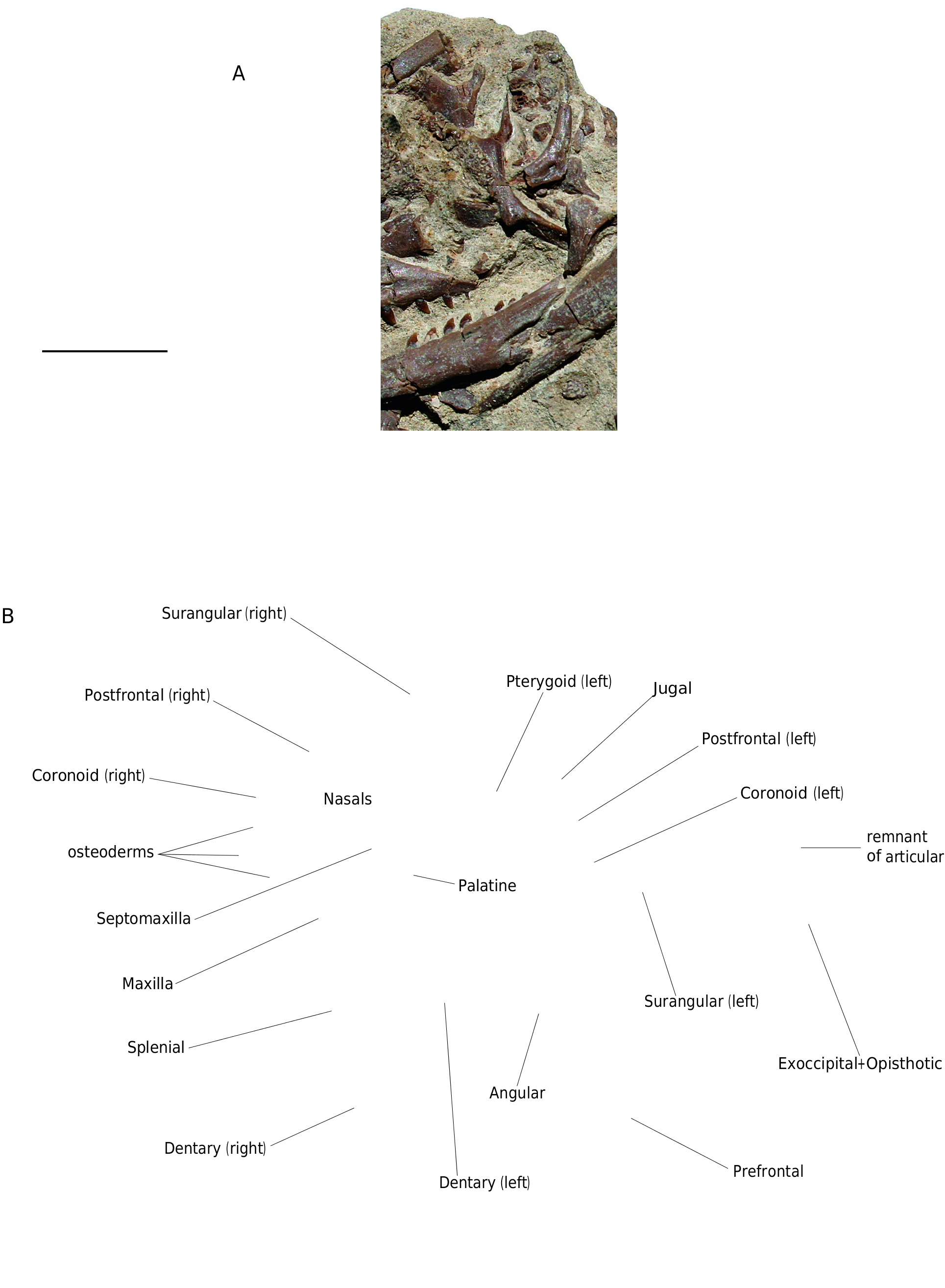
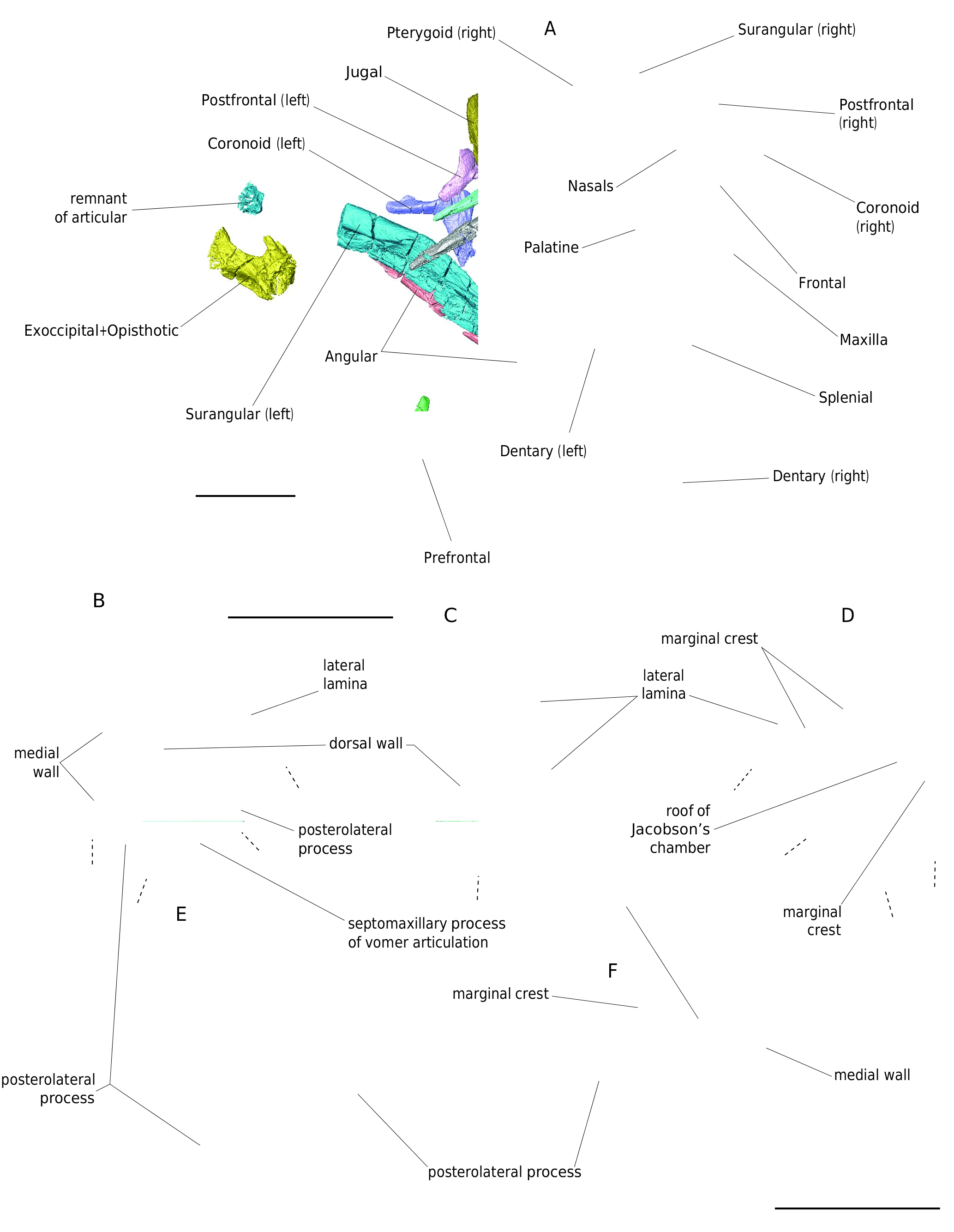
HOLOTYPE. — SMNK-PAL.8561, almost complete and partially disarticulated skull and lower jaws ( Figs 1 View FIG ; 2A View FIG ).
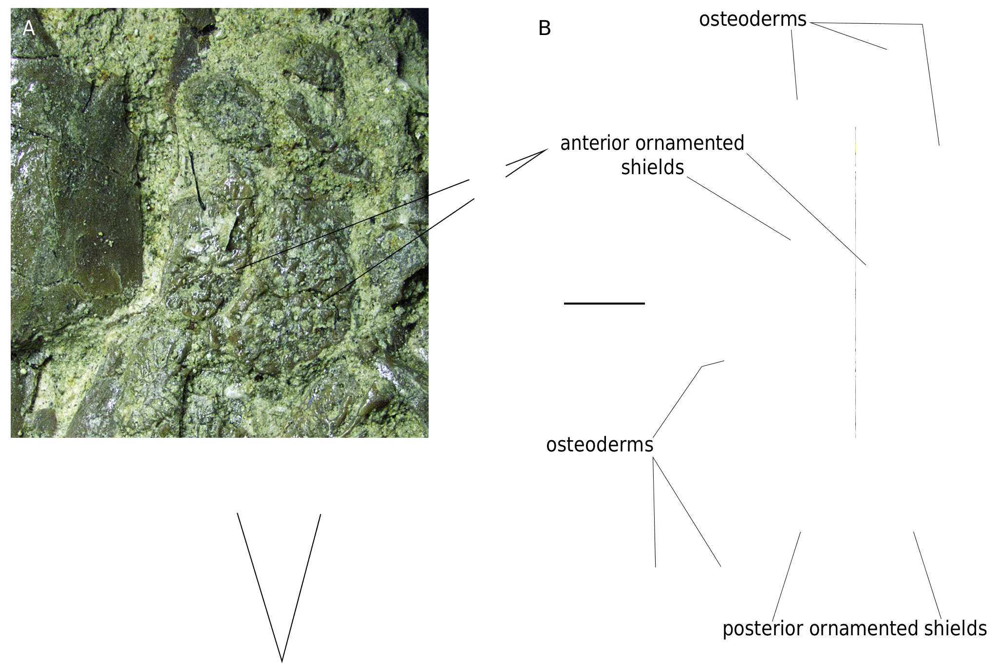
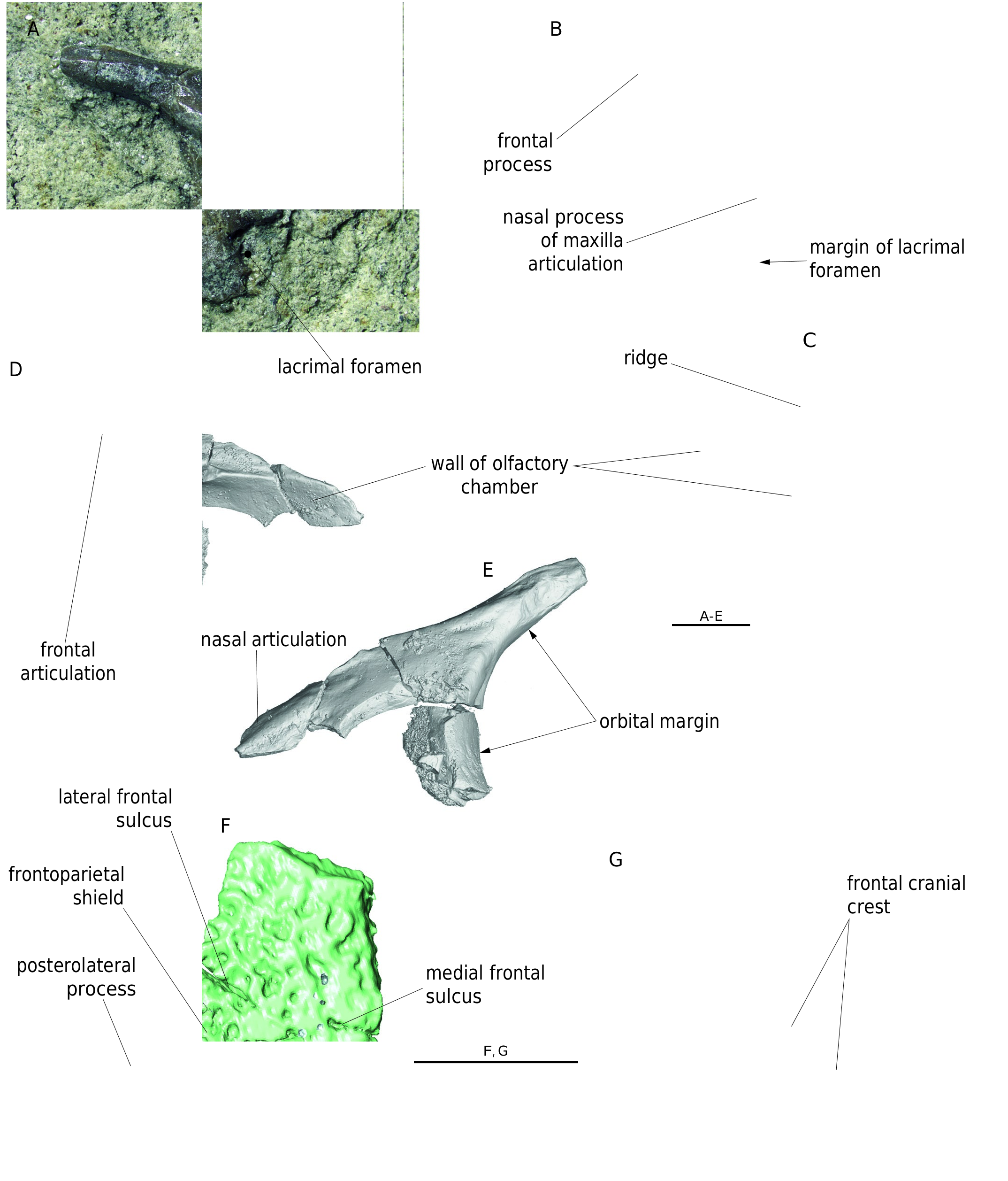
REFERRED SPECIMENS. — SMNK-PAL.8690, left isolated prefrontal ( Fig. 5D, E View FIG ); SMNK-PAL.8610, ten osteoderms ( Fig. 14 View FIG ).
LOCALITY AND AGE. — Höwenegg/Hegau (near the city Öhningen), Germany. Late Miocene (MN 9).
DIFFERENTIAL DIAGNOSIS. — On the basis of the present study, the species O. acuminatus differs from all fossil and Recent species of Ophisaurus by the following distinguished features: 1) distinct ornamentation of nasal and frontal bones (relatively broader and massively developed ridges and tubercles); 2) mediolaterally almost straight posterior margins of posterior ornamented shields of nasals; and 3) on the medial surface of the lower jaw, anterior margin of anterior mylohyoid foramen lies only slightly posterior to the level of posterior margin of anterior inferior alveolar foramen.
DESCRIPTION
Jörg (1965) described the following bones of skull and lower jaw of O. acuminatus : prefrontal, maxilla, jugal, pterygoid, dentary, angular, coronoid, surangular, articular and osteoderms. Below, the correctness of the descriptions of these bones is confirmed or modified in the light of a better accessibility to the anatomical data using high-resolution X-ray microcomputed tomography. All other elements described below are new.
Skull
Septomaxilla. Most of the body of the right septomaxilla is preserved ( Figs 1B View FIG ; 2 View FIG ). Its dorsal wall is smooth. The root portions of the posterolateral and posteromedial processes are preserved. The margin of the septomaxillary process of vomer articulation is rounded. The roof of Jacobson’s chamber is smooth and is surrounded by a distinct marginal crest. The lateral lamina is well-developed and extends ventrolaterally.
Nasal. The nasal is an elongated, paired bone ( Figs 1 View FIG ; 3 View FIG ; 4 View FIG A-C). The median suture is straight. The premaxillary process narrows anteromedially. Only the posterior portions of the anterolateral processes are preserved. Their surfaces are smooth. Both processes form a rounded posteromedial margin of the exonarial fenestra. The dorsal surface of nasals is covered with two pairs of ornamented shields. The anterior ornamented shields are slightly smaller than the posterior shields and the right anterior ornamented shield is larger than the left one. The ornamentation consists of distinctly developed ridges and broad intervening grooves. On all four shields the ossification centres are well recognizable. They lie in about the centres of the shields. The right anterior shield is of about semilunar shape; the left anterior shield is of about oval shape. The posterior portion of the right anterior shield extends medially, slightly posterior to the left anterior shield. The posteromedian portions of both anterior shields have a form of a wedge; the wedge fits between the anterior portions of the posterior shields. The anteromedial margin of the right posterior shield is perfectly preserved. It is straight and runs in anterolateral-posteromedial direction. Its anterolateral margin has an anteromedial-posterolateral course and its posterolateral margin runs in anterolateral-posteromedial direction. The right and left posterior ornamented shields meet in more-or-less straight median suture. The posterior margins of the posterior ornamented shields are lateromedially almost straight; this is in contrast to all other anguines ( Fig. 4 View FIG , see below).
The ventral surface of the nasals is finely coarse ( Fig. 4B View FIG ). The lateral margin of the anterior half of the nasal is distinctly developed and flexed ventrolaterally, thus both nasals form a shallow and wide trough anteriorly.
Remarks. The only fossil species of Ophisaurus in which the nasal is preserved is O. holeci ( Čerňanský & Klembara 2017) . This specimen is much smaller than that of O. acuminatus . The ornamentation of its nasal differs substantially from that of O. acuminatus : it consists of distinct pits of different size and very short grooves. Such ornamentation as present in O. holeci is not present in any other species of Ophisaurus .
Prefrontal
The right and left prefrontals are preserved, however, in both bones most of the wall of the olfactory chamber is damaged ( Fig. 5 View FIG A-E). The prefrontal is of about triangular shape and posteriorly extends into a stout frontal process. The frontal process is well preserved and is almost as long as the rest of the preserved portion of the prefrontal. Along the medial margin of the frontal process, a distinct frontal articulation is present. Only a small portion of the nasal articulation is present at the left prefrontal ( Fig. 5E View FIG ). The ventrolateral portion of both prefrontals is broken on several places, but the outline of the lacrimal foramen is well recognizable on the right prefrontal ( Fig. 5A, B View FIG ). The anteriormost portion of the olfactory chamber is deep and well demarcated by a sharp rounded ridge ( Fig. 5C View FIG ).
posterior ornamented shields
Frontal
The posterior portion of the left frontal is preserved ( Fig. 5F, G View FIG ). The dorsal ornamented surface is distinct. The ornamentation reaches the orbital margin of the preserved portion of the frontal. The lateral frontal sulcus is slightly longer than the medial frontal sulcus.The lateral frontal sulcus runs in anterolateral-posteromedial direction.The medial frontal sulcus is shorter and has a mediolateral course. The place of junction of both sulci is overlapped by ornamentation. The territory of the dorsal surface of the frontal, posterior to both sulci, is also distinctly ornamented, so the junction of the frontoparietal and interfrontal shields is obscured. The posterolateral process is distinct. Most of its dorsal surface is covered by the frontoparietal shield. Only posteriormost surface of the posterolateral process is smooth ( Fig. 5F View FIG ). Most of the ventral surface of the frontal is smooth. A low ridge runs close to the lateral margin of the bone and fades out at the posterolateral process of the frontal. This ridge represents the posteriormost portion of the frontal cranial crest ( Fig. 5G View FIG ).
Remarks.Although the potential junction of the lateral and medial frontal sulci is partially obscured,it is possible to estimate that the lateral sulcus is longer that the medial sulcus. Such condition is present in the frontal bones in several modern species of Ophisaurus , as well as those found in the Cenozoic of Europe and North Africa (e.g., Delfino et al. 2011; Blain et al. 2013; Klembara 2015).
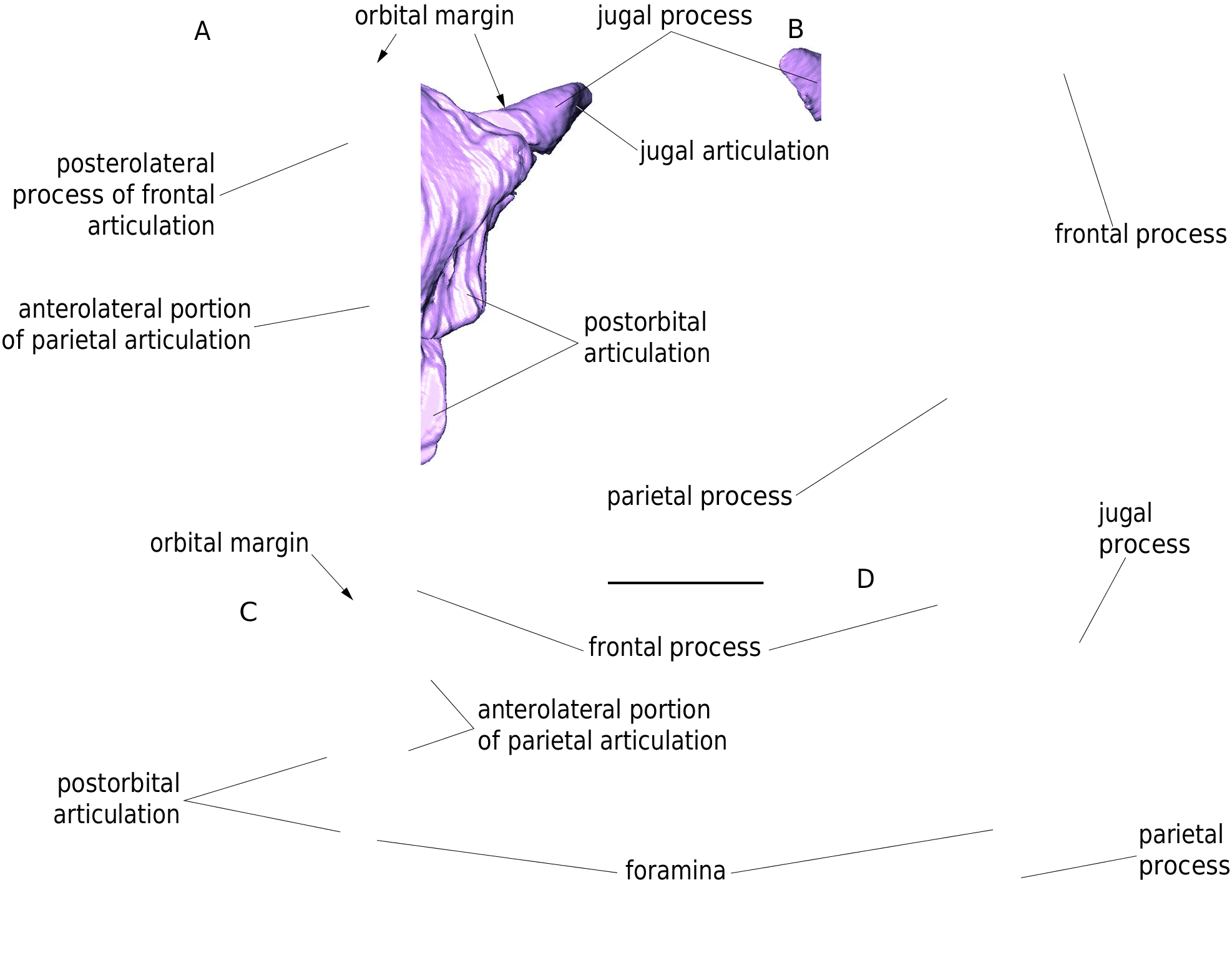
Postfrontal
The right complete postfrontal and almost complete left postfrontal are preserved ( Fig. 6 View FIG ). The postfrontal is a triradiate bone consisting of the frontal, jugal and long parietal processes. The jugal process is slightly shorter than the frontal process. The rounded orbital margin is the deepest at the level of the long ventrolateral margin of the parietal process. The anteriorly tapering frontal process bears a deep groove for the articulation with the frontal. The groove gradually continues posteriorly and forms the articulating surface for the parietal. Short jugal and long postorbital articulating surfaces are better developed on the right postfrontal. The parietal process is of rectangular shape and bears a foramen at its posterior end (present only on the left postfrontal). The left postfrontal shows a dorsoventrally broad articular surface for the anterolateral process of the parietal.
Maxilla
The left maxilla is almost completely preser ved ( Figs 1 View FIG ; 2A View FIG ; 7 View FIG A-D). Its nasal process is prominent and of trapezoidal shape. The dorsal portion of the nasal process is slightly curved medially. The external surface of the maxilla is rather smooth; a weak rugosity is present only in its anterior portion. The surface ventral to the nasal process
is pierced by seven labial foramina of various size. They are arranged in a single line. A shallow groove is associated with the posteriormost foramen which is located at the level of the 7th tooth position (counted from posterior). The posterior portion of the maxilla gradually dorsoventrally narrows into a pointed process. Its dorsal margin, gradually passing into the posterior margin of the nasal process, is almost straight. The anterior margin of the maxilla is concave and forms the posterodorsal margin of the exonarial fenestra. Unfortunately, both rami of the premaxillary process are broken. In medial aspect, the maxilla bears a slightly dorsally convex supradental shelf ( sensu Rage & Augé 2010). It bears 14 tooth positions (nine teeth are preserved). The palatine articulation lies at the level of 5th and 6th tooth positions (counted from posterior). Immediately anterior to it, a large superior alveolar foramen is located. On the medial surface of the nasal process, a distinct rugosity marks the prefrontal articulation.
Jugal
Most of the left jugal is preserved ( Figs 1 View FIG ; 2A View FIG ; 7E, F View FIG ). In the central portion, the external surface of the jugal is pierced by two foramina – a large suborbital foramen and an additional smaller foramen located slightly posterodor-sally to it. The posterior portion of the suborbital process is robustly constructed. The internal surface of the jugal bears a medial ridge. The base of the posteroventral process of the jugal is well preserved and indicates the presence of a distinct process.
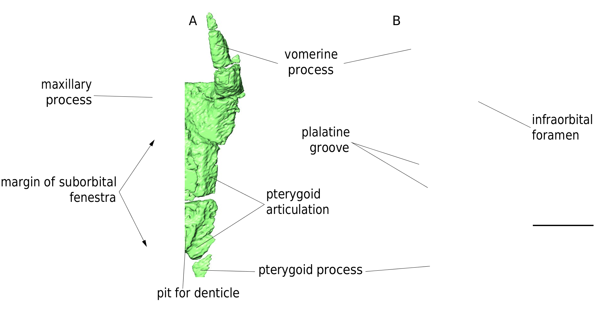
Palatine
Almost completely preserved right palatine is present ( Fig. 8A, B View FIG ). It is an anteroposteriorly elongated and flat bone. The proximal portion of the maxillary process is preserved and contains the infraorbital foramen. The vomerine process is straight, slender, much longer than the maxillary process and tapers anteriorly.The pterygoid process extends posteriorly, and the pterygoid articulation is distinct. On the ventral surface of the palatine the lateral margin of the pterygoid articulation forms a crest medially limiting the rows of teeth positions. The teeth are not preserved, but a short anteroposteriorly running series of pits indicating their positions is clearly recognizable ( Fig. 8A View FIG ). Most of the dorsal surface is smooth. On the dorsal surface the palatine groove is distinct and runs in anteromedial-posterolateral direction.
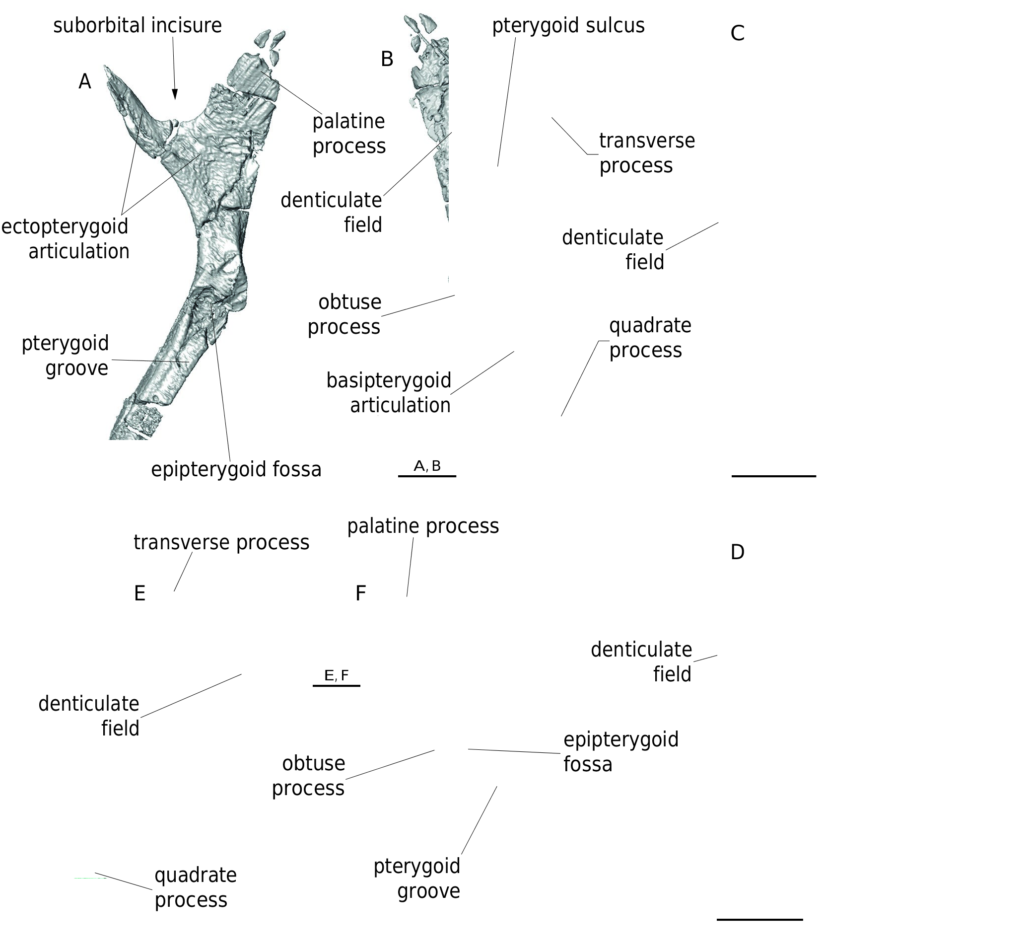
Pterygoid
Both pterygoids are preserved ( Figs 1 View FIG ; 2 View FIG ; 9 View FIG ). The pterygoid is a triradiate bone. The anteriormost portion of the palatine process is missing. However, most of the denticle field is well preserved. The pterygoid sulcus is deep and broad and gradually narrows posteriorly ( Fig. 9B, E View FIG ). The anterolaterally extending transverse process bears a well-developed ectoptery-goid articulation. The quadrate process is long and straight and bears a long pterygoid groove at its dorsal surface. The groove is anteriorly confluent with the epipterygoid fossa. The basipterygoid articulation is shallow. The obtuse process is distinct and rounded. The epipterygoid fossa lies at the level of the posterior portion of the obtuse process. The body of the pterygoid is narrowest in a short distance anteriorly to the obtuse process.
Remarks. The pterygoids of the fossil Ophisaurus are known from several Miocene and Pliocene localities in Europe (e.g., Klembara 1981, 2015; Delfino et al. 2011). The general morphology of these fossil pterygoids is very similar to that described here for O. acuminatus . The distinctive feature of all these fossil species of Ophisaurus is a relatively large denticulated field mostly composed of more or less distinct anteroposteriorly running rows of denticles; the most robust denticles lie in the most lateral row. The pterygoids of early Miocene Pseudopus ahnikoviensis Klembara, 2012 are similarly built as those of contemporaneous specimens of Ophisaurus , however, the pterygoid of P. ahnikoviensis bears a distinct feature - the oblong crest lying on the ventral surface of the transverse process of the pterygoid ( Klembara 2012). The presence of the oblong crest is a shared feature by three species of Pseudopus : P. ahnikoviensis , P. laurillardi ( Lartet, 1851) and P. pannonicus ( Kormos, 1911) ( Klembara et al. 2010) .
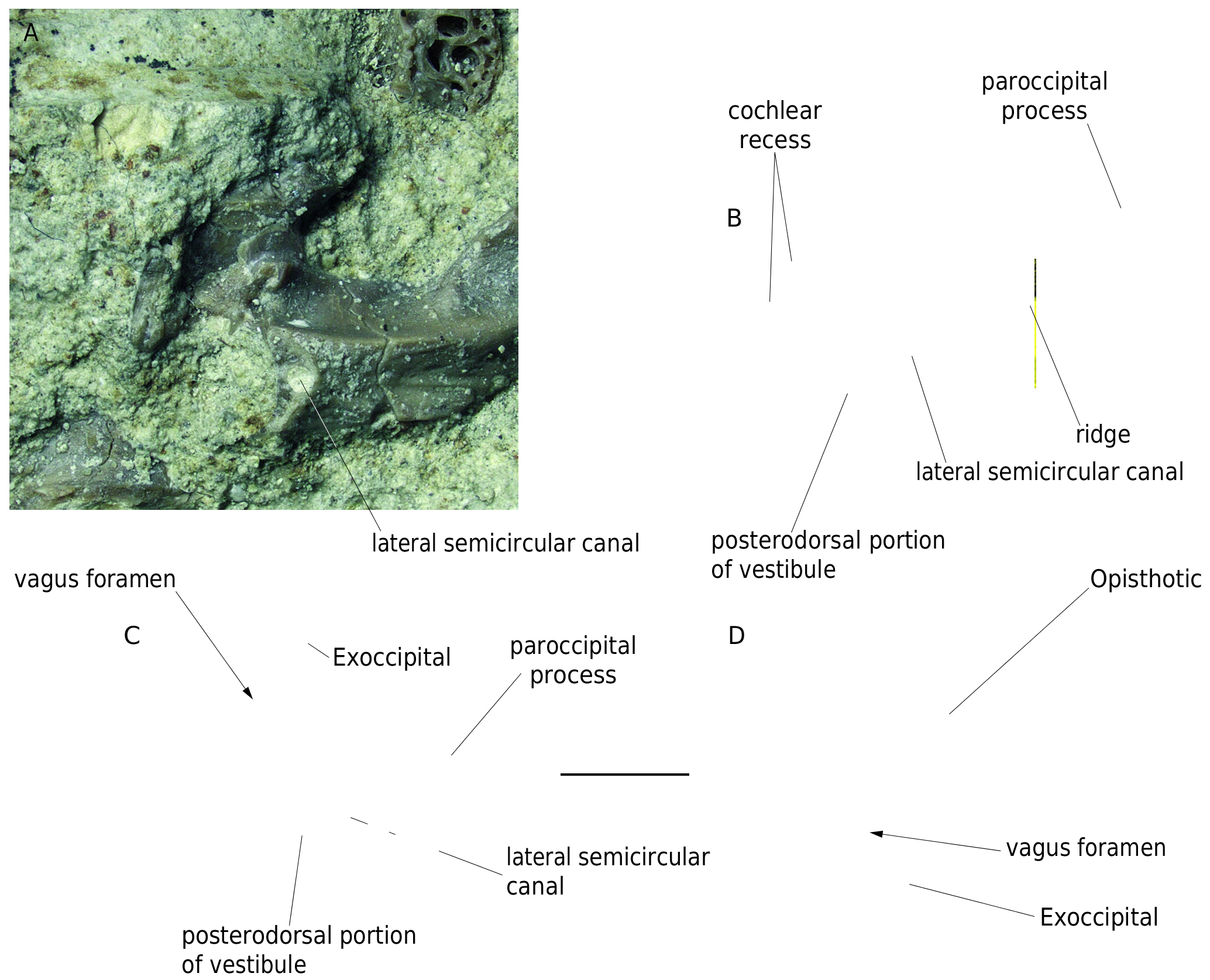
Exoccipital and opisthotic
A substantial portion of the right exoccipital and opisthotic is preserved and both bones are fused together ( Figs 1 View FIG ; 2 View FIG ; 10 View FIG ). The paroccipital process is fan-like at its distal end. On the posterior surface of the paroccipital process, a distinct and sharp ridge passes posterolaterally. The ridge gradually diminishes posteriorly and is confluent with the articular surface of the paroccipital process. Dorsally to the proximal portion of the ridge, the opening for the lateral semicircular canal is present ( Fig. 10 View FIG A-C). Immediately anterior to the
opening of the semicircular canal is a rounded margin limiting a deep excavation; the excavation represents a posterodorsal portion of the vestibule ( Fig. 10B, C View FIG ). Immediately ventral to it, a rounded depression marks the wall of the cochlear recess ( Fig. 10B View FIG ). The proximal portion of the fused exoccipital-opisthotic is on several places damaged, but the vagus foramen marking the original exoccipital-opisthotic suture is present ( Fig. 10C, D View FIG ).
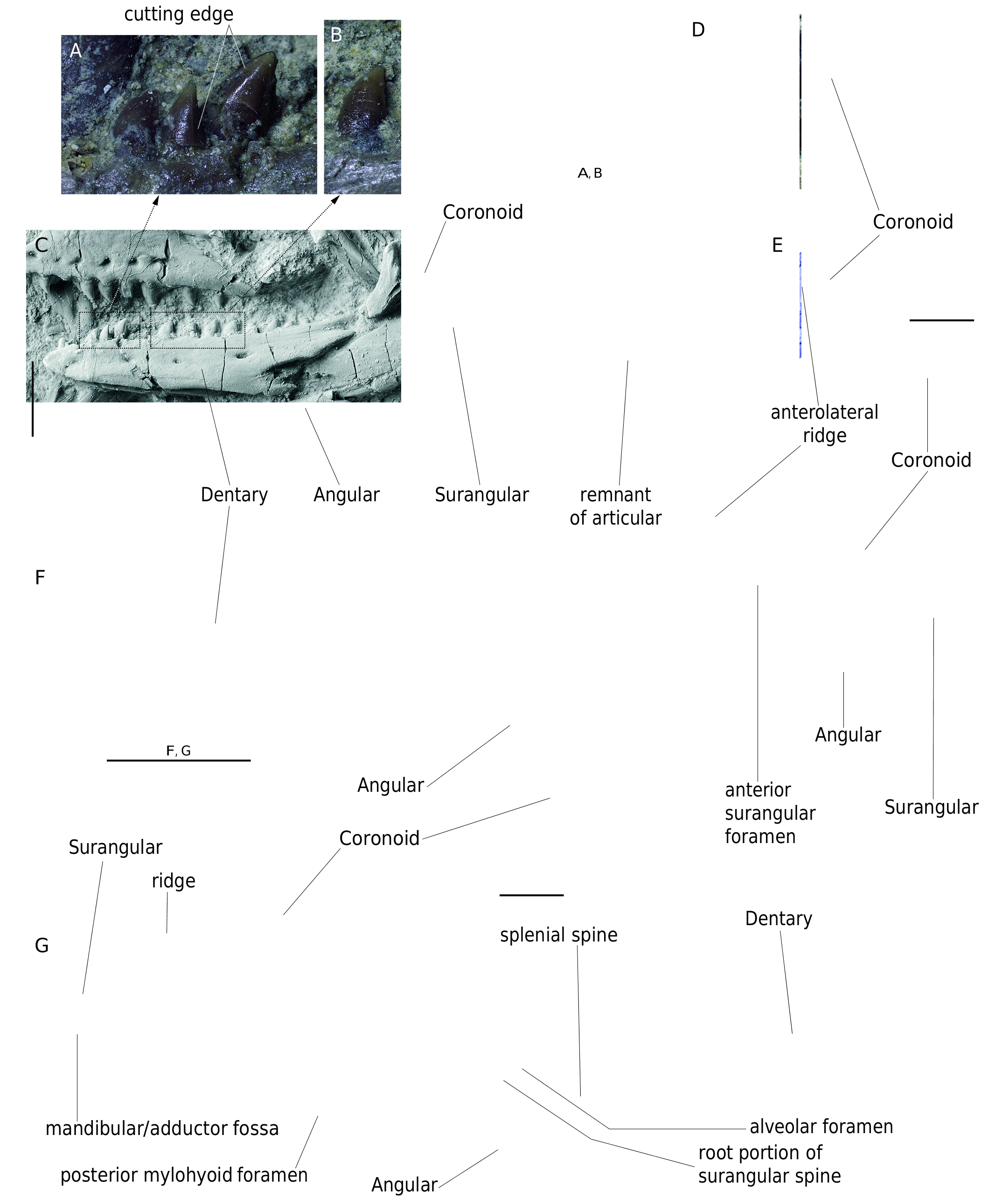
LOWER JAW ( FIGS 1 View FIG ; 2A View FIG ; 11-13 View FIG View FIG View FIG )
Dentary
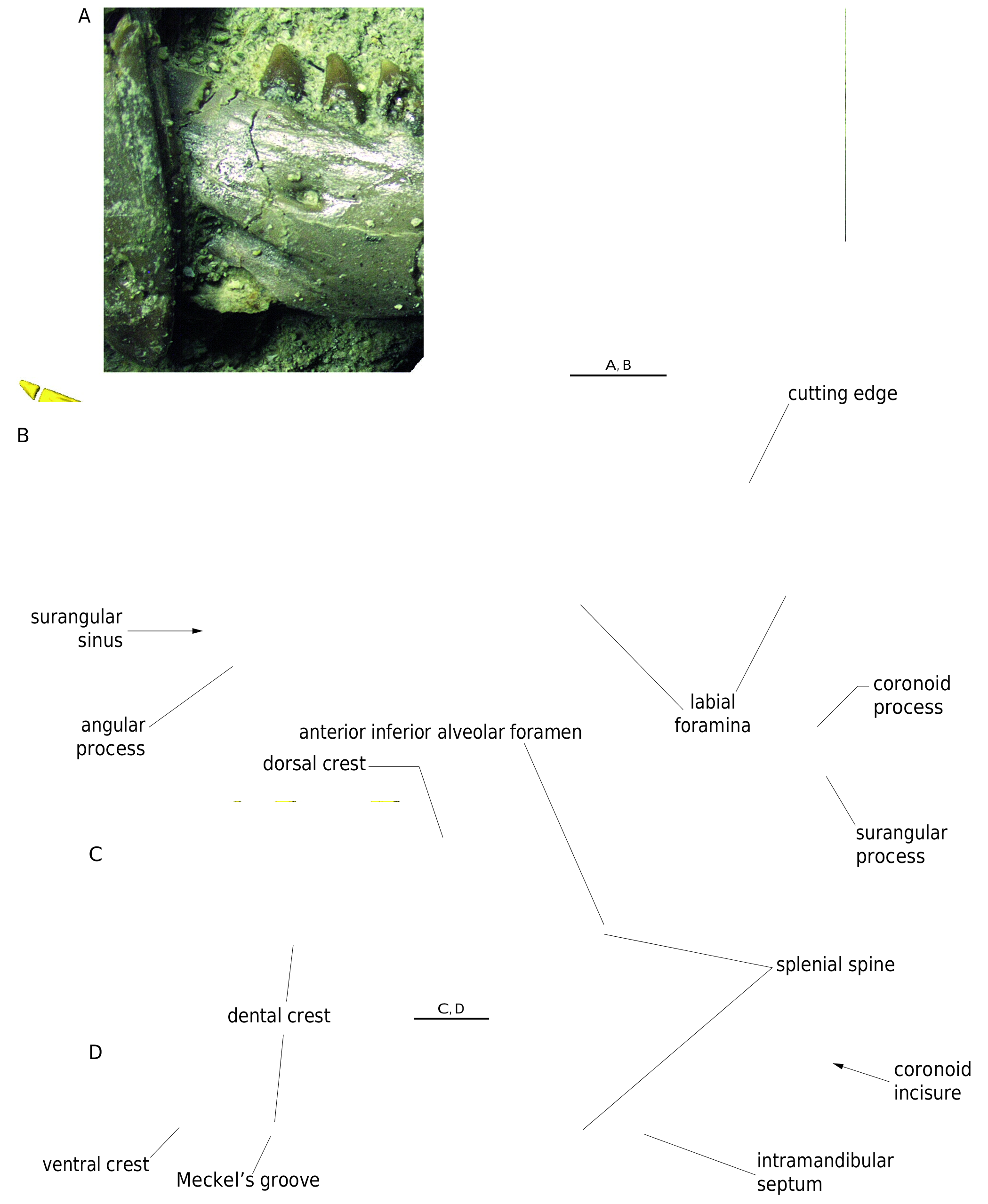
Both dentaries are preserved ( Figs 1 View FIG ; 2A View FIG ; Fig. 11C, F, G View FIG ; 12 View FIG ). The dentary is anteroposteriorly elongate and gradually narrows anteriorly. The smooth lateral surface of the bone is pierced by six mental foramina (well preserved in the left dentary). A high dorsal crest supports fifteen tooth positions (eleven teeth are preserved in each dentary). The dental crest is concave and anteriorly passes into a small symphyseal facet. The crest is flexed ventrally along its entire length. It bears a splenial spine at the level between the fourth and fifth tooth positions (counted from posterior). The splenial spine forms an anteroventral margin of the anterior inferior alveolar foramen ( Figs 11G View FIG ; 12C View FIG ). Posteriorly to it, the dental crest is curved dorsally, being thinner than its anteriorly located portion. Meckel`s groove is narrow, opens ventrally rather than medially ( Fig. 12D View FIG ). The alveolar foramen is located at the level of the third tooth position (counted from posterior) ( Fig. 11G View FIG ). Posteroventrally to this foramen, the surangular spine is present; however, only its root portion is preserved in the left dentary ( Fig. 11G View FIG ). The intramandibular septum is better preserved on the right dentary and has about a vertical position ( Fig. 12D View FIG ). The posteroventral region of the dentary ends by a distinct angular process. It is of triangular shape. Its posterior termination reaches the level of the penultimate tooth position ( Fig. 12 View FIG ). The posterodorsal portion of the dentary is elevated dorsally. The surangular and coronoid processes are pointed. The coronoid process reaches slightly more posteriorly than the surangular process. A shallow coronoid incisure is located between both processes ( Fig. 12 View FIG B-D).
Splenial
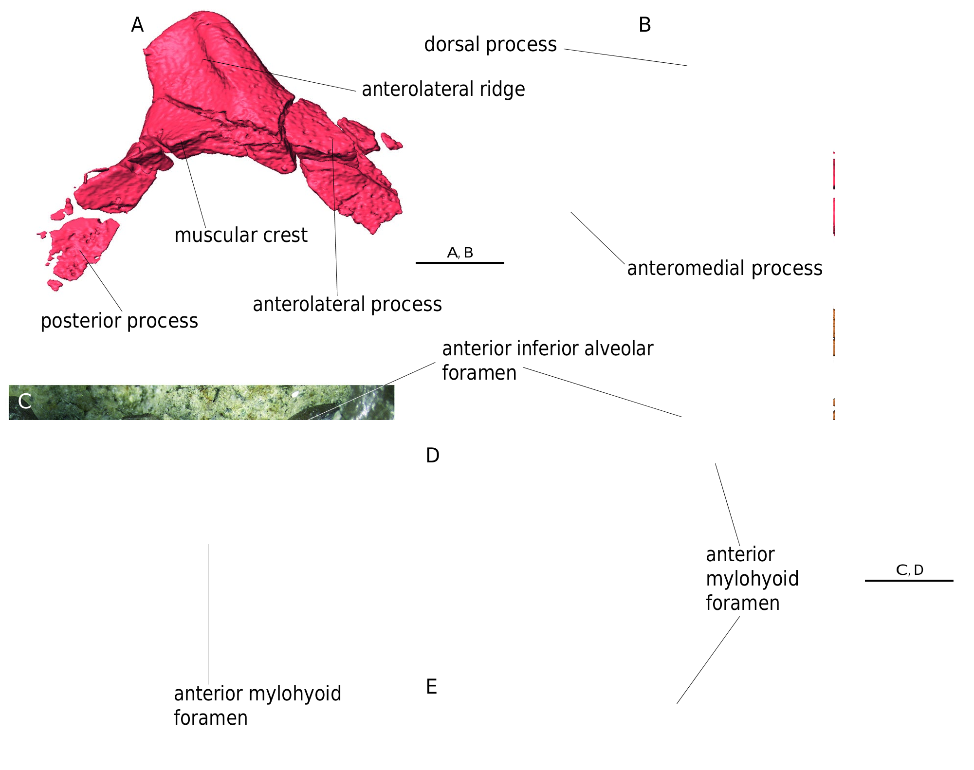
The right splenial is well preserved ( Figs 1 View FIG ; 2A View FIG ; 13 View FIG C-E). Anteriorly it extends into a pointed process. Posteriorly it gradually dorsoventrally broadens. Posteroventrally, the splenial extends into a long, narrow and pointed process. The dorsally to it lying portion of the splenial is partially damaged, but the presence of two other processes is indicated. At the half of the anteroposterior length of the splenial, the anterodorsal
margin is stepped forming a roughly triangular small process. The process is hooked anteriorly. This structure forms the posterior and ventral margins of the anterior inferior alveolar foramen. In the central portion of the bone, a large mylohyoid foramen is present. It lies only slightly posterior to the level of the posterior margin of the anterior inferior alveolar foramen.
Angular
The left angular is preserved; it is an elongate bone lying at the ventral wall of the lower jaw ( Figs 1 View FIG ; 2A View FIG ; 11C, F, G View FIG ). The anterior portion of the angular contacts the posteroventral portion of the dentary. An anteroposteriorly elongated depression on the medial surface of the angular indicates the presence of the posterior mylohyoid foramen ( Fig. 11G View FIG ). The posterior portion of the angular supports the surangular partly ventrally and medially, and mostly laterally.
Coronoid
Both left and right coronoids are preserved ( Figs 1 View FIG ; 2A View FIG ; 11 View FIG C- G; 13A, B). The coronoid is a chevron shaped bone, with four processes: the dorsal, anterolateral, anteromedial and posterior processes. The dorsal process lies in the mid-length of the bone. It is of quadrangular shape. Although it is shorter than other two processes, its overall appearance is robust. The dorsal process is slightly inclined posteriorly. Its anterolateral portion bears a distinct ridge (or keel) for muscle attachment ( Figs 11E View FIG ; 13A View FIG ). The medial side of the process is flat. The posterior process is longer than the anteromedial process. It is bent laterally to form a contact with the surangular. This articulation is bordered dorsally by a sharp muscular crest ( Fig. 13A View FIG ). The medial side of the posterior process bears a low ridge. This ridge forms the anterior border of the mandibular/adductor fossa ( Fig. 11G View FIG ).
Surangular and articular
The left and right surangulars and a small remnant of the left articular are preserved ( Figs 1 View FIG ; 2A View FIG ; 11C, F, G View FIG ). Only the left surangular is partially articulated with the neighbouring bones of the mandible ( Fig. 11F, G View FIG ). The surangular is an elongate and massive element forming the posterodorsal portion of the lower jaw. In the anterodorsal region and close posterior to the dentary, the dorsolateral surface of the bone is pierced by a large anterior surangular foramen ( Figs 1 View FIG ; 11F View FIG ). On the medial wall of the surangular, a deep mandibular/adductor fossa is present immediately posterior to the posterior process of coronoid articulation. ( Fig. 11G View FIG ). The posterior portions of both mandibles are broken, but the impression of the left posterior portion of the mandible is well-preserved. Here, a small remnant of the articular is preserved ( Figs 1 View FIG ; 2A View FIG ; 11C View FIG ).
TEETH
There are four elements bearing dentition: dentary, maxilla, pterygoid and palatine. The denticles on the palatine are not preserved, but a row of rounded pits indicates their presence ( Fig. 8A View FIG ). The denticles on the pterygoid are arranged in four longitudinal rows. The denticles are conical and pointed. Their apices are posteriorly curved. The denticles lying in the most lateral row are the largest ( Fig. 9 View FIG B-E).
Implantation of the marginal teeth is pleurodont. The teeth are large, well exposed over the dorsal crest which supports them laterally ( Figs 1A View FIG ; 7 View FIG A-D; 11A-C, F, G; 12), There are 14 tooth positions in the maxilla and 15 tooth positions in dentary. They are conical and curved distally. Their tips are pointed. The mesial and distal cutting edges are well developed ( Figs 11A, C View FIG ; 12 View FIG ). The mesial surfaces of apices are smooth. The tooth bases are mesiodistally broad. The largest maxillary teeth are present in the mid-region of the tooth row what corresponds to the size of teeth in the dentary.
Remarks. The marginal teeth of O. acuminatus are morphologically similar to those in some extant species of Ophisaurus , like O. koellikeri , however, in contrast to O. koellikeri , the internal surface of their apices is not striated in O. acuminatus . The teeth of O. acuminatus are very similar to those designated as Anguinae indeterminate 1 from early Miocene deposits of North Bohemia ( Czech Republic) ( Klembara 2015). The teeth of Anguinae indeterminate 1 are posteriorly curved, pointed and have well-developed anterior and posterior cutting edges. The medial surface of their apices is not striated ( Klembara 2015: figs 5, 6A). The teeth in O. acuminatus are also similar to those in the Oligocene Ophisaurus roqueprunensis Augé, 1992 ( Augé & Smith 2009).
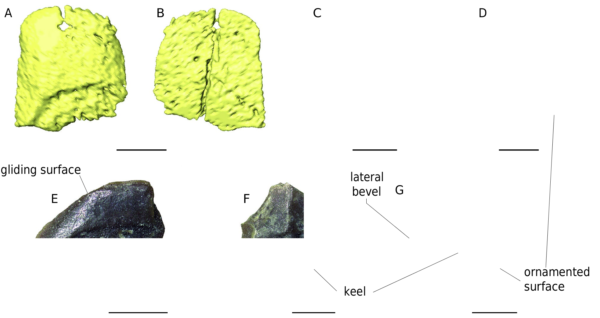
OSTEODERMS
Almost all osteoderms are incompletely preserved ( Figs 1 View FIG ; 14 View FIG ). Some of them are directly associated with the holotype being embedded together in the sediment.Additional ten osteoderms are preserved as isolated elements ( Fig. 14 View FIG ). Unfortunately, the internal surface of the isolated osteoderms is not accessible, because the osteoderms had been fixed by the glue on a sheet of paper in the past. The osteoderms studied here can be divided into two morphotypes.
The first morphotype is represented by a slender, flat and more oval (or elliptical) osteoderms ( Fig. 14 View FIG A-E). This morphotype has no longitudinal keel and the external side bears a very large gliding surface. The lateral bevel appears to be highest close to this surface. Posteriorly located external surfaces of the osteoderms are ornamented. The ornamentation consists of short ridges, grooves, tubercles and shallow pits. The ridges have a vaguely radial orientation from the anterior portion of the osteoderm. The internal surface is smooth and bears several foramina ( Fig. 14B View FIG ).
Osteoderms of the second morphotype are roughly rectangular ( Fig.14F, G View FIG ). This type possesses a low medial keel, as well as the ornamented surface and lateral bevel. These osteoderms bear a similar type of ornamentation as that in the first morphotype.
Remarks. This type of morphology of osteoderms corresponds to that of Ophisaurus (e.g., Čerňanský & Klembara 2017). The presence of two morphotypes most likely reflects the position of the osteoderms in the different portion of the body armour.


No known copyright restrictions apply. See Agosti, D., Egloff, W., 2009. Taxonomic information exchange and copyright: the Plazi approach. BMC Research Notes 2009, 2:53 for further explanation.