
Pleuroxus trigonellus ( O.F. Müller, 1776 ), Garibian & Neretina & Klimovsky & Kotov, 2018
|
publication ID |
https://doi.org/ 10.11646/zootaxa.4532.4.1 |
|
publication LSID |
lsid:zoobank.org:pub:F69AFA65-2777-499D-82D1-1EC7327A798B |
|
DOI |
https://doi.org/10.5281/zenodo.5950052 |
|
persistent identifier |
https://treatment.plazi.org/id/D92B87B6-0B60-FFE6-B2F7-FE70FBC2404E |
|
treatment provided by |
Plazi |
|
scientific name |
Pleuroxus trigonellus ( O.F. Müller, 1776 ) |
| status |
|
Pleuroxus trigonellus ( O.F. Müller, 1776) View in CoL
( Figs. 1–7 View FIGURE 1 View FIGURE 2 View FIGURE 3 View FIGURE 4 View FIGURE 5 View FIGURE 6 View FIGURE 7 )
Lynceus trigonellus O.F. Müller, 1776 in Müller (1776) : p. 199; Müller (1785): p. 74–75, pl. 10, figs. 5–6.
Pleuroxus trigonellus (O.F. Müller) in Sars (1861) View in CoL : p. 124, pl. 91; Lilljeborg (1901): p. 534–537, pl. 74: figs. 13–23; Šrámek- Hušek et al. (1962): p. 376–378, fig. 141; Flössner (1972): p. 355–357, figs. 165–166; Negrea (1983): p. 240–242, fig. 96; Margaritora (1985): p. 211–213, fig. 84; Flössner (2000): p. 250–253, fig. 93; Smirnov (1966): figs. 8, 15, 16, 24, 25; Frey (1993): table 1, figs. 1–8; Røen (1995): 281–284, fig. 128; Hudec (2010): p. 421–423, fig. 104; Szeroczyńska et al. (2007): p. 63–64, figs. 322–332.
Pleuroxus trigonellus trigonellus (O.F. Müller) in Smirnov (1971) View in CoL : p. 226, figs. 208–215.
Pleuroxus ornatus Schödler, 1862 in Schödler (1862) View in CoL : p. 25, pl. 2: fig. 32? (no captions in the paper, fig. 38 in text, this is a mistake!); Schödler (1863): 47–48, pl. 2: fig. 32.
Pleuroxus Bairdii Schödler, 1863 in Schödler (1863): p. 50–51.
Type material. Lost, as whole material of O.F. Müller (Müller 1867; Frey 1980).
Type locality. Denmark. The type locality was not specified, but it is known that O.F. Müller collected at least some samples in the ponds at his summer house in Frederiksdal ( Frey 1980) .
Material examined. 10 parthenogenetic females, 3 ephippial females, 5 males from Dobrasee, Brandenburg, Germany, coll. by M. Belyaeva, AAK M-0919; 5 parthenogenetic females from Lake Glubokoe (N 55.75361°, E 36.50417°), Moscow Area, Russia, coll. 15.08.2005 by N.N. Smirnov, AAK 2006-109; 5 parthenogenetic females from the same location, coll. 16.08.2016 by A.A. Kotov, AAK M-3476; 2 parthenogenetic females from the same location, AAK M-3477; 1 parthenogenetic female from the same location, AAK M-3486; 2 parthenogenetic females from the same location, AAK M-3487; 3 parthenogenetic females from a small pond (N 55.56242°, E 39.82352°), Moscow Area, Russia, coll. 0 4.08.2014 by A.A. Zharov, AAK M-2730; 2 parthenogenetic females from the same location, AAK M-2731; 2 parthenogenetic females from Sen’ga River (N 55.8839°, E 39.52012°), Vladimir Area, Russia, coll. 0 6.08.2014 by A.A. Zharov, AAK M-2740; 5 parthenogenetic females from a small pond 50 m from the shore of Chudskoe Lake, near the village Zapol'je (N 58.63642°, E 27.78997°), Pskov Area, Russia, coll. 19.08.2007 by A.A. Kotov, AAK M-0596; 2 parthenogenetic females from a small lake ffi 1 in the vicinity of Penza, Sosonivka, (N 53.1739°, E 45.0767°), Penza Area, Russia, coll. in May of 2009 by E.I. Bekker, AAK M-0934; 2 parthenogenetic females from a small lake ffi 2 in the vicinity of Penza (N 53.212°, E 45.088°), Penza Area, Russia, coll. 0 5.05.2010 by E.I. Bekker, AAK M-1807; 2 parthenogenetic females from Cherepovets Reservoir, Vologda Area, Russia, coll. 17.07.1966 by N.N. Smirnov, NNS 1998-183; 3 parthenogenetic females from Gor’kovskoe Reservoir (N 56.8°, E 43.3°), Nizhny Novgorod Area, Russia, coll. 28.08.1961 by N.N. Smirnov, NNS 1999-056.
Diagnosis (adapted after Frey 1993). Body transparent. Parthenogenetic and ephippial female with length up to 0.60 mm. Median dorsal keel absent. Sculpture of valves and head moderately or strongly developed. Rostrum sharply pointed, directed downwards. Labral keel short (not projected behind tip of rostrum), triangular, its anterior margin slightly convex, distal angle acute. Posteroventral angle of valve with few denticles. Postabdomen moderately elongated, tapered distally, postanal margin almost straight, with strong postanal teeth, on postabdominal claw proximal basal less than half size of distal one. Antenna I in female elongated, with a basal peg and nine aesthetascs of unequal size. Apical swimming setae of antenna II subequal in size. Filter plate of gnathobase II with eight setae; filter plate III with 8 setae; filter plate IV with 6 setae; filter plate V with 4 setae. Male with length up to 0.45 mm. Rostrum shorter as compared to the female. Distal portion of the postabdomen shaped as a tube lacking setules, postanal teeth transformed into bunches of long, thin setules. Gonopore opens ventrally at level of postabdomen narrowing (base of the tube). Antenna I in male without a basal peg and with nine aesthetascs of unequal size.
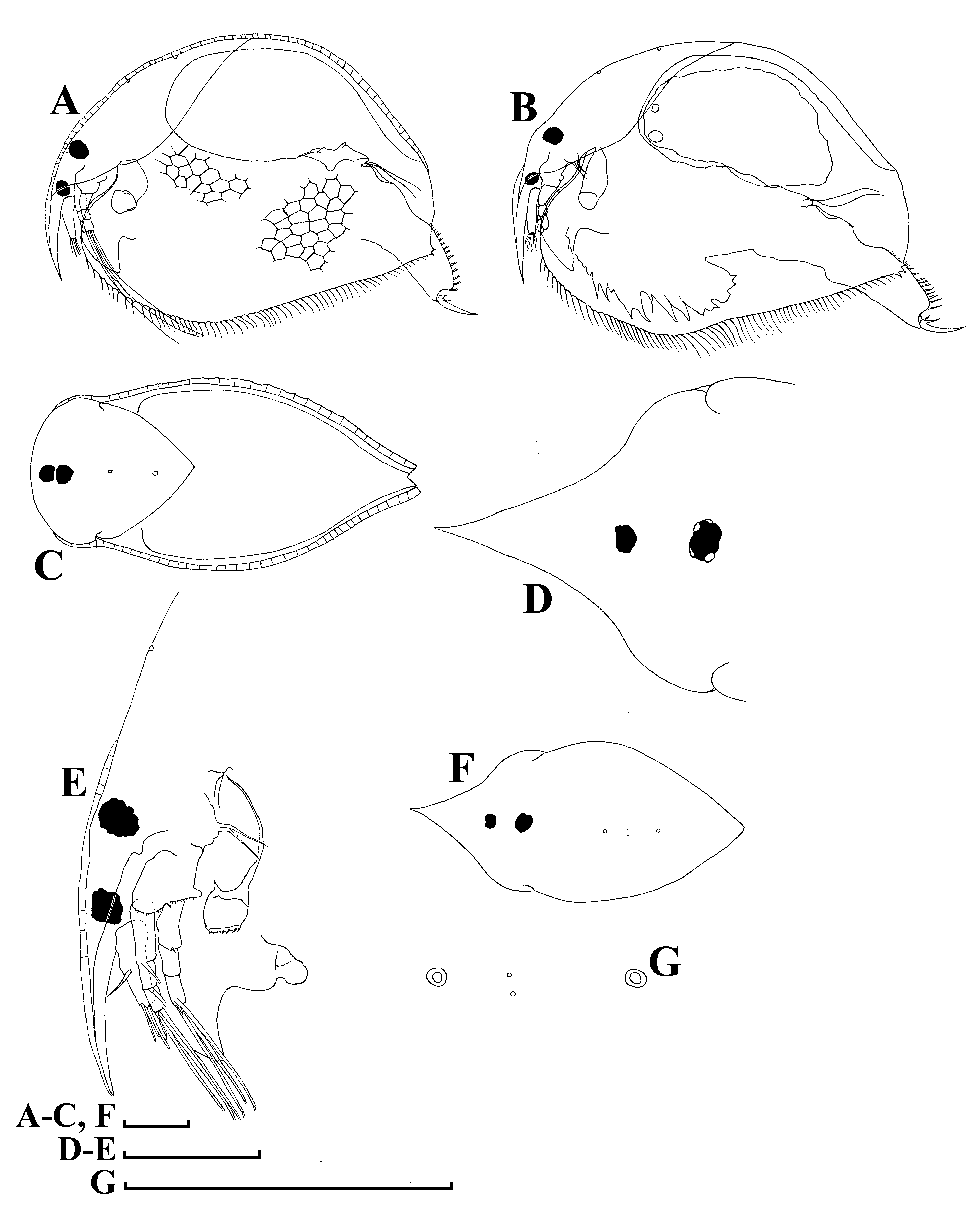
Redescription. Parthenogenetic female. General. Body transparent. In lateral view body subovoid, high (body height/ body length = 0.71–0.73 in adults). Maximum height in middle of body ( Figs. 1 View FIGURE 1 A–B, 2A–B). In dorsal and anterior view body compressed laterally, lacking a median dorsal keel ( Figs. 1C View FIGURE 1 , 2 View FIGURE 2 C–F). Dorsal margin evenly arched from tip of rostrum to posterodorsal angle, without a depression between head and the rest of body. Posterior margin relatively straight, posteroventral angle almost straight, with teeth, ventral margin with a slight prominence in thirst half of body behind head ( Figs. 1 View FIGURE 1 A–B, 2A–B), with a row of marginal setae ( Figs. 1 View FIGURE 1 A–B, 2A, 4C–E). Obscure striation as hexagons on all body surface ( Figs. 1A View FIGURE 1 , 2A View FIGURE 2 , 4 View FIGURE 4 A–B).
Head with a long, pointed rostrum, protruding downward and posteriorly ( Figs. 1 View FIGURE 1 D–E, 2A–F, 4A–B, F). Eye only slightly larger than ocellus, distance from tip of rostrum to ocellus greater than that between ocellus and eye ( Figs. 1 View FIGURE 1 A–B, 1E). Head shield elongated, with maximum width immediately behind mandibular articulation ( Figs. 1D, 1F View FIGURE 1 , 2F View FIGURE 2 , 4 View FIGURE 4 G–H). In dorsal view rostrum elongated, with acute apex ( Figs. 1D View FIGURE 1 , 2 View FIGURE 2 C–F). Two major head pores ( Figs. 1 View FIGURE 1 F–G, 4G–H), PP = 1.6 IP. Lateral head pores minute, located at the headshield midline between major pores ( Fig. 1G View FIGURE 1 ), usually these are positioned somewhat asymmetrically to the midline.
Labrum with a fleshy main body and a small distal labral plate ( Fig. 1E View FIGURE 1 ). Labral keel short (not projected behind rostrum tip), triangular, its anterior margin slightly convex, distal angle acute.
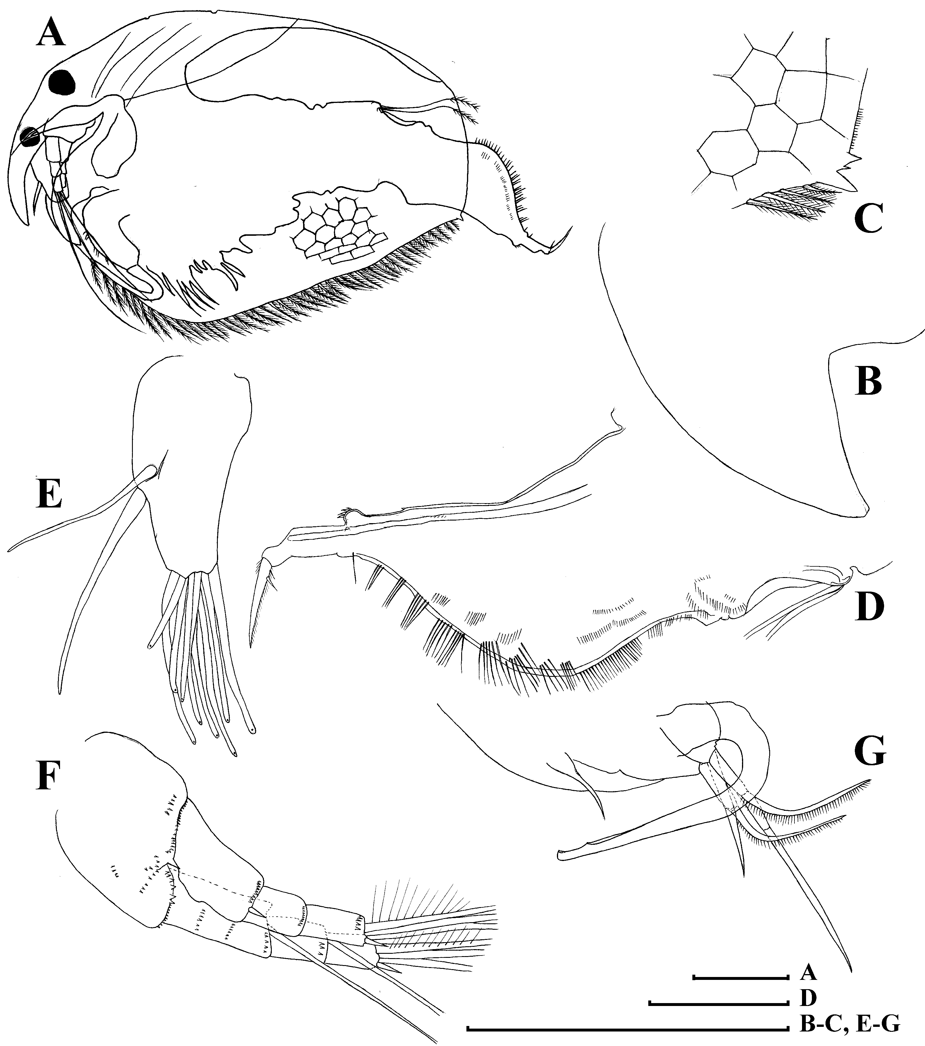
Valves large, ventral margin armed with numerous setae of different size in different regions, all plumose and located exactly marginally ( Figs. 1 View FIGURE 1 A–B, 2A–B, 3C–E, 4B–E). Posterior margin of valves with few (1–3, mainly 2) teeth ( Figs. 1 View FIGURE 1 A–B, 2A–B, 3D–E, 4D–E). Ventral margin bears setae covered by long prominent setules ( Fig. 3 View FIGURE 3 C–E, 4C–D). Anterior valve margin broadly rounded ( Fig. 3A View FIGURE 3 , 4 View FIGURE 4 A–B).
Postabdomen elongated, subrectangular, its ventral margin almost straight ( Figs. 3 View FIGURE 3 F–I, 6A–B). Basis of postabdominal setae surrounded by a prominent chitin ring ( Fig. 3H View FIGURE 3 , 6D View FIGURE 6 ). Preanal margin slightly concave ( Figs. 3 View FIGURE 3 F–H, 6A–C), and slightly longer than anal margin, preanal and postanal angle well-defined, postanal margin clearly longer than anal margin, dorsodistal angle almost right ( Figs. 3I View FIGURE 3 , 6 View FIGURE 6 E–G). Basis of claws inflated ( Figs. 3 View FIGURE 3 H– I, 6E–F). Each side of the postanal portion with a row of long and thickened postanal teeth, increasing in size distally ( Fig. 3I View FIGURE 3 , 6G View FIGURE 6 ). Laterally to marginal denticles, a row of fascicles consisting of bunches of short, fine setules ( Figs. 3 View FIGURE 3 H–I). Postabdominal seta longer than postanal margin, with basal segment approximately as long as preanal margin and distal segment shorter than basal one, implanted with delicate setules ( Fig. 3G View FIGURE 3 ).
Postabdominal claw massive and long (subequal in length to anal and preanal margins), slightly curved ( Figs. 3I View FIGURE 3 , 6 View FIGURE 6 E–G). Dorsal edge of claw armed with a row of fine setules decreasing in size distally ( Fig. 6G View FIGURE 6 ). Two basal spines, distalmost basal spine long (slightly longer than the base of claw), proximalmost basal spine relatively short, two times shorter than distal spine ( Figs. 3 View FIGURE 3 H–I, 6E–F).
Antenna I not reaching tip of rostrum, narrowing distally, with a well-defined basal peg ( Figs. 3 View FIGURE 3 J–K). Antennular sensory seta slender, longer than half of antenna I, arising at half of antennular body. Nine short aesthetascs unequal in length ( Figs. 3 View FIGURE 3 J–K).
Antenna II relatively short, coxal part with two sensory setae, basal segment robust, with a small spine ( Figs. 1 View FIGURE 1 A–B, 3L, 4F). Antennal branches elongated, exopod and endopod subequal in length, all segments cylindrical, covered by transverse rows of fine spinules ( Fig. 4F View FIGURE 4 ). Antennal formula: setae 0-0-3/1-1-3, spines 1-0-1/0-0-1. Proximal segment of exopod with a short spine. Exopod and endopod apical swimming setae, as well as lateral endopod setae covered by fine long setules; distal segments of apical setae with chitin insertions ( Figs. 3 View FIGURE 3 L–M). Apical spines of exopod and endopod subequal in size ( Fig. 4F View FIGURE 4 ).
Limb I ( Figs. 5 View FIGURE 5 A–B, 6H): accessory seta, or corm seta in Frey (1993a), present (not represented in Fig. 5A View FIGURE 5 , because it is located on the other side of the limb corm), ODL relatively small, bears a long seta with a naked distal segment, and a short seta with short, setulated distal segment ( Figs. 5 View FIGURE 5 A–B). IDL larger than ODL, covered by transverse rows of setules ( Fig. 6H View FIGURE 6 ). First IDL seta short, naked, second and third IDL setae not equal in size and similarly armed distally with short, fine setules ( Figs. 5 View FIGURE 5 A–B). Endite 3 with three soft posterior setae (a–c) and stiff anterior seta 1 of similar length. Endite 2 with short seta anterior (seta d), long (setae e and f), and delicate posterior seta 2 armed with minute setules. Endite 1 with long, slender posterior setae (g–i), a very short seta (j), and anterior seta 3 (it significantly shorter than seta 2). Fascicles of thin setules on inner face of limb, plus bunches of longer thicker setules at ventral margin of limb. Two slender ejector hooks of remarkably different size. A short seta, a remnant of maxillar process, on limb base.
Limb II ( Fig. 5C View FIGURE 5 ) subrectangular. Exopodite subquadrangular, with a short seta. Inner portion of limb with eight setae: setae 1–3 especially long, setae 4–5 shorter, subequal in size, setae 6–8 short, also subequal in size. A series of small projections posteriorly to distal setae, and a small sensillum near scraper 4. Distal armature of gnathobase with a bunch of plumose setules, unique for the anomopods, and four setae. Filter plate with eight setae, two distalmost setae subequal in size and shorter than the rest; basal most seta of filter plate with inflated basal segment.
Limb III ( Figs. 5 View FIGURE 5 D–E) with ovoid elongated epipodite. Exopodite subrectangular, with four distal setae (1–4), and three lateral setae (5–7). Distal endite with three anterior setae ( Figs. 5 View FIGURE 5 D–E), all with minute setules distally, of them two distal setae (1–2) long, basalmost seta (3) short. Small sensillae near bases of setae 2 and 3. Basal endite with four anterior setae (4–7), slightly increasing in size basally, not armed, small bottle-shaped sensillum near seta 4. On the posterior surface, six soft setae (a–f) subequal in length, bilaterally armed with sparse, fine setules. Gnathobase not clearly separated from basal endite. Distal armature of gnathobase with large, bottle-shaped sensillum, three setae, and a bunch of setules. Filter plate with eight setae.
Limb IV ( Figs. 5 View FIGURE 5 F–G) with ovoid elongated epipodite and rounded preepipodite. Exopodite wide, subovoid, with seven setae of unequal size. Innerdistal portion of limb IV with four marginal setae. Distalmost seta (1) slender and uncovered with minute setules on distal segment, setae 2–4 with thick basal segments and slender, setulated distal segments. On posterior surface, four soft setae (a–d). Gnathobase well-separated, its distal armature with four setae. Filter plate with six setae.
Limb V ( Fig. 5H View FIGURE 5 ) with ovoid elongated epipodite and rounded preepipodite. Exopodite large, subovoid, with a single distal seta 1 and three lateral setae (2–4), distally to seta 1 there are two projections bearing long setules. Inner limb portion as truncated, flat lobe, with setulated inner margin, supplied with setulated setae 1 and 2, the latter bears specially robust setules. Distal armature of gnathobase as a single projection. Filter plate with four long setae.
Ephippial female. Identical to parthenogenetic female in size and shape, but anterodorsal portion of valves modified into an ephippium. The ephippium is not bordered from the rest of body, brownish, contains a single resting egg.
Adult male. General. Body transparent. In lateral view body elongated (height/length ratio about 0.69 in adults) ( Fig. 7A View FIGURE 7 ). Dorsal margin slightly convex, posterodorsal angle expressed, posteroventral angle broadly rounded. Ventral margin strongly convex.
Head relatively small, narrow. Rostrum relatively short, pointed, only a little bit longer than antenna I ( Fig. 7A View FIGURE 7 ). Compound eye slightly larger than ocellus. Distance from tip of rostrum to centre of ocellus somewhat larger than distance between centers of ocellus and eye.
Labrum with a relatively short, triangular labral keel, similar in proportion to that in female ( Fig. 7B View FIGURE 7 ).
Armature of anteroventral angle of valve similar to female ( Fig. 7C View FIGURE 7 ).
Postabdomen elongated, tapering distally ( Fig. 7D View FIGURE 7 ). Postabdomen length/height ratio about 2.8. Ventral margin broadly concave, somewhat undulated in postanal portion. Preanal and anal margins slightly concave. Postanal margin broadly convex. Postanal margin about 2 times longer than anal and preanal margins. Postanal and preanal angles smooth. Postanal margin armed by bunches of long setules of similar size through all the margin length, following by clusters of short setules on anal margin. Bunches of fine setules on lateral surfaces of postanal and anal margins. Distal portion of postabdomen as a tube, no setules on it. Gonopores open ventrally on the level of postabdominal narrowing. Some short spinules near them.
Postabdominal claw almost straight, 2 times shorter than anal margin, with a thin basal spine subequal in length to claw diameter at base, and a tiny second basal spine ( Fig. 7D View FIGURE 7 ).
Antenna I conically narrowing, short (thicker than in female), without a basal peg, with nine terminal aesthetascs of different size. Male seta long, arising at the middle of antenna I, slightly shorter than antennular sensory seta ( Fig. 7E View FIGURE 7 ).
Antenna II similar to female ( Fig. 7F View FIGURE 7 ).
Limb I with a U-shaped copulatory hook ( Fig. 7G View FIGURE 7 ). Inner distal lobe carries four setae (one of them especially long, three setae shorter). Male seta slender, slightly curved.
Size. Maximum length of adult parthenogenetic females up to 0.60 mm, maximum height 0.45 mm. Maximum length and height of ephippial females subequal to adult parthenogenetic females. Maximum length of adult males up to 0.45 mm, height 0.30 mm.
Variability. No significant variability between investigated individuals was found. Some variability was found in armature of posteroventral angle of valve and concerns both the number of denticles and their shape.
Distribution. In the course of our investigation, adult males were found only in a sole population from Germany (relatively close to terra typica in Denmark). We assume that typical Pleuroxus trigonellus is widely distributed in North and Central Europe and in the European part of Russia. Confirmation of this fact could be found in previous literature (keeping in mind differences between two species only in male characters: basal peg on antenna I and distal portion of postabdomen). Our analysis of previous literature suggests that P. trigonellus s.str. is common in Denmark ( Frey 1993, see male antenna I in fig. 10; male postabdomen in fig. 8), present in Norway (Sars 1861, see male postabdomen in fig. 91(7)), Germany ( Flössner 1972, see male postabdomen in fig. 165D; Flössner 2000, male antenna I in fig. 93I; male postabdomen in fig. 93K), Italy ( Margaritora 1985, see male antenna I in fig. 84c; male postabdomen in fig. 84E), Czech Republic ( Šrámek-Hušek et al. 1962, see male antenna I in fig. 141e; male postabdomen in fig. 141f), Slovakia ( Hudec 2010, see male antenna I in fig. 104M; male postabdomen in fig. 104L), Poland ( Szeroczyńska et al. 2007, see male postabdomen in fig. 323), Romania ( Negrea 1983, male postabdomen, see fig. 96H), it is common in different regions of European Russia ( Smirnov 1966, male antenna I and postabdomen, see fig. 16; Smirnov 1971, male antenna I and postabdomen, see fig. 213). At least similar, if not conspecific, forms are present in Greenland ( Røen 1995, see male postabdomen in fig. 128c).
According to Smirnov (1971), P. ornatus Schödler, 1862 , P. Bairdii Schödler, 1863 and Pleuroxus trigonellus var. Entzi Kottász, 1913 are junior synonyms of P. trigonellus . In reality, the latter was established as a nomen nudum, and could not be discussed. Most probably, former two taxa are junior synonyms of P. trigonellus , e.g. taking into consideration that we found only a single taxon from this group in Europe.


No known copyright restrictions apply. See Agosti, D., Egloff, W., 2009. Taxonomic information exchange and copyright: the Plazi approach. BMC Research Notes 2009, 2:53 for further explanation.
|
Kingdom |
|
|
Phylum |
|
|
Class |
|
|
Order |
|
|
Family |
|
|
Genus |
Pleuroxus trigonellus ( O.F. Müller, 1776 )
| Garibian, Petr G., Neretina, Anna N., Klimovsky, Alexey I. & Kotov, Alexey A. 2018 |
Pleuroxus trigonellus trigonellus (O.F. Müller)
| in Smirnov 1971 |
Pleuroxus ornatus Schödler, 1862 in Schödler (1862)
| Schodler, 1862 in Schodler 1862 |
Pleuroxus trigonellus (O.F. Müller)
| in Sars 1861 |
Lynceus trigonellus O.F. Müller, 1776 in Müller (1776)
| O. F. Muller, 1776 in Muller 1776 |