
Labulla undetermined
|
publication ID |
https://doi.org/ 10.1111/j.1096-3642.2005.00147.x |
|
persistent identifier |
https://treatment.plazi.org/id/D97887B6-FFD6-D95F-F50C-CC93911CF8A2 |
|
treatment provided by |
Diego |
|
scientific name |
Labulla undetermined |
| status |
|
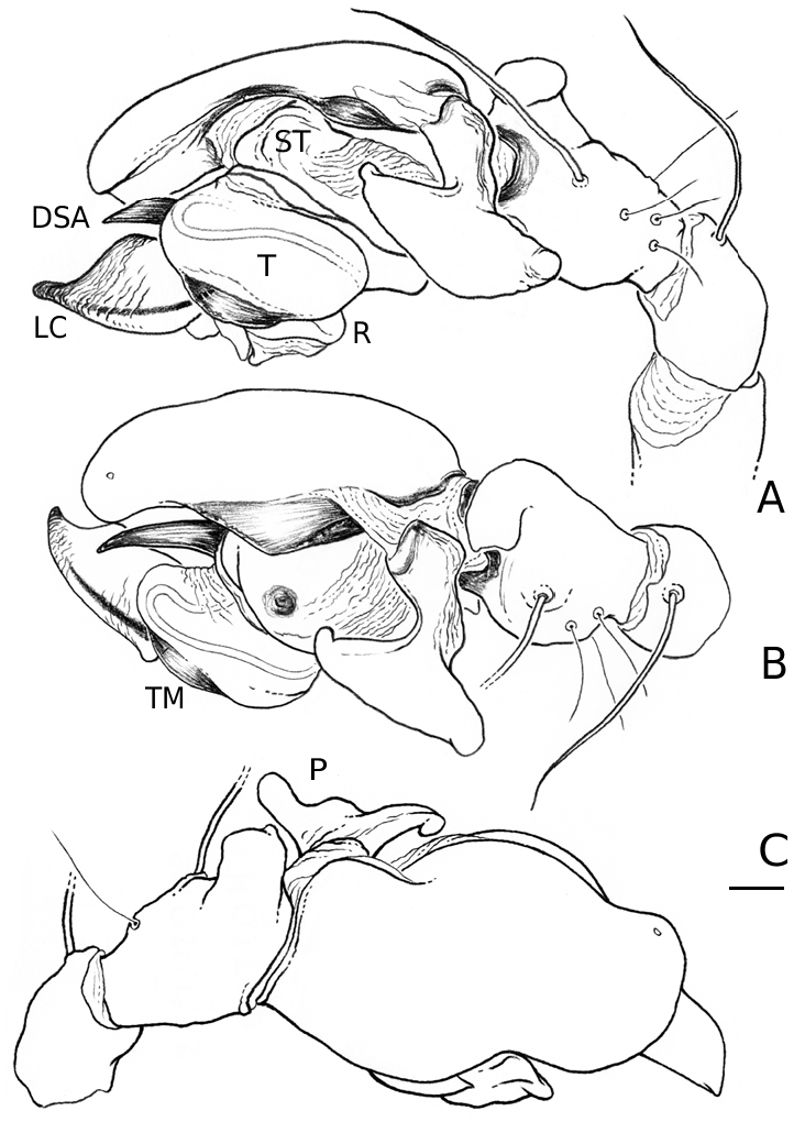
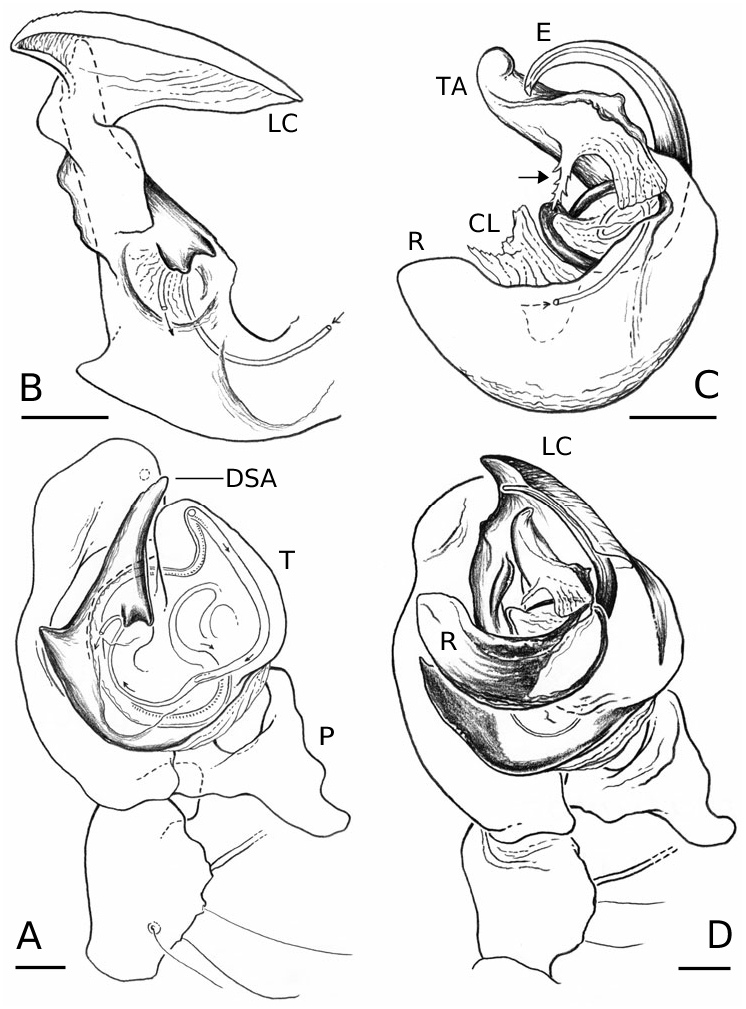
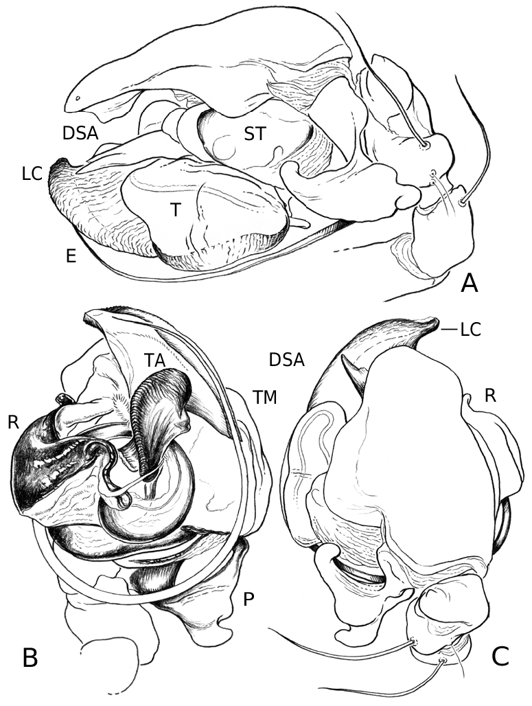
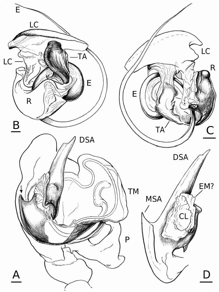
Male palp
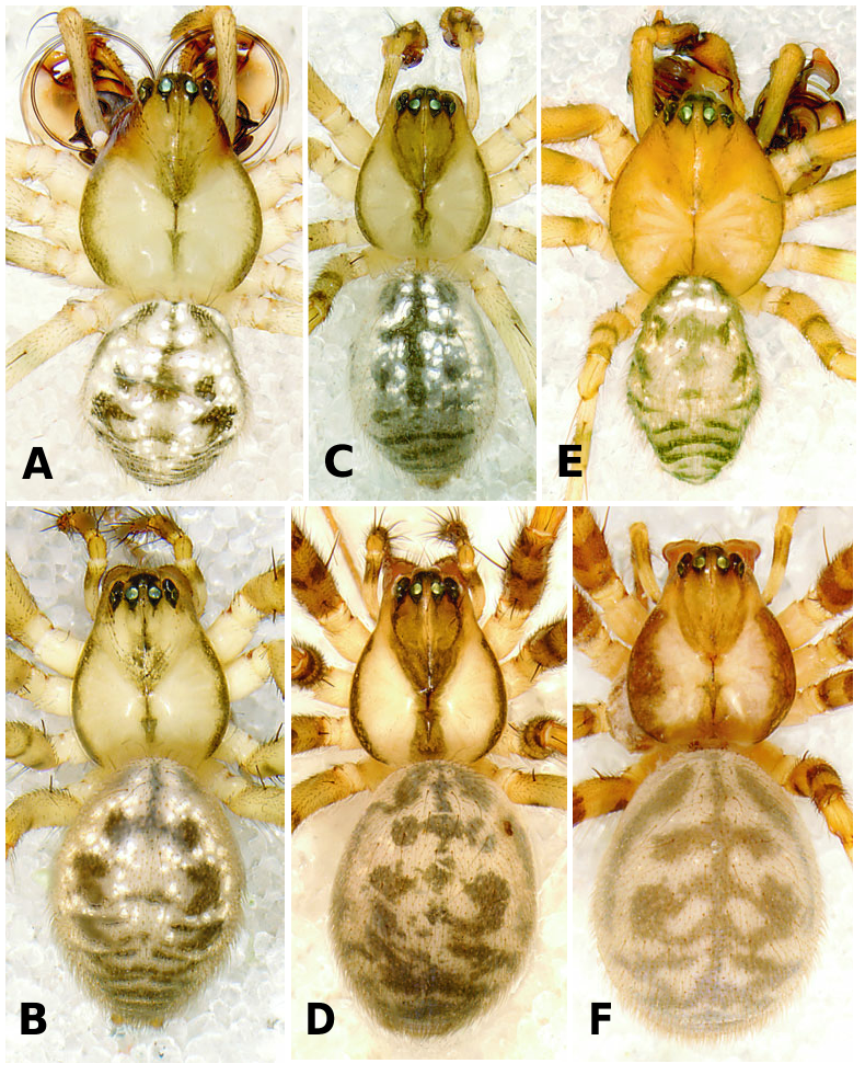
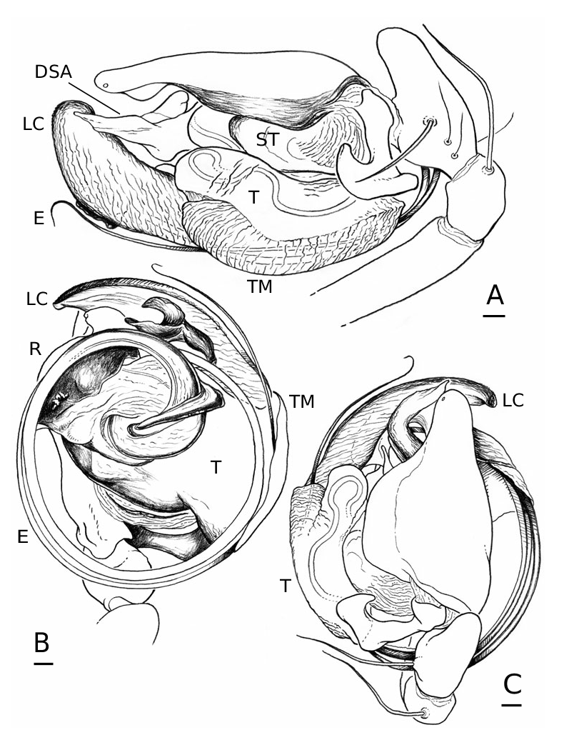
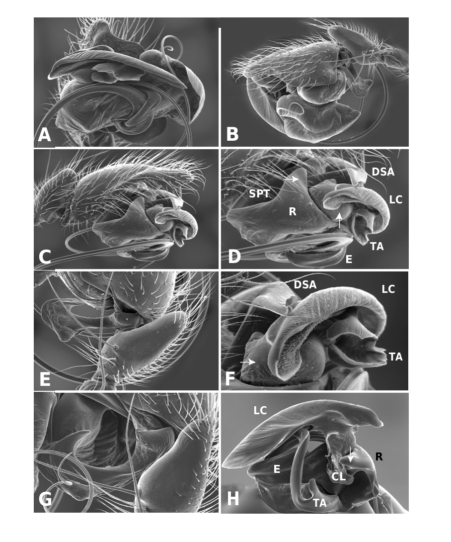
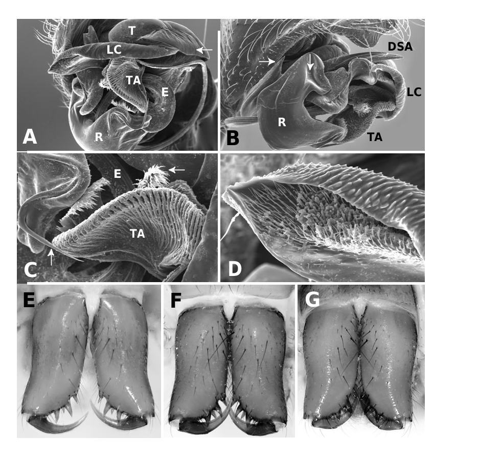
In L. thoracica ( Fig. 1A View Figure 1 ) and L. machadoi sp. nov. ( Fig. 1E View Figure 1 ) the size of the male palps, relative to body size, is distinctively large; in L. flahaulti ( Fig. 1C View Figure 1 ) they are considerably smaller. Palpal patella unmodified. Tibia with a dorsal apophysis whose morphology varies across the species ( Figs 5A View Figure 5 , 8A View Figure 8 , 11A View Figure 11 ). Cymbium, as seen in dorsal view ( Figs 5C View Figure 5 , 8C View Figure 8 , 11C View Figure 11 ), sinuous and with a thin membranous ectal margin that overhangs the subtegulum and the basal part of the tegulum (e.g. Fig. 5C View Figure 5 ). Tarsal organ ectoapical. Paracymbium connected to the cymbium-tibia membrane at the base of the cymbium ( Fig. 5C View Figure 5 ) via a membrane at the apex of the proximal paracymbial branch. Paracymbium with a caudally projecting apophysis in its median curvature whose morphology is species specific ( Figs 5A View Figure 5 , 8A View Figure 8 , 9A View Figure 9 , 11A–C View Figure 11 , 16B View Figure 16 , 21F). Subtegulum circular. Tegulum elongated, provided with a membranous and transparent flap (termed here ‘tegular membrane’) running along its ectal margin, most easily seen in an ectal view, and present in all species but particularly conspicuous in L. thoracica ( Figs 5A View Figure 5 , 6A View Figure 6 , 17A – arrow left). In ventral view, the tegulum appears ectally projected (especially in L. thoracica and L. machadoi sp. nov.), with an anteriorly directed lobe on its apical region ( Figs 6A View Figure 6 , 9A View Figure 9 , 12A View Figure 12 ). Spermduct running along the external margin of the tegulum, with a loop in the anterior lobe ( Figs 6A View Figure 6 , 9A View Figure 9 , 12A View Figure 12 ). Spermduct diameter decreases as it enters the suprategulum; in L. machadoi sp. nov. the trajectory also shows a kink ( Fig. 12A View Figure 12 ).
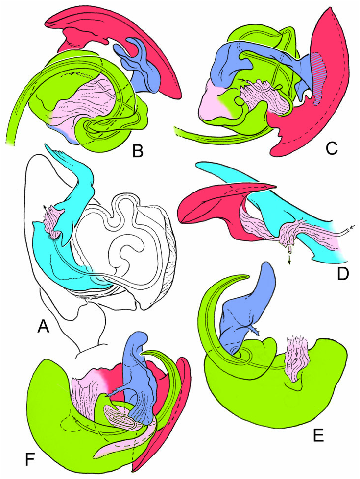
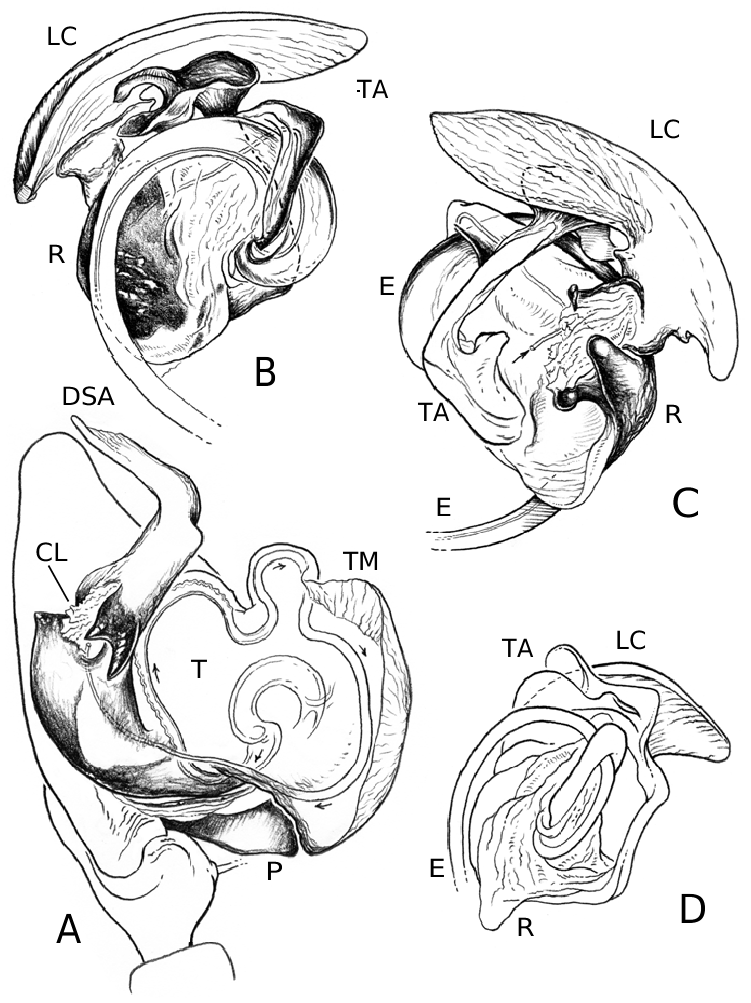
Suprategulum large and conspicuous (e.g. Fig. 4A, D View Figure 4 – blue) with a pointed distal apophysis that reaches the cymbial apex (in ectal view). Suprategulum heavily sclerotized, except for a small region, anterior to the suprategular foramen, that is only slightly sclerotized and shows a clear posterior margin ( Figs 6A View Figure 6 , 9A View Figure 9 , 12A View Figure 12 ). All species provided with a small pointed apophysis adjacent to the column, mesal to the suprategular foramen (marginal suprategular apophysis; Figs 6A View Figure 6 , 9A View Figure 9 , 12A, D View Figure 12 – arrow, 17A). On its ectal margin, also adjacent to the column, all species have a distinctive and morphologically similar bifid suprategular apophysis (Fig. 17F). No embolic membrane can be discerned as such (but see Discussion). Radix large and with a highly sclerotized core surrounded by membranes connecting it to other embolic division sclerites. All species have a sclerotized radical process that projects mesally ( Figs 6C View Figure 6 , 9D View Figure 9 , 11B View Figure 11 , 12C View Figure 12 , 16D, H View Figure 16 , 21D, 22B). Embolus length and morphology highly variable across species, but always turning anticlockwise (left palp, ventral) in a plane parallel to the cymbium. In L. thoracica the embolus makes about two and a quarter turns ( Fig. 5B View Figure 5 ), in L. machadoi sp. nov. about one complete turn ( Fig. 11B View Figure 11 ) and in L. flahaulti ( Fig. 9D View Figure 9 ) less than half a turn; in the first two species the embolus is filiform and has an internal membranous margin. A minute filiform membrane projects from the membranous region of the base of the embolus in L. thoracica (Fig. 17B, D) and L. machadoi sp. nov. ( Figs 11B View Figure 11 , 22A, C View Figure 22 ). In the latter species this filiform process can be easily discerned with a dissecting stereoscope; in the former this is barely possible and SEM is required. A homologous structure was not found in L. flahaulti .
Lamella characteristica large and conspicuous, of semi-membranous and partially transparent appearance ( Figs 5A View Figure 5 , 6B–D View Figure 6 , 8A View Figure 8 , 9B View Figure 9 , 11A View Figure 11 , 12B, C View Figure 12 , 16H View Figure 16 ). The ectal margin of the lamella is folded ventrally and in the unexpanded palp it contains along its length the distal part of the embolus (Fig. 21H). The apical end of the embolus is housed in the apical end of the lamella; the internal surface of the latter is covered with piliform papillae, densely distributed in L. thoracica (Fig. 17G) and L. machadoi sp. nov. ( Fig. 22D View Figure 22 ) and sparsely in L. flahaulti (Fig. 21H). The external surface of the apical region of the lamella is covered with small denticles (Fig. 17G). The membranous connection between the lamella and the radix is in the anterior radical region ( Figs 6C View Figure 6 , 9B View Figure 9 , 16C View Figure 16 ). The morphology of the terminal apophysis is highly variable across species (Figs 17C, 21G, 22C), but it always connects to the radix in its dorsal anterior region. The terminal apophysis consists of an arm that runs along the longitudinal axis (dorsally) of the palp and that has a distinctive process at its apical end. It also has a membranous connection to the radix at the proximal end. In L. thoracica ( Fig. 6C View Figure 6 ) and L. machadoi sp. nov. ( Fig. 12C View Figure 12 ) the distal process is larger than in L. flahaulti ( Fig. 9C View Figure 9 ) but in all three species the distal process has a membranous or blade-like posterior part. In addition, the latter two species have a plumose process in the terminal apophysis (Figs 21G, 22C).
No known copyright restrictions apply. See Agosti, D., Egloff, W., 2009. Taxonomic information exchange and copyright: the Plazi approach. BMC Research Notes 2009, 2:53 for further explanation.
|
Kingdom |
|
|
Phylum |
|
|
Class |
|
|
Order |
|
|
Family |
|
|
Genus |