
Micropora mawatarii, Arakawa, 2016
|
publication ID |
https://doi.org/10.12782/sd.21.1.009 |
|
publication LSID |
lsid:zoobank.org:pub:61308999-8455-4892-8464-423FFBACF0A1 |
|
DOI |
https://doi.org/10.5281/zenodo.5526911 |
|
persistent identifier |
https://treatment.plazi.org/id/E0C328AA-8157-4BBB-8C52-7278A3F1735E |
|
taxon LSID |
lsid:zoobank.org:act:E0C328AA-8157-4BBB-8C52-7278A3F1735E |
|
treatment provided by |
Felipe |
|
scientific name |
Micropora mawatarii |
| status |
sp. nov. |
Micropora mawatarii sp. nov.
( Figs 2–3 View Fig View Fig )
Micropora coriacea View in CoL (not of Esper in Johnston, 1847): Sakakura 1935: 11, pl. 2, fig. 8; Mawatari 1952: 274, fig. 9; Arakawa 1995: 81 (only listed); Nishizawa 1997: 152 (only listed); Arakawa 1999: 56 (in part).
Material examined. Holotype: NMNS PA 16827 View Materials (five fragments, one colony), Jizodo Formation, Pleistocene, Nishiyatsu , Chiba Prefecture, Japan . Paratypes: NMNS PA 16828 View Materials (on razor clam shell), Jizodo Formation, Pleistocene, Nishiyatsu , Chiba Prefecture, Japan; NMNS PA 16829 View Materials (eight fragments), Station 1748, Hakurei-Maru cruise GH80-2 . Other material examined: NMNS PA 16830 View Materials (two fragments), and SGBC-0371 (nine fragments), Yabu Formation , Pleistocene , Neriki , Chiba Prefecture, Japan; NMNS PA 16831 View Materials (on mollusc shell), Jizodo Formation , Pleistocene , Oi , Chiba Prefecture, Japan; NMNS PA 16832 View Materials (on barnacle plate), Shimoda , Shizuoka Prefecture, Japan; SGBC-0403 (not coated with metal), Station 1746, Hakurei-Maru cruise GH80-2; SGBC-0404 (on large gastropod, not coated with metal), Ohara , Chiba Prefecture, Japan; SGBC-0406 (three fragments), Yabu Formation , Pleistocene , Semata , Chiba Prefecture, Japan; SGBC-0408 (not coated with metal), Jizodo Formation , Pleistocene , Kawazai , Chiba Prefecture, Japan; SGBC-0409 (not coated with metal), Jizodo Formation , Pleistocene, Nagara Dam, Chiba Prefecture, Japan . See Table 1 View Table 1 for coordinates and depths of cruise samples.
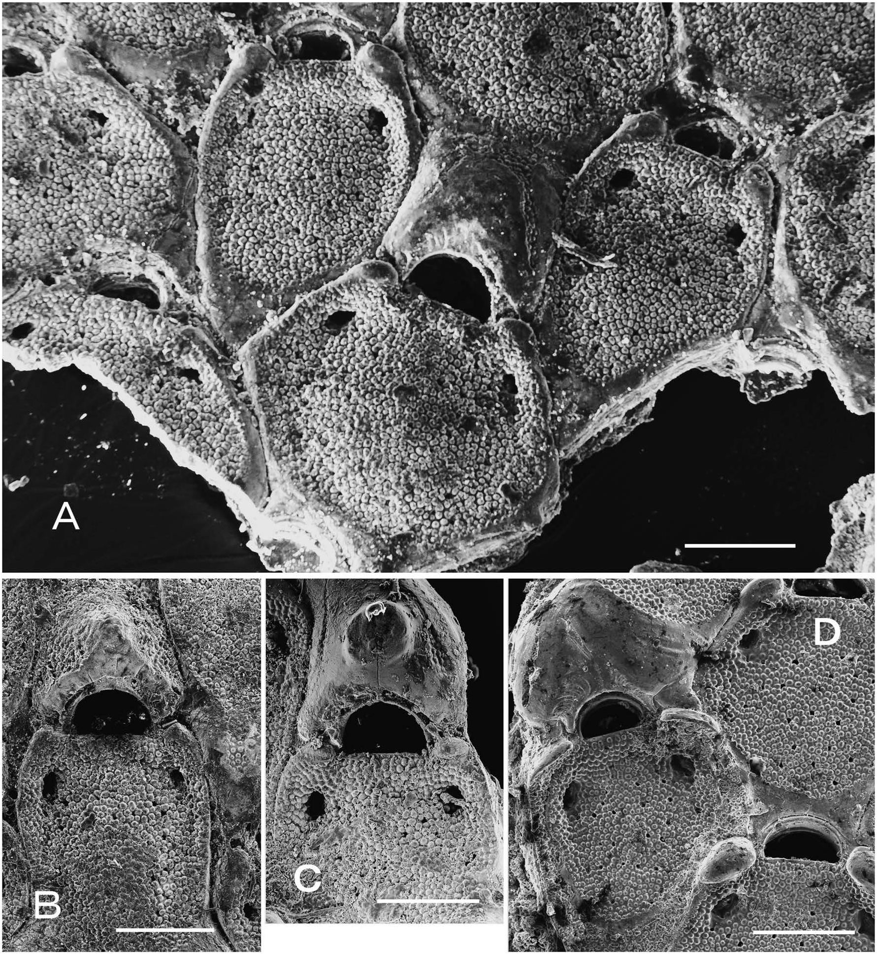
Diagnosis. Zooids with small zone of proximal gymnocyst, salient knobs beside proximolateral corners of orifice, and elliptical or circular opesiules. Orifice dimorphic, larger in ovicellate zooids. Spines and avicularia lacking. Ovicell distally granulate, with smooth proximal area; sometimes almost entirely smooth, with large umbo.
Etymology. Named for the Japanese bryozoologist Dr Shizuo Mawatari, who reported and illustrated this species from the Kii Peninsula ( Mawatari 1952).
Measurements (in milimetres). NMNS PA 16827, 16828, 16829, 16830, 16831, 16832, and SGBC-0371. Autozooids ( n =475, 27): ZL, 0.30–0.86 (0.574±0.100); ZW, 0.23–0.72 (0.437±0.071); OrL, 0.04–0.11 (0.067±0.010); OrW, 0.09–0.17 (0.126±0.015). Ovicellate zooids ( n =43, 16): ZL, 0.51–0.81 (0.661±0.075); ZW, 0.35–0.62 (0.457±0.061); OrL, 0.06–0.11 (0.087±0.012); OrW, 0.12– 0.19 (0.150±0.015). Ovicells ( n =47, 4): OvL, 0.21–0.33 (0.275±0.032); OvW, 0.23–0.38 (0.314±0.031).
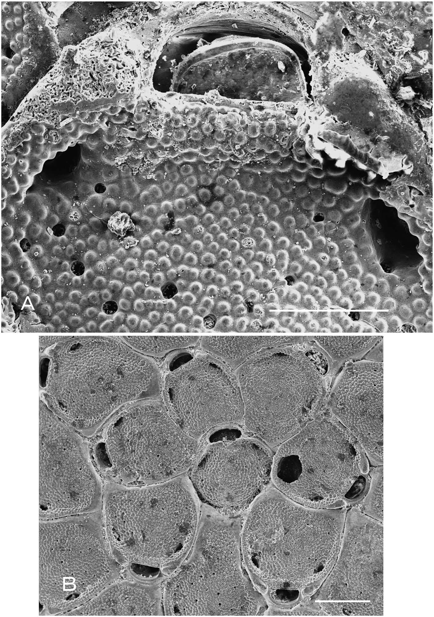
Description. Colony encrusting, unilaminar, multiserial sheet; maximum observed size in largest dimension 7.9 mm (NMNS PA 16831). Zooids elliptical to subhexagonal, irregular in outline ( Fig. 2A View Fig ); zooidal size also much variable, smaller in zone of changing growth direction (NMNS PA 16831, 16832) except for astogenetic change zone (NMNS PA 16828). Frontal shield slightly convex, with granular surface, surrounded by mural rim; frontal pores small, scattered, generally with radiating spicules ( Fig. 3A View Fig ). Paired smooth, elliptical knobs situated beside proximolateral corners of orifice, up to about 0.13 mm high ( Fig. 2D View Fig ). Opesiules small, elliptical or circular, generally situated at each distolateral corner of sunken cryptocystal area. Orifice semielliptical, wider than long, dimorphic, larger and more semicircular in ovicellate zooids ( Fig. 2A View Fig ). Oral spines lacking. Surface of operculum granular, rimmed distally ( Fig. 3A View Fig ). Ovicell raised, oπen with large umbo, initially with granular cryptocystal texture except in smooth, thickened, chevron-shaped or semilunar proximal area; with age, sometimes covered almost entirely by smooth calcification, being continuous with proximal gymnocyst of next zooid ( Fig. 2 View Fig A–D). Avicularia lacking. Two basal pore chambers in each distolateral wall and one in transverse wall, each leading to several septula. Ancestrula ( Fig. 3B View Fig ) like autozooids but smaller (0.30× 0.31 mm), wider than long, budding one zooid distally and two distolaterally, surrounded by total of seven zooids. Reparative intramural buds inside damaged or degenerated zooids occurred in all of measured specimens except NMNS PA 16828, with frequency of 9.3% repaired zooids among all zooids measured.
Distribution. This species occurs subtidally off the Kii, Izu, and Boso Peninsulas along the Pacific coast of Japan. Kataoka (1960) and Mawatari (1963) also reported Micropora coriacea from the Japan Sea, and so M. mawatarii may be more widely distributed, if these authors’ determinations are correct. Recent specimens from the continental shelf in this study ranged in depth from 65 m (NMNS PA 16829, Station 1748) to 154 m (SGBC-0403, Station 1746), although the latter specimen was poorly preserved and may have been transported.
The geological range of this species is Pleistocene to Recent. Hayami (1970, 1975, 1976) reported M. coriacea from Miocene and Pliocene marine deposits in northern Japan, but only described and illustrated material from the Miocene Kaigarabashi Sandstone in Hokkaido ( Hayami 1970). Although Hayami’s species is similar to M. mawatarii , the identity of her material remains unclear, as she did not mention orifice dimorphism or illustrate the ovicells ( Hayami 1970: 324, pl. 36, fig. 19). The orifice measurements (L= 0.12 mm, W= 0.20 mm) in her specimens are larger than those of M. mawatarii .
Remarks. This species resembles European Micropora coriacea in lacking avicularia, and in having latero-oral knobs and umbonate ovicells ( Hayward and Ryland 1998: figs 98, 99C and D), and was previously identified as such by Japanese researchers. However, Micropora mawatarii differs from M. coriacea in having zooids typically with a zone of smooth proximal gymnocyst, marked dimorphism in orifice size and shape between ovicellate and non-ovicellate zooids, and more widely separated frontal pores; and in occasionally lacking an umbo on the ovicell. The ancestrula of M. mawatarii also appears to differ from that of M. coriacea ; Waters (1925: pl. XXI, fig. 3) illustrated an ancestrula (or first-generation periancestrular zooid) for the latter from Hastings, England, that is unlike later autozooids and instead has a large opesial opening without opesiules.
Among the species of Micropora from the Pacific and adjoining seas ( Table 2 View Table 2 ), Micropora rimulata Canu and Bassler, 1929 and Micropora plana sp. nov. (both described herein) resemble M. mawatarii in having dimorphic orifices and a triangular or chevron-shaped proximal area on the ovicell, but M. rimulata differs in having avicularia. Differences from M. plana are given in the Remarks for that species. Micropora selknami Moyano, 1994 from the Strait of Magellan has similarly well-developed latero-oral knobs and a triangular proximal area on the umbonate ovicell, but it bears avicularia ( Moyano 1994a).
| NMNS |
National Museum of Natural Science |
No known copyright restrictions apply. See Agosti, D., Egloff, W., 2009. Taxonomic information exchange and copyright: the Plazi approach. BMC Research Notes 2009, 2:53 for further explanation.
|
Kingdom |
|
|
Phylum |
|
|
Class |
|
|
Order |
|
|
SubOrder |
Neocheilostomina |
|
InfraOrder |
Flustrina |
|
SuperFamily |
Microporoidea |
|
Family |
|
|
Genus |
Micropora mawatarii
| Arakawa, Shinji 2016 |
Micropora coriacea
| Arakawa, S. 1999: 56 |
| Nishizawa, Y. 1997: 152 |
| Arakawa, S. 1995: 81 |
| Mawatari, S. 1952: 274 |
| Sakakura, K. 1935: 11 |