
Crassiplana albatrossi Hyman 1955
|
publication ID |
https://doi.org/10.5281/zenodo.201139 |
|
DOI |
https://doi.org/10.5281/zenodo.6189881 |
|
persistent identifier |
https://treatment.plazi.org/id/DA758791-6814-FFEE-FF30-FF1B3010A687 |
|
treatment provided by |
Plazi |
|
scientific name |
Crassiplana albatrossi Hyman 1955 |
| status |
|
Crassiplana albatrossi Hyman 1955 View in CoL
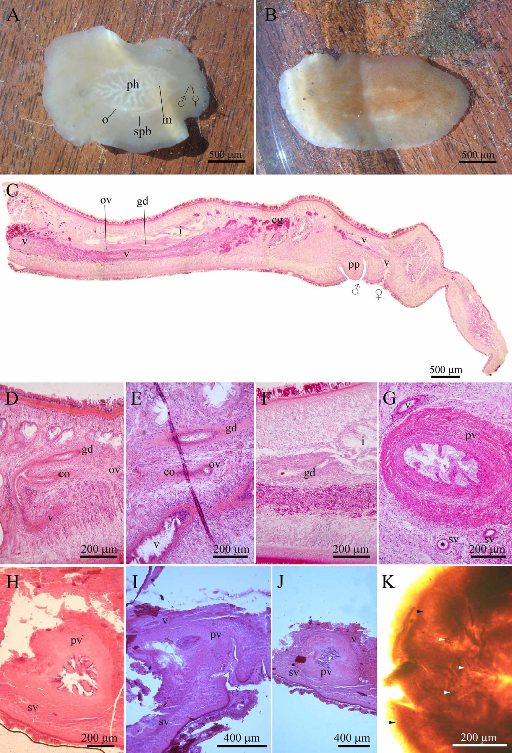
Studied material: Holotype, USNM Catalog number 26928. Three slides. Other material: Specimens from the northern coast of Patagonia, Argentina, MLP (Table 1, Figure 1 View FIGURE 1 ).
Description. Live specimens are of a light brown color ( Figure 3 View FIGURE 3 A–B). Body ovate, with frilled edges. Fixed specimens studied from Patagonia are approximately 23 mm long and 16 mm wide. Epidermis is ciliated and carries high cells with a strong basal membrane. Body wall contains four muscular layers from outside to inside: a thin external circular layer, a longitudinal layer, a diagonal layer formed of fibers in two orientations, and an inner circular fasciculate layer. Parenchyma showing bundles of dorsoventral muscles.
One pair of tentacles in brain region ( Figure 3 View FIGURE 3 B). Tentacular, brain, and marginal eyes present; the latter abundant in the anterior region of body and decreasing in number posteriorly to disappear beyond mid-length of body. Sparse eyes present in region anterior to brain.
Large folded pharynx placed at mid-length of body ( Figure 3 View FIGURE 3 A). Mouth placed near posterior end of pharynx.
Gonopores: separate, placed near posterior end of body and very close to each other.
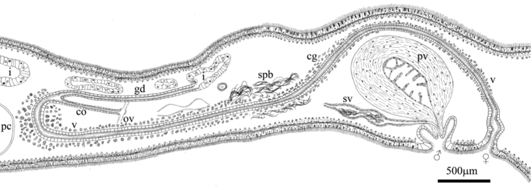
Deferent ducts ventral, widening towards their distal end, where they twist and form the spermiductal vesicles; spermiductal vesicles thin-walled, and full with spermatozoids in adult specimens. Tubular seminal vesicle with muscular wall ventral to prostatic vesicle ( Figures 2 View FIGURE 2 , 3 View FIGURE 3 G). Seminal vesicle merging into ejaculatory duct, which joins prostatic duct. Penis papilla blunt and strongly muscular, opening into male gonopore ( Figure 3 View FIGURE 3 C). Large free prostatic vesicle strongly muscular; wall projecting towards lumen, rendering a tubular shape. Prostatic glands crossing muscular wall ( Figure 3 View FIGURE 3 G).
Female gonopore behind male gonopore, originating a muscular vagina inlaid with numerous glands throughout its length; inner surface ciliated ( Figures 2 View FIGURE 2 , 3 View FIGURE 3 C). Distal portion of vagina vertical, turning forward and then dorsal to prostatic vesicle. Anterior to the prostatic vesicle, the vagina is placed ventrally and stretches anteriorly almost to the pharynx ( Figures 2 View FIGURE 2 , 3 View FIGURE 3 C–D); at this level it turns dorsally and then backwards, in a half-moon shape. Vagina receiving common oviduct ( Figures 2 View FIGURE 2 , 3 View FIGURE 3 D–E). Lang’s vesicle absent. Genito-intestinal duct dorsal to common oviduct. Junction of genito-intestinal duct and lumen of intestine close to prostatic vesicle ( Figures 2 View FIGURE 2 , 3 View FIGURE 3 F).
Remarks. The holotype, as mentioned in the original description by Hyman (1955), is poorly preserved. However, diagnostic structures such as the presence of nuchal tentacles, tentacular, brain and marginal eyes, the structure (shape, size, lumen) of the prostatic vesicle, the dorsal vagina and the ventral seminal vesicle in the holotype ( Figure 3 View FIGURE 3 H–K) confirm that the newly collected specimens are Crassiplana albatrossi . The holotype also shows spermiducal vesicles.
The internal lining of the large prostatic vesicle is not smooth —neither in the holotype ( Figure 3 View FIGURE 3 H–J) nor in the Patagonian material ( Figure 3 View FIGURE 3 G)— as depicted in the original description (p. 15, fig. 22), but instead projects into the lumen. The presence of a smooth lining ( Hyman, 1955) originally allows the placement of this genus in the Callioplanidae ( Faubel, 1983) . However, the presence of well-developed crests in the holotype and the material from Patagonia suggests that Crassiplana does not belong to that family.
Of the female genitalia, the only part preserved in the holotype is the independent gonopore and the vagina that is dorsal and anterior to the male copulatory apparatus ( Figure 3 View FIGURE 3 I–J). The anterior part of the vagina and its junction with the oviducts are not observed. Hyman (1955) doubtfully mentioned the junction of oviducts and the presence of Lang´s vesicle. These two structures are not observed in the holotype. The material from Patagonia allows describing the anterior tract of the vagina, and also the opening of oviducts into the vagina and the presence of a genito-intestinal duct.
Contrary to what can be observed in the Patagonian material, Hyman (1955) described marginal eyes completely surrounding the body. This feature could not be observed in the holotype, as only the anterior part of the body is mounted in toto .
The shape and position of the pharynx as well as the male copulatoty apparatus and prostatic vesicle, allow C. albatrossi to be placed confidently in the family Pseudostylochidae . Within this family, the genus Idioplana Woodworth 1898 , has female genitalia dorsal to the male one. This peculiar feature is also observed in Crassiplana . However, Idioplana has an anchor-shaped Lang vesicle, while this vesicle is missing in Crassiplana , which has a conspicuous genito-intestinal duct.

No known copyright restrictions apply. See Agosti, D., Egloff, W., 2009. Taxonomic information exchange and copyright: the Plazi approach. BMC Research Notes 2009, 2:53 for further explanation.
|
Kingdom |
|
|
Phylum |
|
|
Class |
|
|
Order |
|
|
Family |
|
|
Genus |