
Auplopus cf. rufipes (Banks, 1946)
|
publication ID |
https://doi.org/ 10.11606/1807-0205/2020.60.55 |
|
persistent identifier |
https://treatment.plazi.org/id/DB044071-861F-FFE0-92DF-EC9CD640F599 |
|
treatment provided by |
Carolina |
|
scientific name |
Auplopus cf. rufipes |
| status |
|
Auplopus cf. rufipes View in CoL
Nests of this species were sparsely collected in the studied area. They were built in bamboo cane traps that ranged from 12 to 16 mm in diameter and were approximately 15 cm long. Five nests were collected between April/2017 and February/2019 ( Table 1). The nest collected in April/2017 spawned two specimens, a male and a female; from the nests collected in May/2017 all wasps died in the pupal stage. From the nests collected in September/2017, three males and a female emerged 20 days after retrieval from the field ( Tables 1 and 2). Additionally, one larva died due to handling and anoth- er one for unknown reasons. In 2018, only one nest was collected in November, from which emerged one male and a specimen of cuckoo wasp Caenochrysis crotonis (Ducke) ( Hymenoptera : Chrysididae ) emerged. Other four immatures died from unknown causes.
Direct observations of female nesting activities were frustrated given their aggressive behaviour whenever attempts to record the foundress activity closely were made,causing them to stop working on the nests.In such events, the females would face the unwelcome observer, flapping their wings against the trap floor and extending their metasoma, as if preparing to sting. This menacing posture behaviour was only witnessed twice, since the observed nests were unattended most of the time. Prey carrying behaviour, unfortunately, could not be seen.
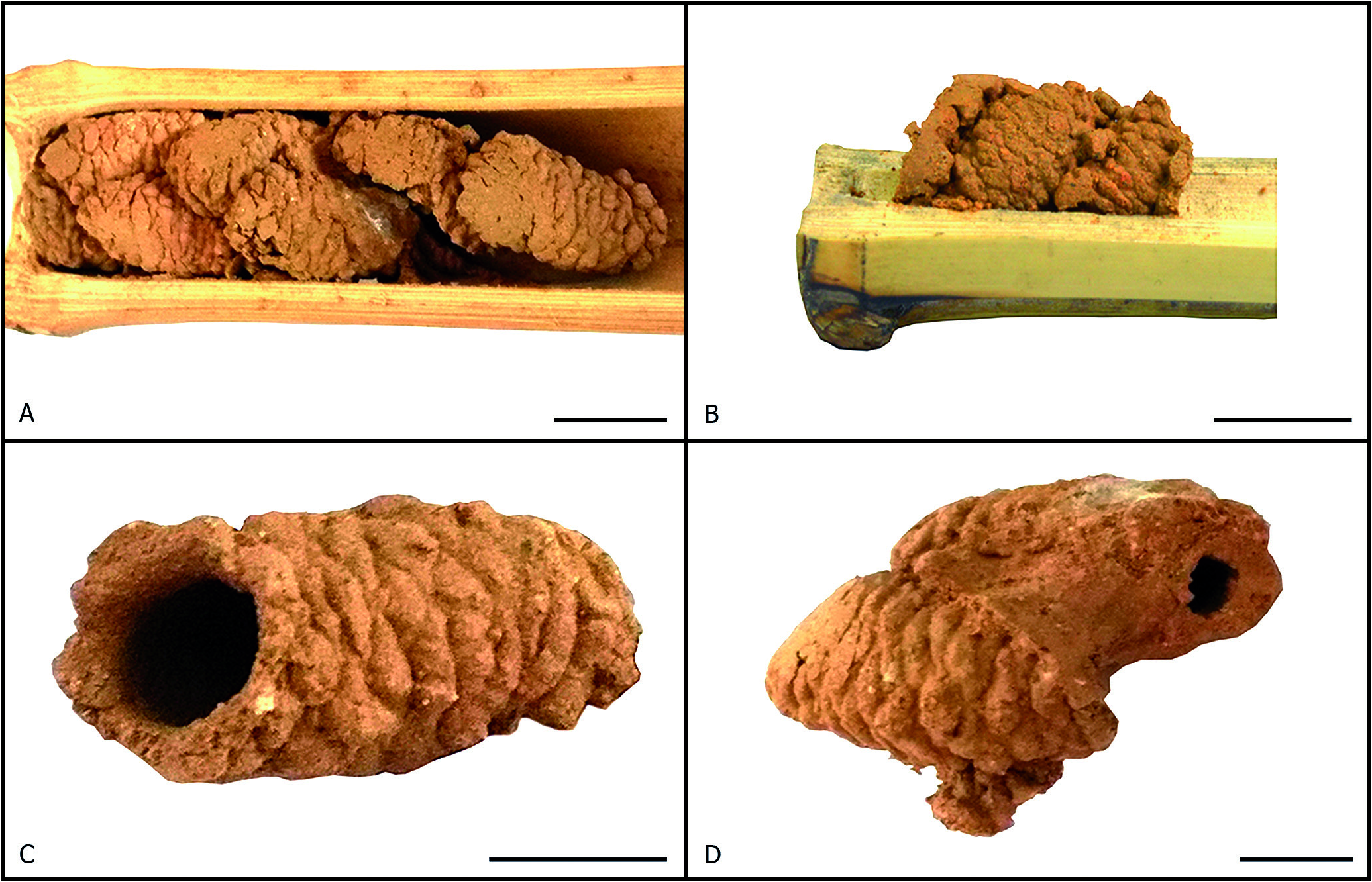
The nests of this species were made of mud. Moist mud droplets were stacked, creating a papillated outer surface for the nest cells. After some time, the mud dried and became cemented. Measurements for brood cells are summarised in Table 3. Most nests were built near the bottom of the trap, except for one that was built detached from the trap bottom by about 4 mm, leaving a small empty space. The first cells were built sequentially, occupying only half of the trap’s width. As the nest grew, other brood cells were added next to the first row, occupying the remaining space. Nests with more than two cells show a typical“V” formation when seen from above ( Fig. 2A View Figure 2 ). The second set of nests, embedded between the first ones, had their entrances directed upward, filling all the space. When the two halves of the trap were separated, part of the mud that formed the nest was adhered to its superior half. In addition, the cells were tightly glued together, in a way that the entire nest may be removed from the trap.
Empty spaces of the nests were covered by one lay- er of mud, which partially enveloped all the brood cells ( Fig. 2B View Figure 2 ) creating an overlay. Although most of the exposed surface of each brood cell was papillated ( Fig. 2C View Figure 2 ), the regions in contact with the trap walls or other brood cells were smooth ( Fig. 2D View Figure 2 ). The inner surface of each brood cell was smooth, with almost no wrinkle. Brood cells were cylindric-oblong and they were tilted about 80° related to the substrate ( Fig. 2B View Figure 2 ). Unfinished cells had a structure binding the entrance orifice, similar to a “lip” ( Fig. 2C View Figure 2 ). Brood cells colour was uniform.
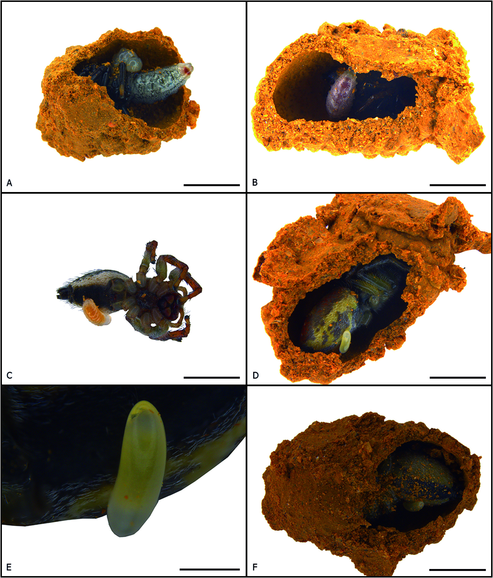
One of the nests collected was opened to identify the prey hunted by the foundress wasp. All spiders stocked as food for the larvae belonged to the species Frigga cf. quintensis (Tullgren) (Salticidae) . Six cells were analysed which contained two males, three females and a juvenile, which could not be sexed. Figure 3 View Figure 3 depicts the different stages of the immatures from the innermost ( Fig. 3A View Figure 3 ) to the outermost cell ( Fig. 3F View Figure 3 ) of the opened nest. The spiders were placed with their abdomen facing the posterior part of the brood cell, the cephalothorax facing the opening of the cell, and only the posterior-most pair of legs were amputated near the coxa-trochanter joint ( Fig. 3C View Figure 3 ). The egg was laid on the ventral surface of the spider’s opisthosoma, dislocated to the right ( Figs. 3 View Figure 3 D-F). The larva started to feed in the region where the egg was laid, sucking the prey’s internal fluids. Upon reaching the third instar, the larva consumed the prey entirely and, after the fourth instar, it pupated, but did not spin a cocoon. Subsequently, the adult emerged by chewing through the cell operculum, making a small hole.
No known copyright restrictions apply. See Agosti, D., Egloff, W., 2009. Taxonomic information exchange and copyright: the Plazi approach. BMC Research Notes 2009, 2:53 for further explanation.