
Tribolonychus collyerae Zhang & Martin, 2001
|
publication ID |
https://doi.org/ 10.11646/zootaxa.3721.4.1 |
|
publication LSID |
lsid:zoobank.org:pub:AC2B4773-FC97-4C43-9A07-4D3C3848FBF4 |
|
DOI |
https://doi.org/10.5281/zenodo.6151559 |
|
persistent identifier |
https://treatment.plazi.org/id/DB303E64-FFEC-FFCA-FF6B-FE6DFD477F00 |
|
treatment provided by |
Plazi |
|
scientific name |
Tribolonychus collyerae Zhang & Martin, 2001 |
| status |
|
Tribolonychus collyerae Zhang & Martin, 2001 ( Figs. 1–37 View FIGURE 1 View FIGURE 2 View FIGURE 3 View FIGURE 4 View FIGURE 5 View FIGURE 6 View FIGURE 8 View FIGURE 9 View FIGURE 10 View FIGURE 11 View FIGURE 12 )
Tribolonychus collyerae Zhang & Martin, 2001: 321 , figs. 24–34.
Material examined
Holotype female (ZQZ990826-1A) and two paratype females (ZQZ990826-1B and C), collected by E. Collyer from Nothofagus sp. on 10 Oct. 1964, Lake Rotoroa, BP, New Zealand. Three females (ZQZ2001273A, B and C), one male (ZQZ20020404-1D) and four deutonymphs (ZQZ2001273D and E, ZQZ20020404-1A and B), collected by N.A. Martin from Nothofagus fusca on 1 Nov. 2001, Hinewai Reserve, Banks Peninsula, MC, New Zealand. Two females (ZQZ02366Z), collected by N.A. Martin from Nothofagus fusca on 14 Dec. 2002, Historic Place Carpark, Lake Rotoaira, Opotaka, TO, New Zealand. Two protonymphs and one larva, collected by N.A. Martin from Nothofagus sp. on 22 Jan 2002, Hinewai Reserve, Banks Peninsula, New Zealand. Three females (ZQZ2003317A, ZQZ2003317B, and ZQZ2003317D) and two males (ZQZ2003317C), collected by N.A. Martin from Nothofagus truncata on 2 Feb. 2003, Parapara Peak Tk, Golden Bay, New Zealand. Five females (05-696ZA, 05-696ZB, 05-696ZC, 05-696ZD and 05-696ZJ), four males (05-696ZE, 05-696ZF, 05-696ZG and 05-696ZH), two deutonymphs (05-696ZK, 05-696ZL), one protonymph (05-696ZKM) and two larvae (05-696ZKI), collected by N.A. Martin from Nothofagus fusca on 17 Aug. 2005, Hinewai Reserve, Banks Peninsula, MC, New Zealand.
Description
Female (n=9).
Dorsum ( Fig. 1 View FIGURE 1 ). Length of body from posterior end of idiosoma to anterior end of gnathosoma 474–517; length of idiosoma 354–411; maximum width at level of setae c 3 285–304. Idiosoma broadly truncate in anterior margin, with longitudinal medial striae anteriorly flanked by a narrow strip of transverse striae ( Fig. 3 View FIGURE 3 B) and posteriorly intercepted by V-shaped striae between setae c 1 ( Fig. 1 View FIGURE 1 ). Longitudinal medial striae beyond setae c 1 level posteriorly and not to d 1. V-shaped striae extending from c 1 to e 1 (Fig. 29). Striae between e 1 and h 1–2 transverse. Striae between e 1– f 1 trapezoidal pattern. Striae near setal base closer to each other and thinner than other dorsal striae (Fig. 29C).
Prodorsal setae well tapered and with small barbs (Fig. 7). Setae v 2 (26–35) the shortest, sc 1 (37–48) the longest, sc 2 (30–46) slightly or much shorter than sc 1 (Figs. 7, 30) among adult specimens. Among specimens from Lake Rotoroa (No. ZQZ990826-1A, B and C) and Golden Bay (No. ZQZ2003317A, ZQZ2003317B, and ZQZ2003317D) setae sc 2 (30–36, n=6) much shorter than sc 1 (37–45, n=6), while among specimens from Banks Peninsula (No. 05-696ZA, 05-696ZB, 05-696ZC, 05-696ZD and 05-696ZJ) setae sc 2 (42–48, n=3) slightly shorter than sc 1 (41–46, n=3)—the variations in the lengths of these setae are here considered intraspecific in nature, and attributed to the geographical position of the samples and different host plant species. Distances between setal bases: v 2– v 2 80 –93, sc 1– sc 1 113–131.
Hysterosoma with nine pairs of setae (c 1–3, d 1 – 2, e 1–2, f 1, h 1–2), dorsal setae on hysterosoma similar to prodorsal setae. Para-anal setae h 1 sub-terminal, h 2 terminal and h 3 located ventrally, setae h 2-3 slightly thinner than h 1 (Fig. 31). Length of setae: c 1 21–32, c 2 21–30, c 3 55–65, d 1 19–31, d 2 29–36, e 1 18 –28, e 2 19 –28, f 1 20–30, h 1 17–32, h 2 16–28, h 3 25–35. Distances between setal bases: c 1– c 1 56–77, d 1– d 1 51–57, e 1– e 1 44 –54, f 1– f 1 37–47, h 1– h 1 31–54, h 2– h 2 18–38.
Venter ( Fig. 2 View FIGURE 2 ). Surface finely striate, mostly transverse, coxal bases I–II smooth or with fine striae, coxal bases III–IV smooth. Setae 1a (43–50) and 3a (41–52) longer than distance 1a–1a (31–35) and 3a–3a (23–30) respectively. Setae 4a (42–50) shorter than distance 4a–4a (58–66). Length of seate: 1b 34–43, 1 c 30–47, 2 b 30– 38, 2 c 32–50, 3 b 37–47, 4 b 32–42.
Membranous plicated genital opening flanked by two pairs of genital setae and an anteromedial flap with transverse striae anterially and longitudinal striae posteriorly ( Fig. 32 View FIGURE 32 ).
Genital flap small with truncate or round posterior edge ( Fig. 32 View FIGURE 32 ). Two pairs of anal setae (ps) nude ( Fig. 3 View FIGURE 3 E), subequal in length (11 – 14). Two pairs of nude genital setae (g 1 & g 2) also subequal in length (16–24) ( Fig. 3 View FIGURE 3 E); setae g 1 about 2/3 as long as distance g 1 – g 1 (28 – 44). Pregenital setae (ag) nude, 35 – 39 long, ag – ag 53–55 ( Fig. 3 View FIGURE 3 E).
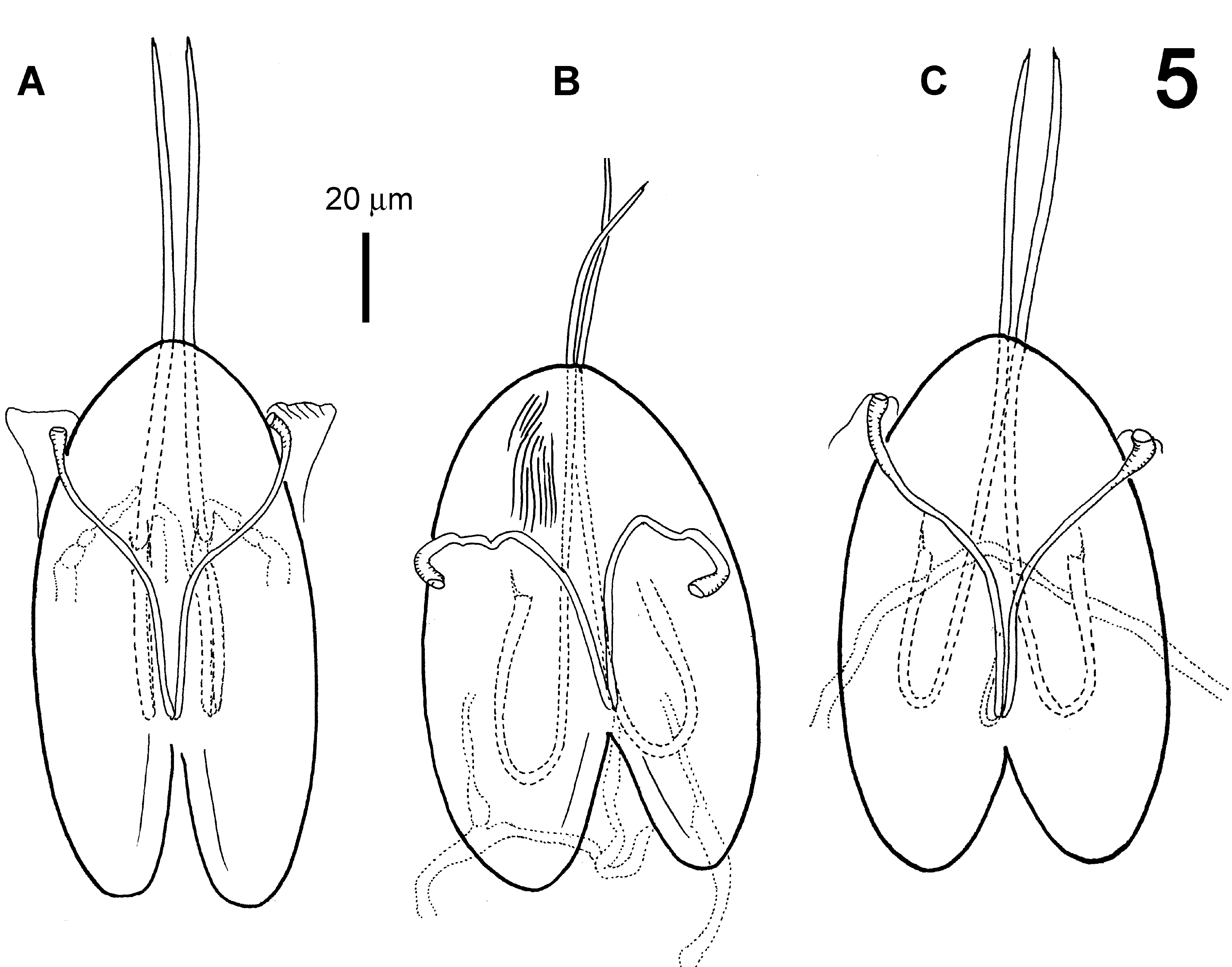
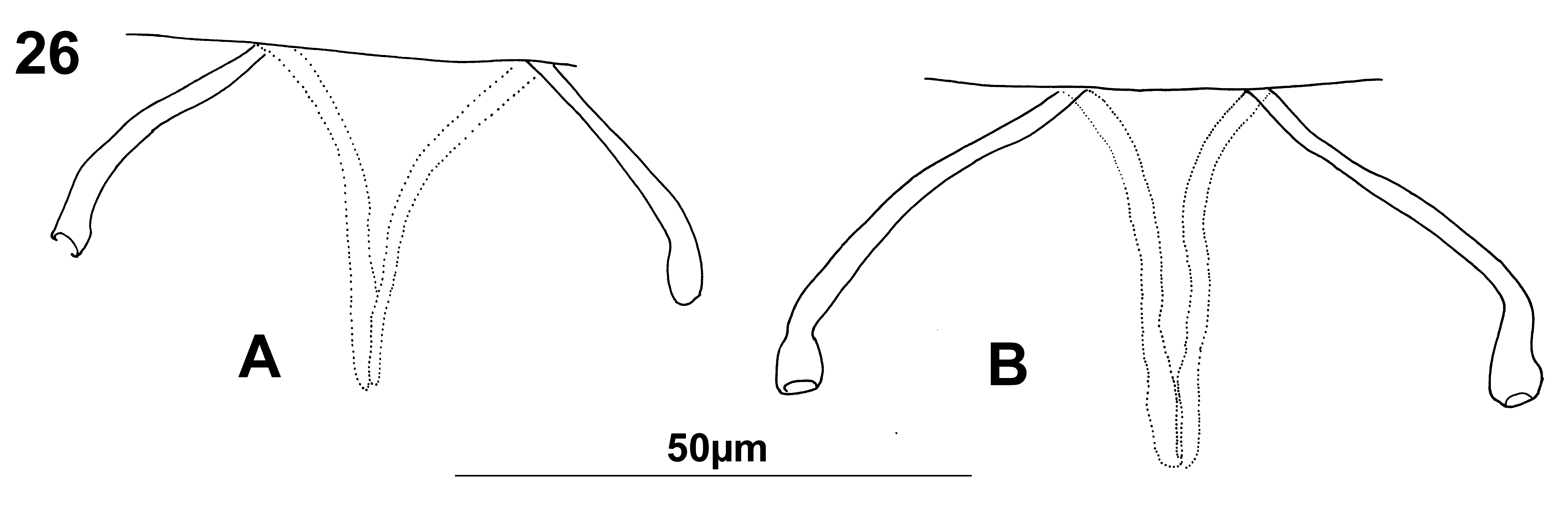
Gnathosoma ( Figs. 3 View FIGURE 3 A–D). Stylophore with longitudinal striae only in its distal third ( Fig. 3 View FIGURE 3 A). Length of chelicera slightly different among specimens ( Fig. 5 View FIGURE 5 ). Peritreme ending in small expansion ( Figs. 5 View FIGURE 5 and 26 View FIGURE 26 b showing various shapes reflecting how the specimens were pressed or extended when prepared). Subcapitular setae m smooth, length (23 – 34) shorter than distance m – m (33–47). Three pairs of adoral setae (or 1–3) conspicuous, spine-like, about 5 long ( Fig. 3 View FIGURE 3 C and 24C).
Palpi strongly built with base as long as combined length of femur-tarsus. Palp striate dorsally, but membranes between palpal segments punctate. Dorsal surface of palp base with a pair of conspicuous supracoxal setae (e), 3 to 4 long ( Fig. 3 View FIGURE 3 B). Seta d PFe well barbed and about twice as long as nearly smooth l″ PGe. All three palptibial setae nude; the shortest l ′ PTi (7 in length) located near base of tibial claw.
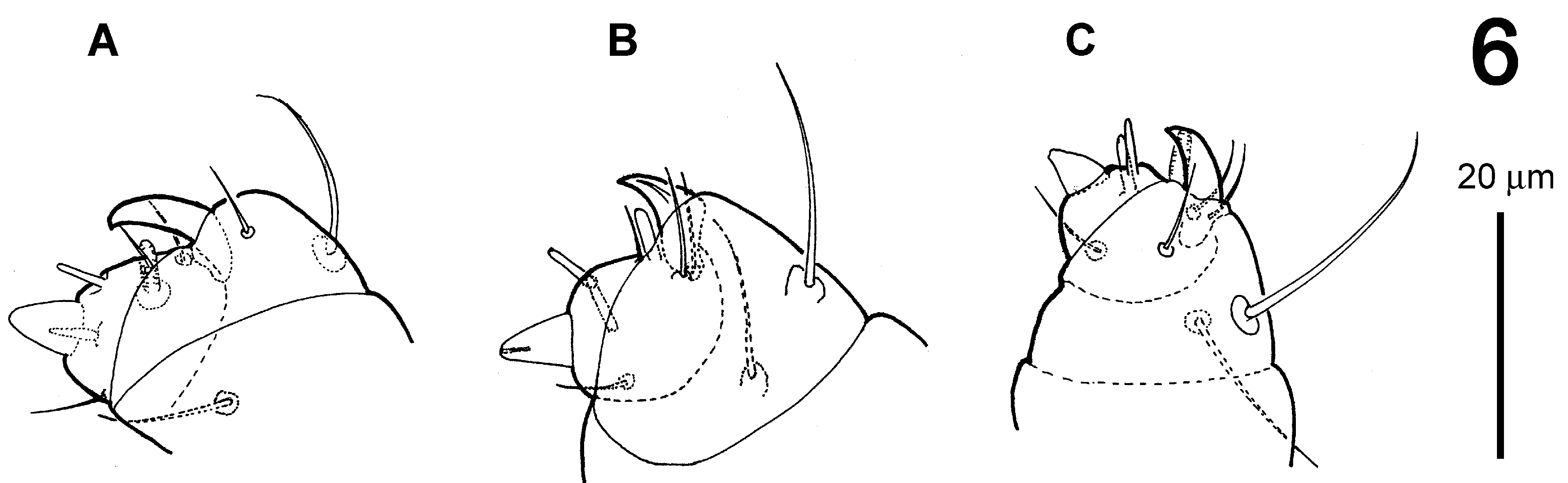
Palptarsus: Terminal eupathidium (suζ) sub-conical, 5–6 long and 4–5 in diameter at base ( Fig. 3 View FIGURE 3 D), and showing slightly difference among specimens ( Fig. 6 View FIGURE 6 ). Two lateral eupathia (ul ′ζ and ul″ζ) subequal in length (4– 5). Solenidion (ω) 5 long, about twice as long as wide. Three normal setae (a 5–6, b 5–6, c 6–7) nude, longer than solenidion.
Legs ( Fig. 4 View FIGURE 4 ). Number of solenidia on tarsi I–IV: 3-2-1-1, two pairs of duplex setae of tarsus I distal and adjacent. Tarsi I: proximal solenidion ω ′ 50–66, distal solenidion ω″ 73–84, ω″ 1 27–39 in length; tarsi II: ω″ 53–66 and ω″ 1 24–34 in length; tarsi III: ω ′ 59–73 in length; tarsi IV: ω ′ 59–71 in length. Number of normal setae on leg (I–IV) segments: trochanters 1-1-1-1; femora 9-6-3-2 but sometimes also 9-5-3-2 on one leg or both; genua 5-5-3- 2; tibiae 7-5-5-5; tarsi 9-7-6-6. Number of eupathidia on tarsi I-V: 3-3-0-0. Setae d of femur I well developed and located on a hump. Legs I–IV setation and notation as showing Fig. 4 View FIGURE 4 .
Number of solenidia (1–3) on tibia I varies among specimens, and differs between right and left legs in the same specimen. Addition of one or two solenidia (φ ′1 and φ ″1) normally present in males. Among 16 adult females including holotype 13 with one solenidia (φ) on both right and left tibiae I, one with two solenidia (φ, φ ′1) on left and two (φ, φ ″1) on right tibia I, one with three solenidia (φ, φ ′1, φ ″1) on left and two (φ, φ ″1) on right tibia I, one with two solenidia (φ, φ ″1) on right and one (φ) on left tibia I. ( Figs. 4 View FIGURE 4 A, 27).
Leg empodia claw-like and three-pronged (two lateral and one dorso-median).
Segments of legs stubby. Length of leg segments: femur I 55 –73, genu I 36 –44, tibia I 35 –46, tarsus I 39 –47; femur II 46 –66, genu II 30–41, tibia II 29–34, tarsus II 35 –44; femur III 48 –57, genu III 29–38, tibia III 35 –44, tarsus III 49 –62; femur IV 48 –63, genu IV 33 –43, tibia IV 37 –50, tarsus IV 51 –66.
FIGURE 7. Tribolonychus collyerae , female, variations of setae sc 2, A (collected from Lake Rotoroa) and C (Golden Bay) sc 2 much shorter than sc 1; B (specimens from Banks Peninsula) sc 2 slightly shorter than sc 1.
Male (n=7).
Dorsum ( Fig. 8 View FIGURE 8 ). Length of body from posterior end of idiosoma to anterior end of gnathosoma 245–315; length of idiosoma 217–235; maximum width at level of setae c 3 (183–225). Longitudinal medial striae ending at level of setae c 1. V-shaped striae weaker than that of female. Striae between c 1 – c 1 and e 1– e 1 transverse. Shape of dorsal setae as in female, h 1 and h 2 on dorsum and h 3 terminal ( Figs. 8 View FIGURE 8 , 34). Length of setae: v 2 22–27, sc 1 31–40, sc 2 22–40, c 1 18–23, c 2 20–23, c 3 34–52, d 1 17–23, d 2 19–25, e 1 15 –20, e 2 15 –19, f 1 15–19, h 1 14–19, h 2 14–19, h 3 15– 21. Distances between setal bases: v 2– v 2 72 –81, sc 1– sc 1 97–108 c 1– c 1 59–66, d 1– d 1 50–63, e 1– e 1 46 –57, f 1– f 1 37–43, h 1– h 1 39–46, h 2– h 2 33–45.
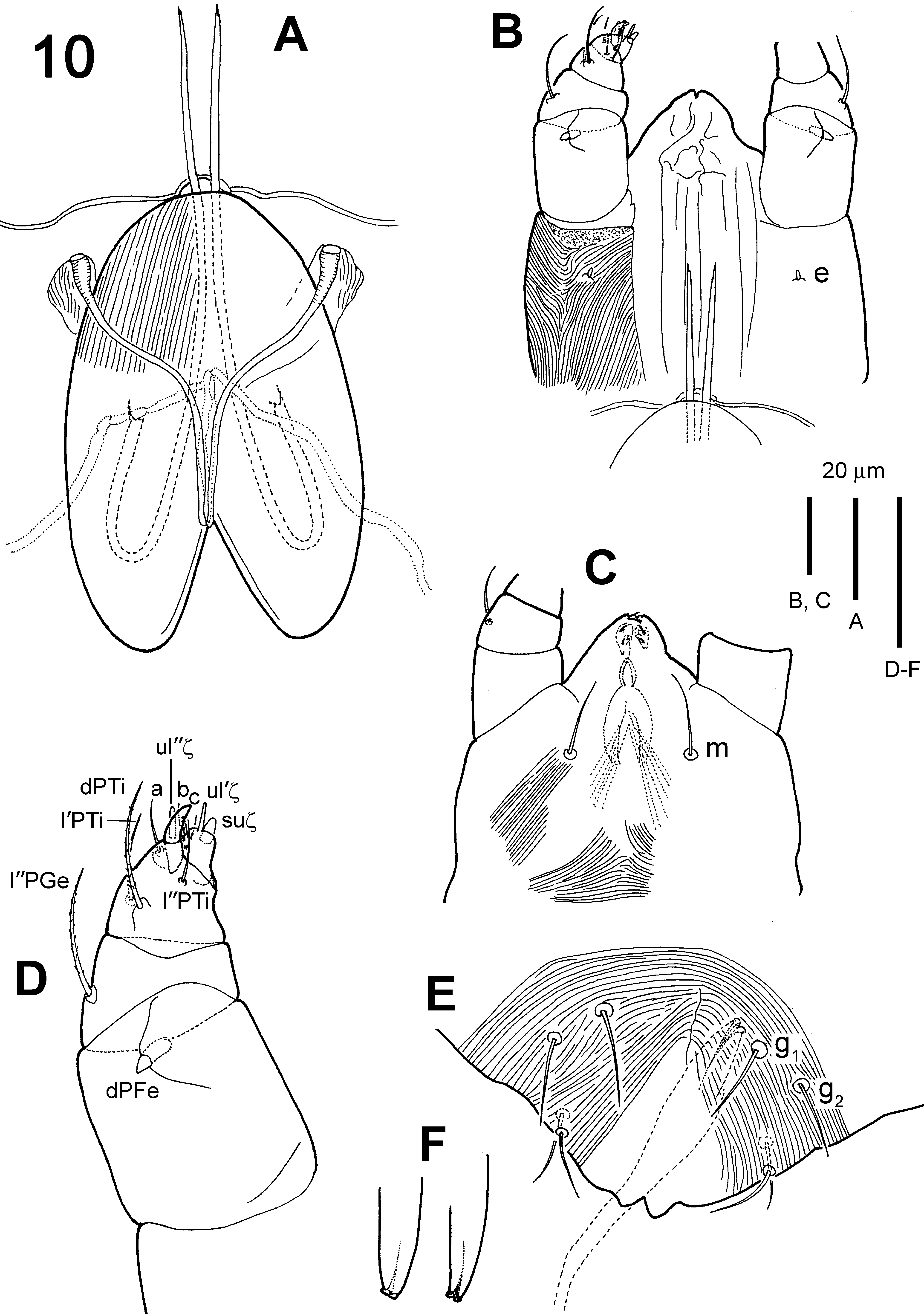
Venter ( Fig. 9 View FIGURE 9 ). Body finely striate with large areas of smooth cuticle around coxal bases; all ventral setae smooth and thin: 1a 38–43, 1 b 29–35, 1 c 27–33, 2 b 27–32, 2c 29–36, 3 a 41–43, 3 b 36–39, 4 a 32–37, 4 b 26–39, ag 22–26, g 1 9–14, g 2 9–13, ps 1 7–11, ps 2 7–11. Distances between setal bases: 1a–1a 19–29, 3 a–3a 19–31, 4 a–4a 41– 47, g 1– g 1 17–28, ag – ag 11–18.
Palp ( Fig. 10 View FIGURE 10 D). Seta d PFe thick spine-like and about 6 in length. Palpal tarsus with cone-shaped spinneret (suζ) smaller than that in female ( Fig. 11 View FIGURE 11 ), 3–4 in length and 1.5– 2 in diameter at base; single solenidion (3–4); two eupathidia (ul ′ζ 3–4, ul″ζ 3–4) and three normal setae (a 5, b 5–6, c 6 in length).
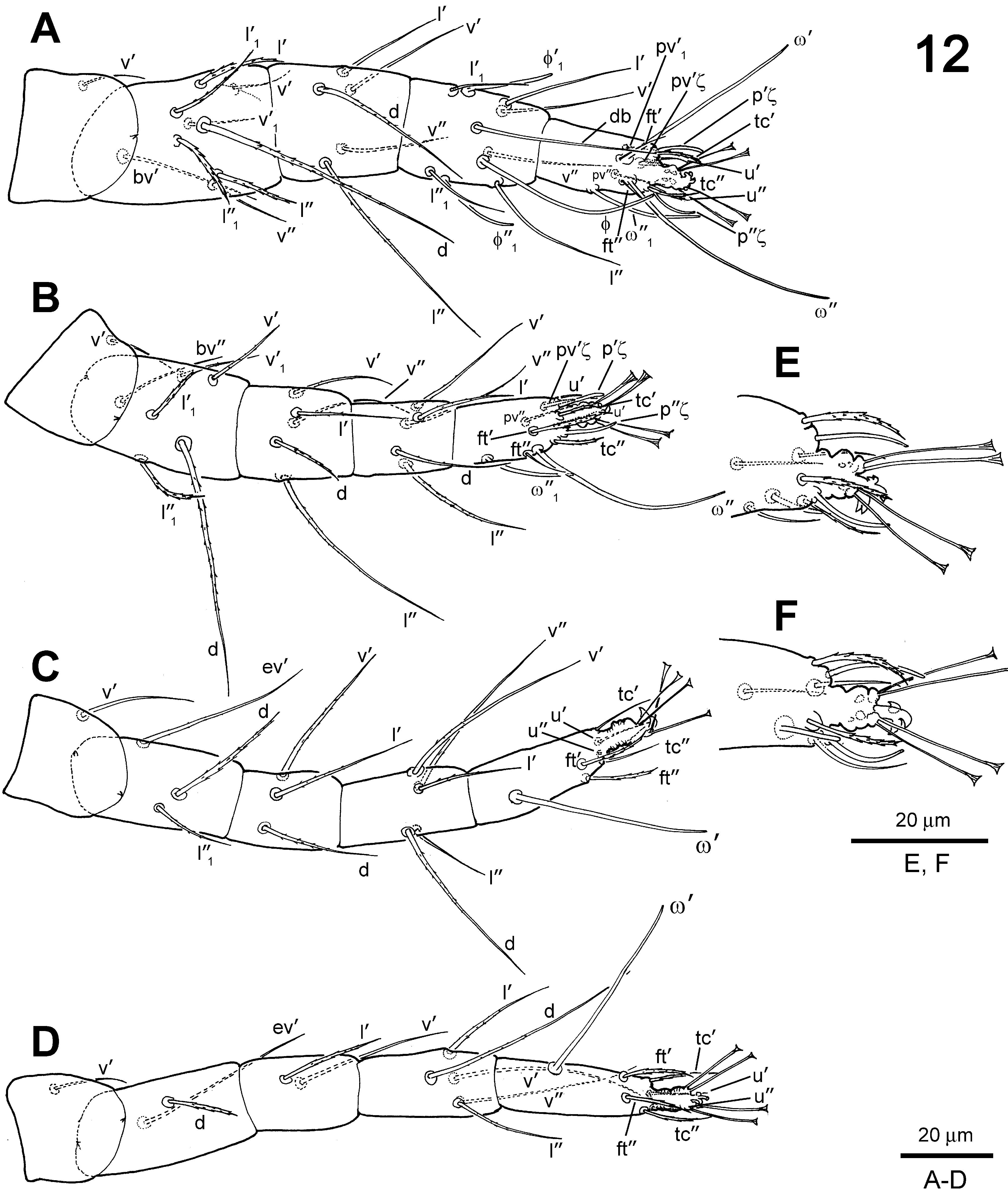
Legs ( Fig. 12 View FIGURE 12 ). Number of solenidia on tarsi I–IV: 3-2-1-1, two pairs of duplex setae of tarsus I distal and adjacent. Tarsi I: ω ′ 49–54, ω ″ 52–71, ω″ 1 25–33 in length; tarsi II: ω″ 56–59 and ω″ 1 20–26 in length; tarsi III: ω ′ 53–61 in length; tarsi IV: ω ′ 53–60 in length. Tibiae I with three solenidia φ (52–55), φ ′1 (23–31) and φ ″1 (22–26). Number of normal setae on leg (I–IV) segments: trochanters 1-1-1-1; femora 9-6-3-2; genua 5-5-3-2; tibiae 7-5-5- 5; tarsi 9-7-6-6. Number of eupathidia on tarsi I–IV: 3-3-0-0.
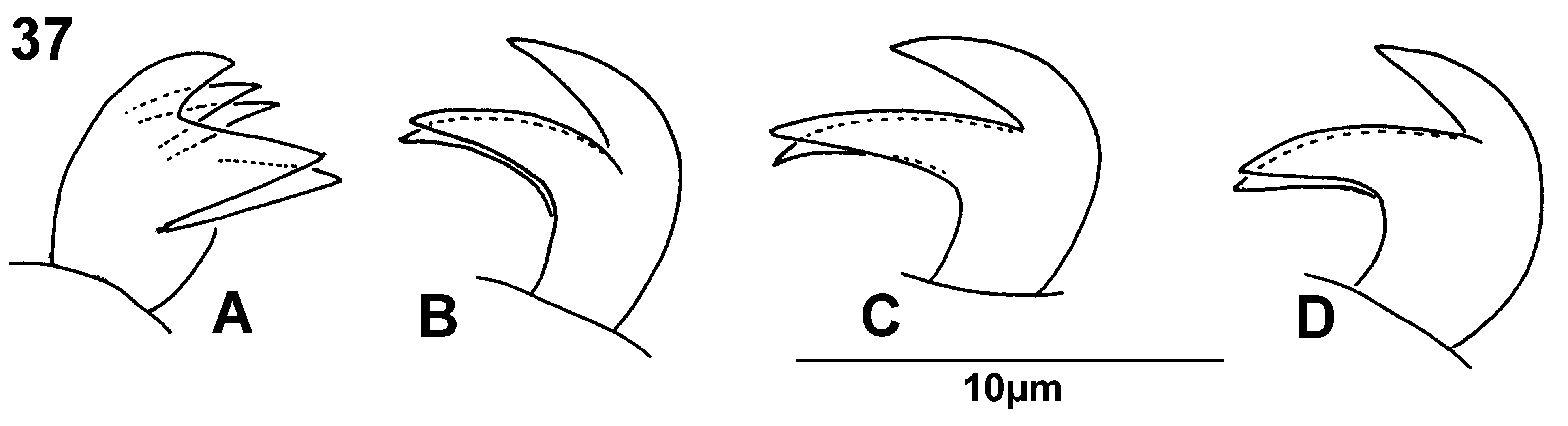
Lateral prongs of leg I empodia each with a strong dorsal hair ( Fig. 37 View FIGURE 37 ), giving the appearance of a fivepronged claw. Leg II–IV empodia claw-like and three-pronged (two lateral and one dorso-median).
Segments of legs stubby. Length of leg segments: femur I 45 –60, genu I 28–36, tibia I 28–40, tarsus I 34 –39; femur II 40 –51, genu II 29–30, tibia II 27–30, tarsus II 34 –37; femur III 39 –50, genu III 27–32, tibia III 33 –37, tarsus III 46 –49; femur IV 43 –51, genu IV 32 –36, tibia IV 35 –40, tarsus IV 47 –55.
Aedeagus ( Figs. 10 View FIGURE 10 F, 13, 36) short, relatively straight and slightly knobbed distially (dorsal view), flanked distally by an accessory structure; a thin, long ejaculatory duct connecting aedeagus to a conspicuous cup-shaped seminal vesicle.
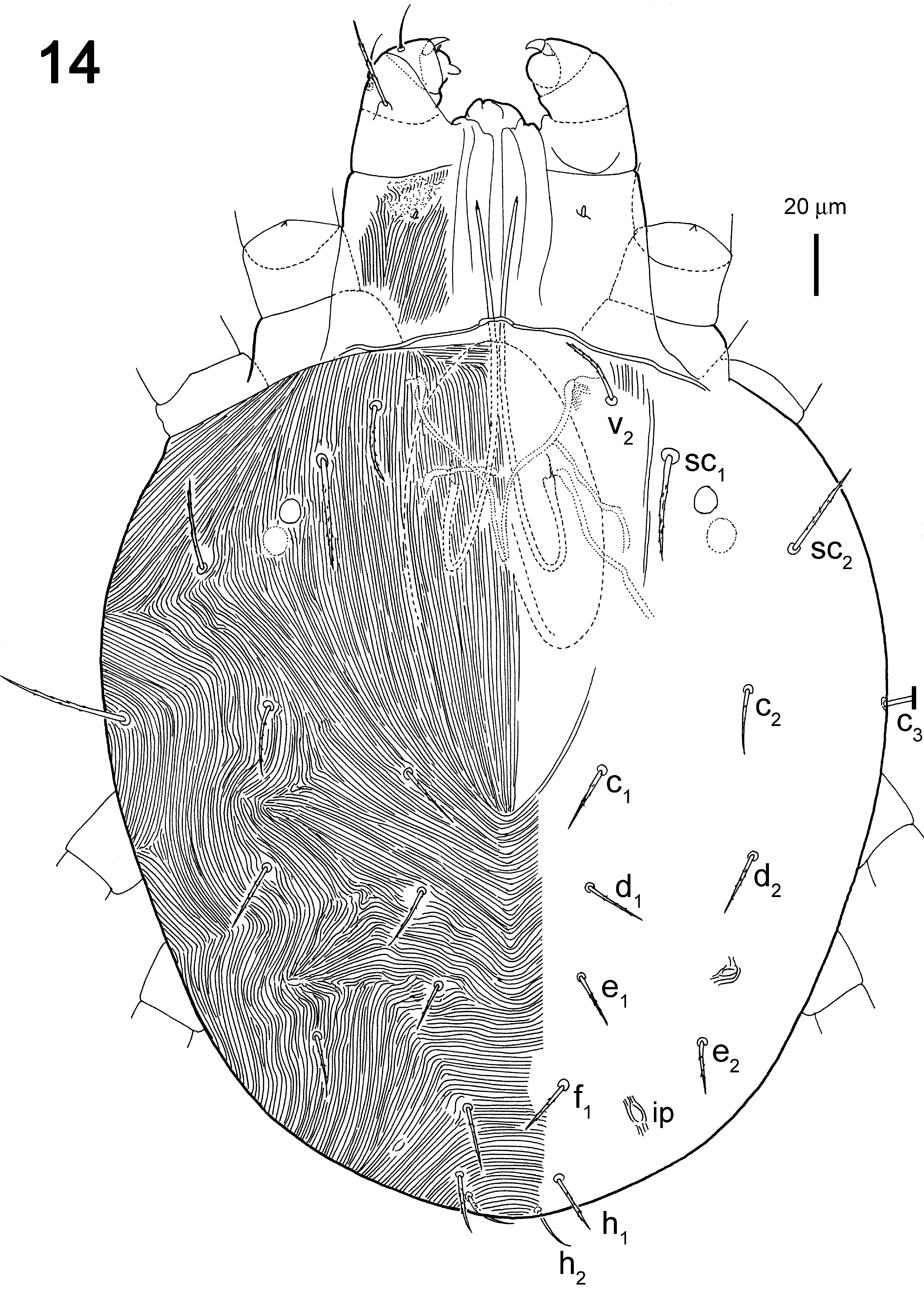
Deutonymph (n=6).
Dorsum ( Fig 14 View FIGURE 14 ). Length of body from posterior end of idiosoma to anterior end of gnathosoma 413–415; length of idiosoma 324–385; maximum width about at level of setae c 3 265–281. Dorsal striation similar to that of female, longitudinal medial striae beyond setae c 1 level posteriorly. Dorsal setae tapered and with small barbs. h 3 far away from h 2, while in larva, protonymph, female and male h 3 close to anal pore and h 2 (Figs. 31, 33, 34). v 2 22– 23, sc 1 34–35, sc 2 33–39, c 1 17–19, c 2 18–23, c 3 41–44, d 1 17–18, d 2 21–22, e 1 18 –20, e 2 17 –24, f 1 16–19, h 1 17–20, h 2 16–19, h 3 19–21. Distances between setal bases: v 2– v 2 68 –75, sc 1– sc 1 107–110, c 1– c 1 47–61, d 1– d 1 48–56, e 1– e 1 49 –51, f 1– f 1 29–39, h 1– h 1 33–40, h 2– h 2 17–31.
Venter ( Fig 15 View FIGURE 15 ). Venter almost entirely transversely striate. Ventral setae thin, smooth: 1a 29–32, 1 b 25–30, 1 c 24–26, 2 b 24–26, 2 c 25–29, 3 a 31–33, 3 b 26–31, 4 a 32–39, 4 b 24–26, ag 21–26, g 1 16–17, ps 1 8–9, ps 2 8–9. Distances between setal bases: 1a–1a 26–27, 3 a–3a 24–30, 4 a–4a 53–65, g 1– g 1 23–29, ag – ag 38–44.
Gnathosoma ( Figs. 16 View FIGURE 16 A–D). Subcapitular setae m smooth, length (17–23) shorter than distance m - m (32–40). Setae l″ PGe 36–41 long, d PTi 12–21, l ′ PTi 7–6 and l″ PTi 9–13 spinneret broadly rounded (4–5 in length and 2–3 in diameter at base); single solenidion (4 in length and 1 in diameter) and two eupathidia (ul ′ζ 4–5, ul″ζ 4–5).
Legs ( Fig. 17 View FIGURE 17 ). Leg I–IV empodia claw-like and three-pronged (two lateral and one dorso-median). Setae d of femur I well developed and located on a hump.
Two pairs of duplex setae of tarsus I distal and adjacent. Tarsi I: ω ′ 39–47, ω″ 59–62, ω″ 1 17–27. Tarsi II with one duplex seta, ω″ 44–55 in length. Tibiae I with one solenidion φ (45–55). Number of normal setae on leg (I–IV) segments: trochanters 1-1-1-0; femora 6-3-2-1; genua 5-5-3-1; tibiae 7-5-5-5; tarsi 9-7-6-6. Number of eupathidia on tarsi I–IV: 3-3-0-0.
Segments of legs stubby. Length of leg segments: femur I 44 –52, genu I 29–33, tibia I 31–36, tarsus I 34 –37; femur II 37 –43, genu II 24–27, tibia II 24–27, tarsus II 26–28; femur III 34 –44, genu III 27–30, tibia III 33 –38, tarsus III 40 –41; femur IV 34 –45, genu IV 24–29, tibia IV 29–37, tarsus IV 41 –46.
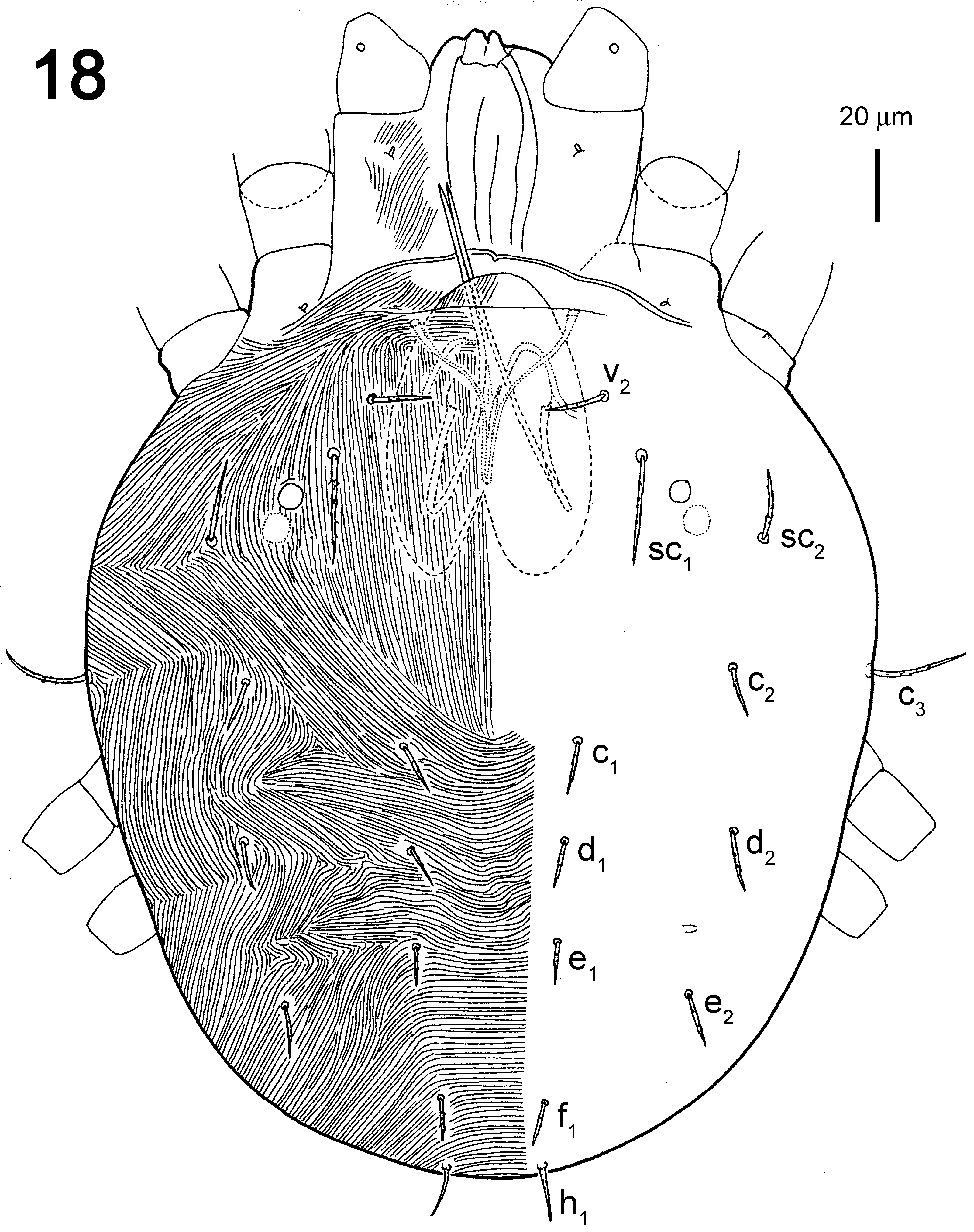
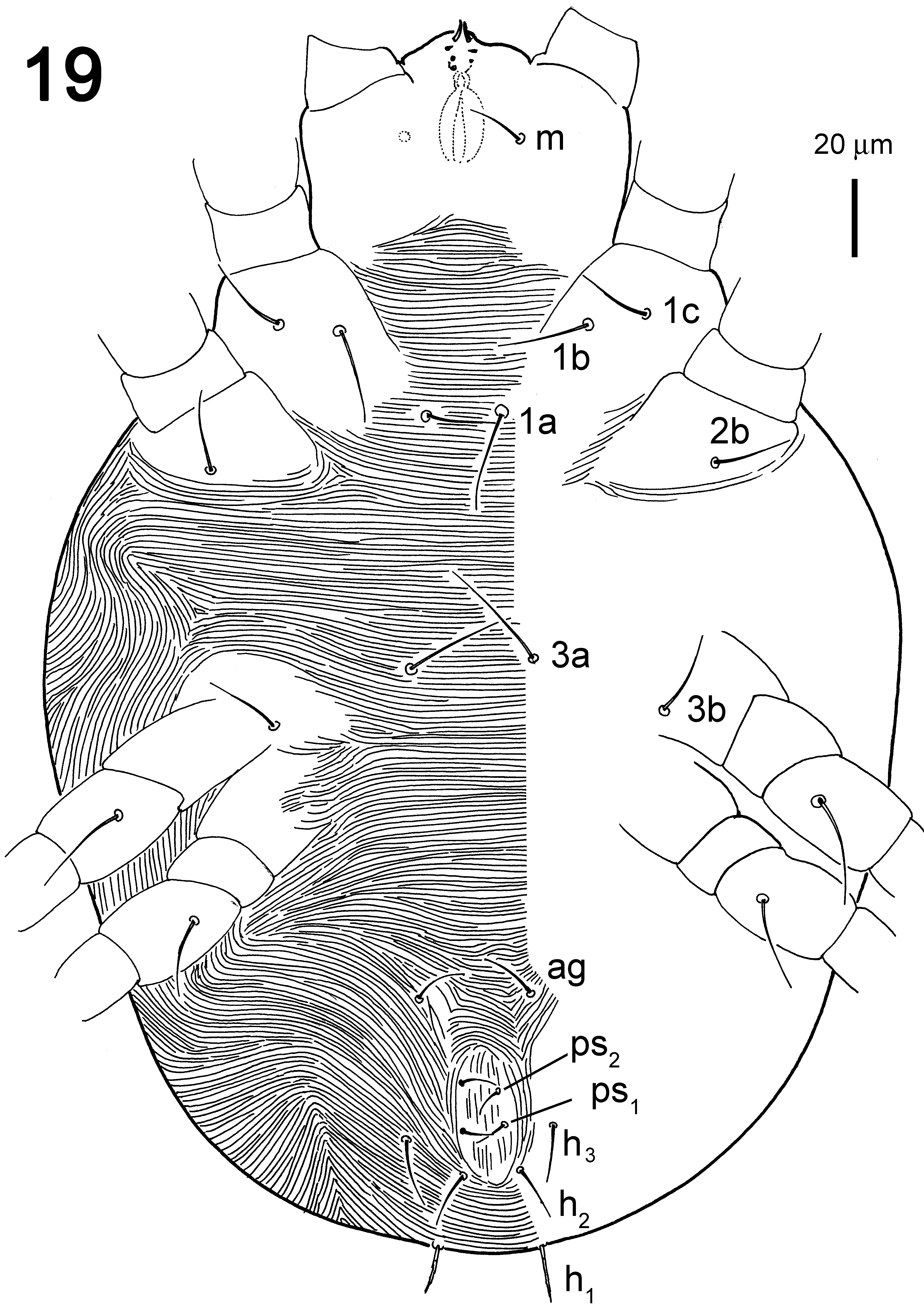
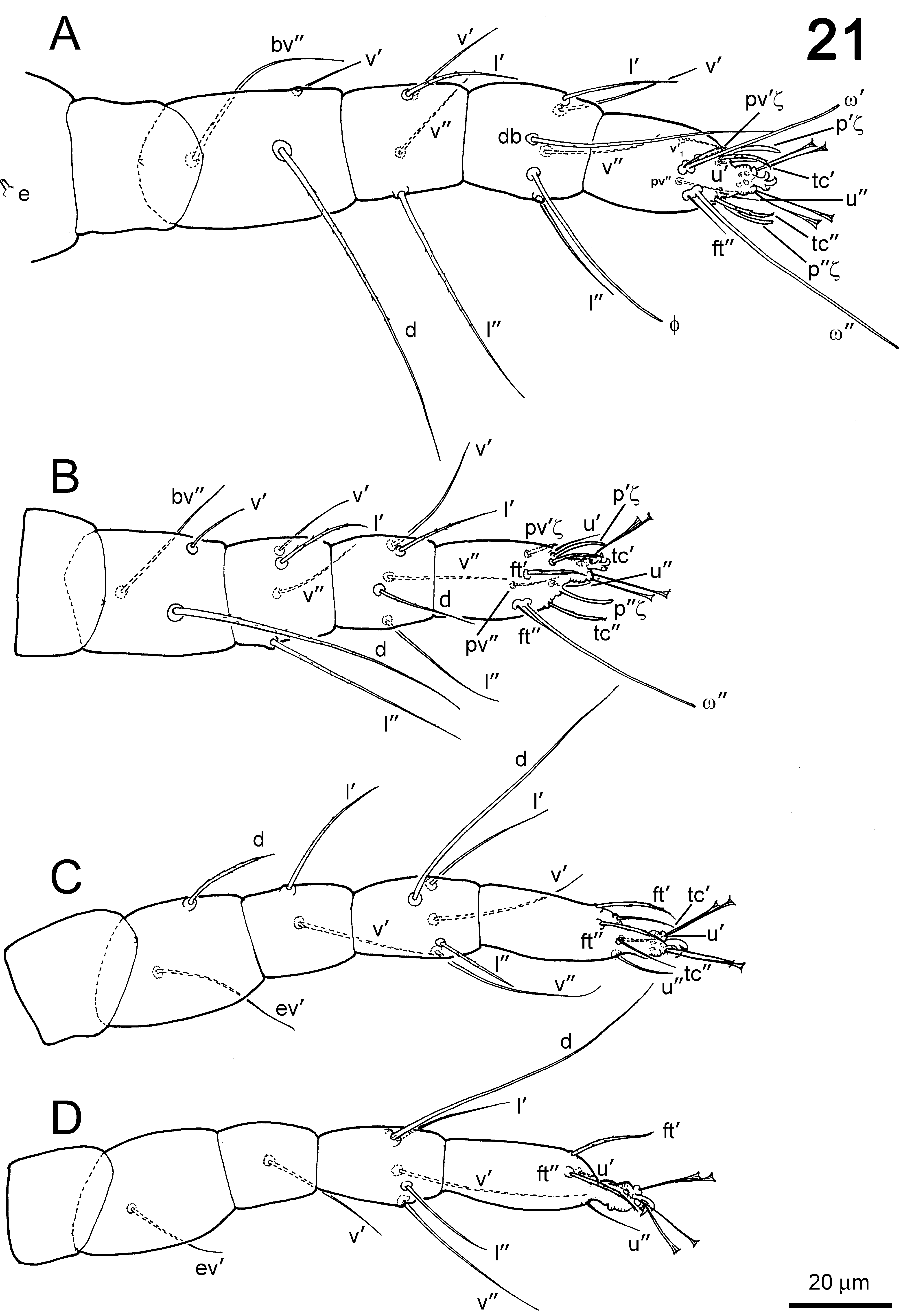
Protonymph (n=3).
Dorsum ( Fig. 18 View FIGURE 18 ). Length of body from posterior end of idiosoma to anterior end of gnathosoma 277–308; length of idiosoma 221–253; maximum width at level of setae c 3 205–221. Longitudinal medial striae ending at level of setae c 1. Except for smooth setae h 2 and h 3, dorsal setae tapered and with small barbs. v 2 18–19, sc 1 27–33, sc 2 21–24, c 1 17–22, c 2 16–22, c 3 33–36, d 1 13–14, d 2 16–19, e 1 12 –24, e 2 16 –17, f 1 13–14, h 1 15–16, h 2 11–14, h 3 14–18. Distances between setal bases: v 2– v 2 65 –66, sc 1– sc 1 86–93, c 1– c 1 49–52, d 1– d 1 37–44, e 1– e 1 33 –40, f 1– f 1 25– 29, h 1– h 1 25–27, h 2– h 2 15–16.
Venter ( Fig. 19 View FIGURE 19 ). Venter almost entirely transversely striated. Ventral setae thin, smooth: 1a 25–26, 1 b 24–27, 1 c 19–20, 2 b 19–22, 3 a 30–30, 3 b 19–21, ag 16–17, ps 1 9–9, ps 2 9–10.
Gnathosoma ( Figs. 20 View FIGURE 20 A–D). Subcapitular setae m smooth, length (17–21) shorter than distance m - m (34). Setae d PFe whip-like, 15–29 in length. Setae l″ PGe 14–22 long, d PTi 11–17, l ′ PTi 7–6 and l″ PTi 9–13; palpal tarsus with slender cone-shaped spinneret suζ 4 in length and 1 in diameter at base; single solenidion ω 3 in length and 1 in diameter, two eupathidia (ul ′ζ 3 in length, ul″ζ 3–4 in length) and three normal setae a 5, b 4, c 5–7 in length.
Legs ( Fig. 21 View FIGURE 21 ). Setae d of femur I well developed and located on a hump. Two pairs of duplex setae of tarsus I distal and adjacent. Tarsi I: ω ′ 34–36, ω″ 53–58. Tarsi II with one duplex setae, ω″ 56–59 in length. Tibiae I with one solenidia φ (41–57). Number of normal setae on leg (I–IV) segments: trochanters 0-0-0-0; femora 3-3-2-1; genua 4-4-2-1; tibiae 5-5-5-5; tarsi 8-7-6-6. Number of eupathidia on tarsi I–IV: 3-3-0-0.
Leg I empodia as that of female. Segments of legs stubby. Length of leg segments: femur I 36 –38, genu I 25– 26, tibia I 25–26, tarsus I 32 –34; femur II 32 –35, genu II 20–21, tibia II 20–21, tarsus II 31–31; femur III 31–31, genu III 20–22, tibia III 24–25, tarsus III 36 –38; femur IV 21–23, genu IV 19–20, tibia IV 19–24, tarsus IV 32 –34.
Larva (n=3).
Dorsum ( Fig. 22 View FIGURE 22 ). Length of body from posterior end of idiosoma to anterior end of gnathosoma 225–256, length of idiosoma 209–221; maximum width about at level of setae c 3 183–206.
Longitudinal medial striae ending at level of setae c 1. Except for smooth setae h 2 and h 3, dorsal setae tapered and with small barbs. v 2 21–22, sc 1 28–33, sc 2 22–26, c 1 14–16, c 2 16–20, c 3 26–31, d 1 14–18, d 2 15–21, e 1 15 –19, e 2 18 –22, f 1 12–17, h 1 18–20, h 2 16–18, h 3 21–23. Distances between setal bases: v 2– v 2 57 –61, sc 1– sc 1 75–89, c 1– c 1 42– 62, d 1– d 1 41–50, e 1– e 1 28 –40, f 1– f 1 17–20, h 1– h 1 22–34, h 2– h 2 10–18.
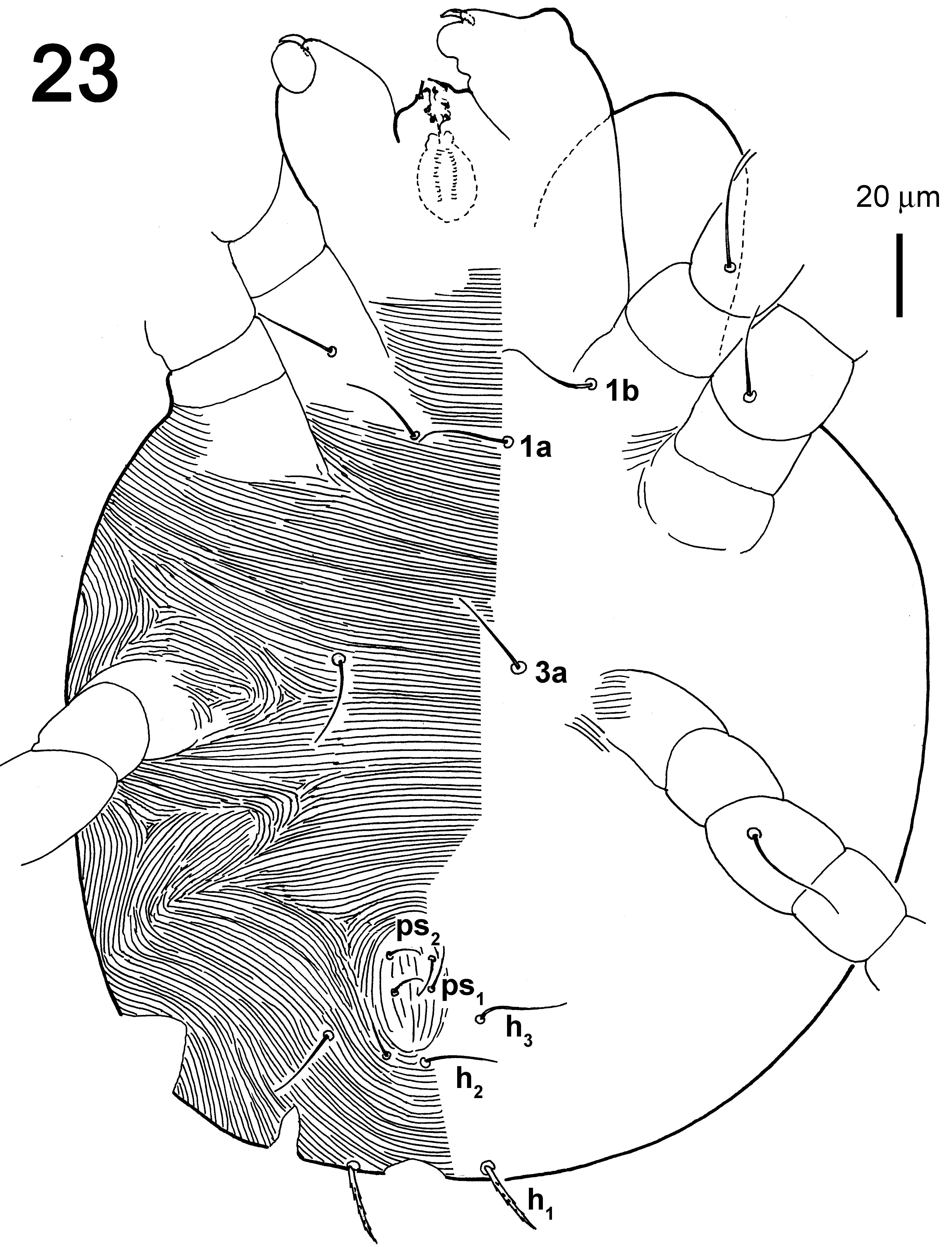
Venter ( Fig. 23 View FIGURE 23 ). Venter almost entirely transversely striate. Ventral setae thin, smooth: 1a 23–26, 1 b 22–25, 3 a 21–26, ps 1 10–11, ps 2 10–11.
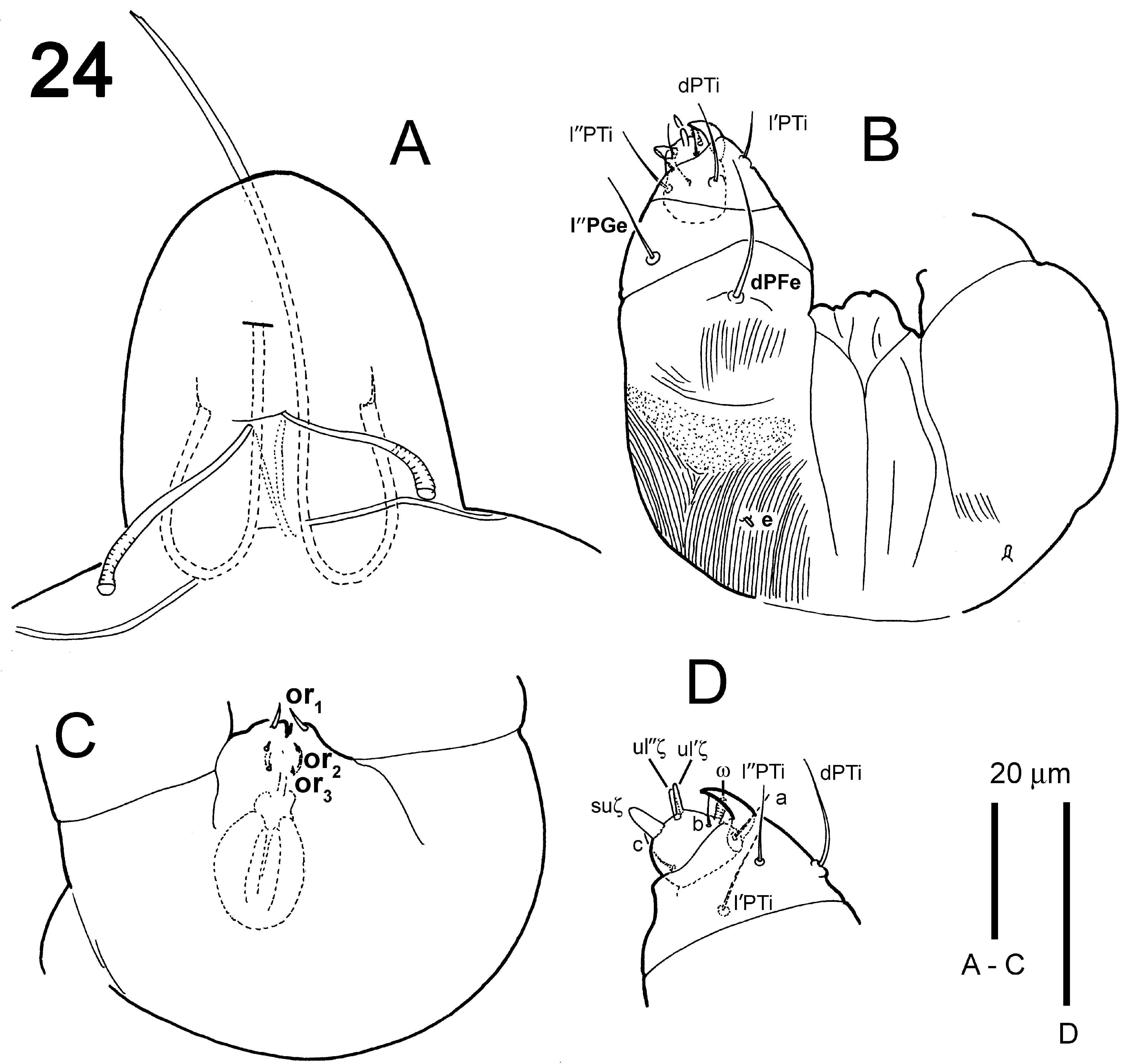
Gnathosoma ( Fig. 24 View FIGURE 24 ). Setae d PFe whip-like, 11–26 in length. Setae l″ PGe 11–16 long, d PTi 11–18, l ′ PTi 6–9 and l″ PTi 10–13; Palpal tarsus with slender cone-shaped spinneret suζ 3–4 in length and 1 in diameter at base; single solenidion ω 3 in length and 1 in diameter, two eupathidia (ul ′ζ 3–4, ul″ζ 3–4 in length) and three normal setae a 5, b 5–6, c 6 in length.
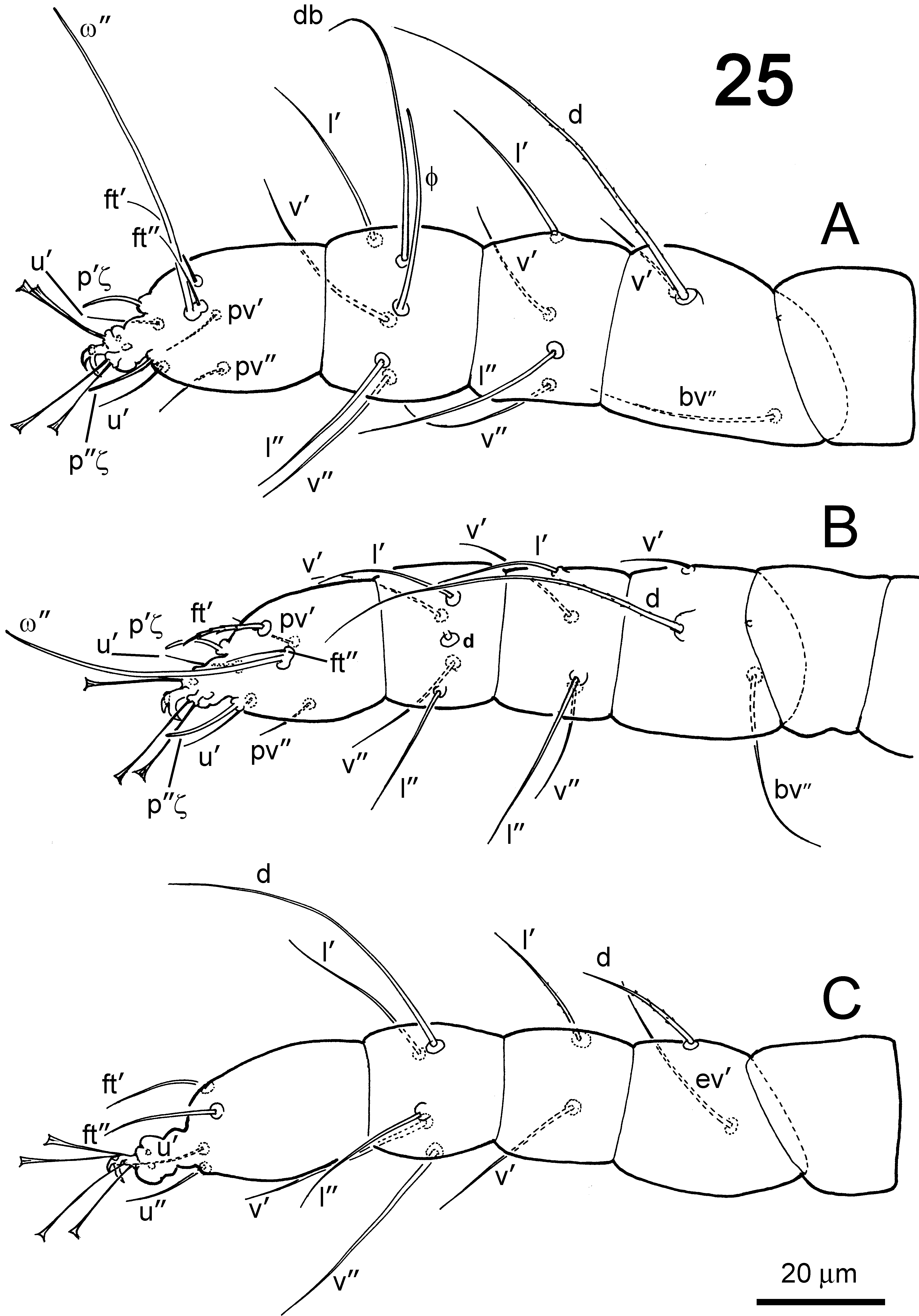
Legs ( Fig. 25 View FIGURE 25 ). Setae d of femur I well developed and located on a hump. One pairs of duplex setae of tarsus I distal. Tarsi I: ω ″ 56–58. Tarsi II with one duplex seta, ω ″ 40–46 in length. Tibiae I with one solenidion φ (39–42). Number of normal setae on leg (I–III): trochanters 0-0-0; femora 3-3-2; genua 4-4-2; tibiae 5-5-5; tarsi 8-7-4. Number of eupathidia on tarsi I–III: 2-3-0.
Leg I empodia as that of female. Segments of legs stubby. Length of leg segments: femur I 29–36, genu I 20– 22, tibia I 19–22, tarsus I 29–31; femur II 22–28, genu II 17–19, tibia II 16–19, tarsus II 22–27; femur III 23–30, genu III 16–20, tibia III 21–23, tarsus III 33 –34.
FIGURE 27. Tribolonychus collyerae , tibia I of female, showing the various chaetotaxy, A, tibia I with two additional solendions (φ ′1, φ ″1); B, tibia I with one additional solendion (φ ′1).
FIGURE 29. Tribolonychus collyerae , female, photomicrographs, showing the variations of seate c 1, d 1, e 1 among different groups. A, specimens from Lake Rotoroa; B, specimens from Golden Bay; C, specimens from Banks Peninsula.
FIGURE 30. Tribolonychus collyerae , female, photomicrographs, showing the variations of seate sc 2 and sc 1 among different groups, A, specimens from Lake Rotoroa; B, specimens from Golden Bay; C, specimens from Banks Peninsula.
FIGURE 31. Tribolonychus collyerae , female, photomicrographs, showing the variations of seate h 1, h 2 and h 3 among different groups, A, holotype, specimens from Lake Rotoroa (two the paratype females show the setae h 1 locating on the terminal of dorsum, h 2 and h 3 locating on venter); B, specimens from Golden Bay; C, specimens from Banks Peninsula.
FIGURE 33. Tribolonychus collyerae , photomicrographs, showing the ontogenetic development of position of setae h 2 and h 3 in the group from Banks Peninsula, A larva; B, protonymph; C, deutonymph.
FIGURE 34. Tribolonychus collyerae , male, photomicrographs, showing the variations of seate f 1 and h 1, A, specimens from Golden Bay; B, specimens from Banks Peninsula.


No known copyright restrictions apply. See Agosti, D., Egloff, W., 2009. Taxonomic information exchange and copyright: the Plazi approach. BMC Research Notes 2009, 2:53 for further explanation.
|
Kingdom |
|
|
Phylum |
|
|
Class |
|
|
Order |
|
|
Family |
|
|
Genus |