
Amphisbaena maranhensis, Gomes, Jerriane O. & Maciel, Adriano O., 2012
|
publication ID |
https://doi.org/ 10.5281/zenodo.208925 |
|
DOI |
https://doi.org/10.5281/zenodo.6166203 |
|
persistent identifier |
https://treatment.plazi.org/id/DD1D878D-0007-617D-C5C6-01F1BF12FE03 |
|
treatment provided by |
Plazi |
|
scientific name |
Amphisbaena maranhensis |
| status |
sp. nov. |
Amphisbaena maranhensis sp. nov.
( Figs. 2–6 View FIGURE 2 View FIGURE 3 View FIGURE 4 View FIGURE 5 View FIGURE 6 )
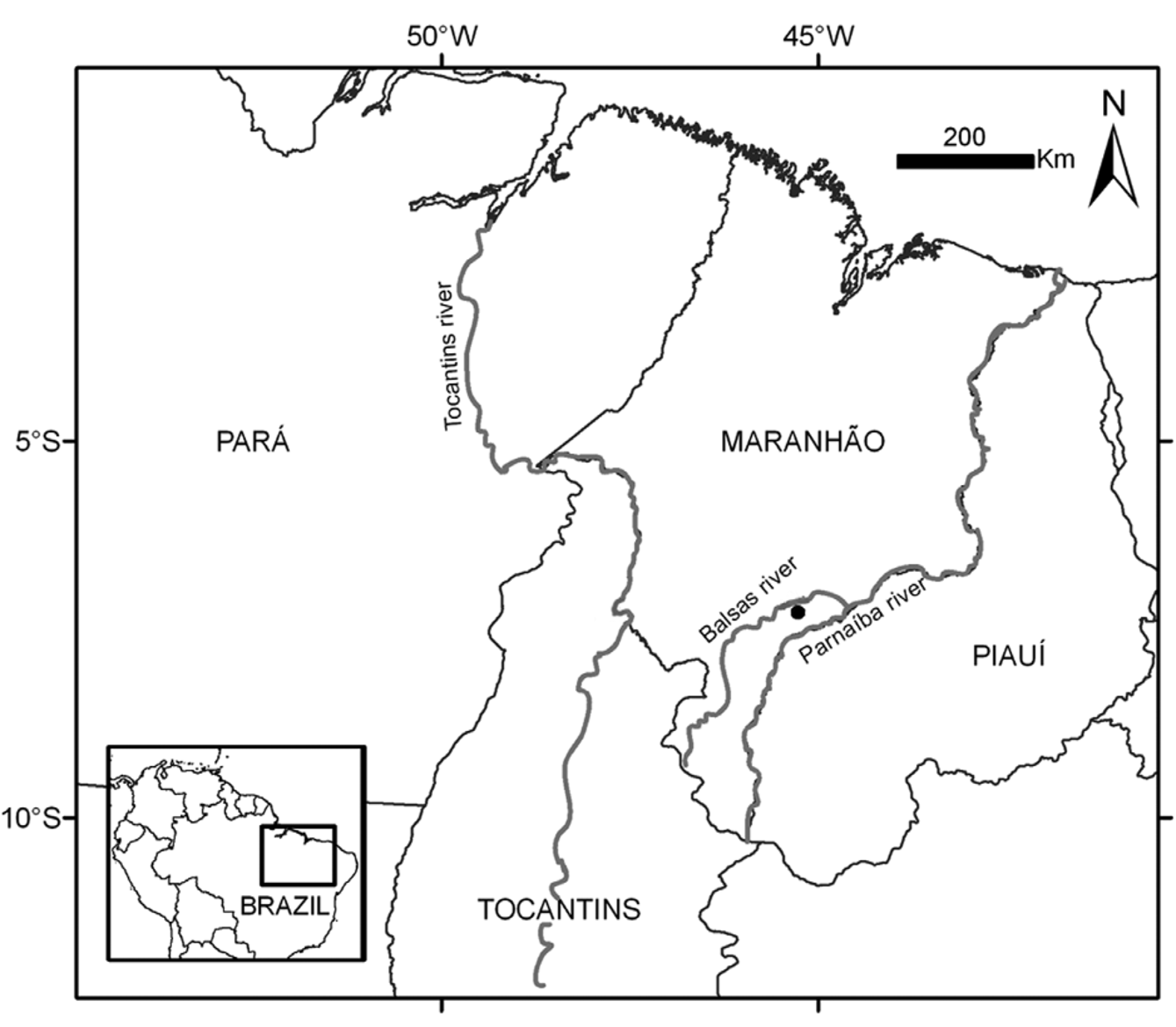
Holotype. MPEG 28562 (field number: DT 178). Specimen after collection broken into two pieces, in the posterior region of the body near the cloaca; both pieces preserved ( Fig. 2 View FIGURE 2 ). Municipality of Loreto (07o09’09” S; 45o18’14” W), Maranhão state, Brazil. Collected in pitfall trap by Dante Pavan, Vinícius de Avelar São Pedro, and José Mario Guelhere on 3 March, 2009.
Etymology. The name of the species refers to Maranhão state, where the new species was collected.
Diagnosis. A small species of Amphisbaena , without major fusion of head shields: rostral, nasals, prefrontals, frontals, parietals and labials are distinct. Three supralabials, irregularly polygonal; first supralabial, rhomboid, longer and lower than the second and third; three infralabials, third one, pentagonal, higher than the first and second. Two rows of postgenials, first with two scales and second with three scales, lacking a postmalar row. Body annuli 306, caudal annuli 21, with the eighth postcloacal annulus more pigmented than adjacent annuli, 10 dorsal segments and 14 ventral segments in a midbody annulus, and no precloacal pores. Tip of tail with a vertical keel. Dorsal sulcus absent, lateral sulci distinct.
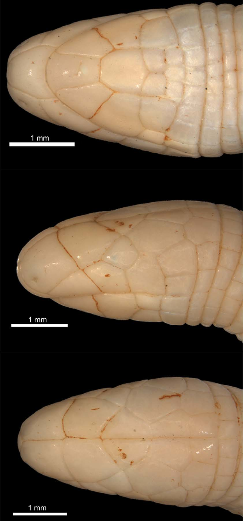
Description of the holotype ( Fig. 3–6 View FIGURE 3 View FIGURE 4 View FIGURE 5 View FIGURE 6 ). A small amphisbaenian [snout-vent length 125 mm (122 mm + 3 mm); caudal length 13.8 mm; midbody diameter 2.7 mm]; head short (2.6 % of snout-vent length), rounded and hardly distinct from the neck; rostrum rounded, projecting beyond the lower jaw (prognathous snout); body with dorsal sulcus absent and lateral sulci clearly marked.
Rostral subtriangular, visible from above ( Fig. 3 View FIGURE 3 ), ventrally expanded ( Fig. 3 View FIGURE 3 ), lateral portion concave, in contact with nasals laterally and first supralabial latero-anteriorly. Nasals quadrangular, paired, with a long middorsal suture, relatively large (suture length ca. 23 % of head length), in contact with the rostral, first and second (only on right side) supralabials and prefrontals posteriorly; nostril near the antero-inferior angle of the nasal ( Fig. 3 View FIGURE 3 ). Prefrontals trapezoid, paired, with a long middorsal suture (suture length ca. 20 % of head length), in contact with nasal, first (only left side) and second supralabial, ocular and frontals. Frontals rhomboid, paired, converging posteriorly, with a long middorsal suture (suture length ca. 30.4 % of head length), in point contact with ocular, in contact with postocular, prefrontal and parietal. Parietals pentagonal, paired, longer than wide, with a long middorsal suture (suture length ca. 20.9 % of head length), with their posterior border straight and parallel to the second body annulus, in contact with postocular, frontal, and second body annulus. Posterior to the parietals is a pair of occipital scales, rectangular, almost as wide as the parietals. Ocular diamond-shaped, in broad contact with second and third supralabials, prefrontal and postocular, in point contact with frontals. Eye barely visible in anterior corner of ocular. Three supralabials, increasing in size posteriorly; first rhomboid, smallest, longer than high, in point contact with prefrontal (only left side), in contact with rostral, second supralabial and in extensive upper contact with the nasal; second one pentagonal, in point contact with nasal (only right side), in contact with prefrontal, ocular, first and third supralabial; third pentagonal, higher than the first and second, in contact with second supralabial, ocular, postocular, temporal and postsupralabial. The anterior edge of the parietals is flush with the level of the angulus oris. Posteriorly of the supralabials there is a row of scales that includes postocular, temporal, postsupralabial and the first segment of the first body annulus.
Mental roughly rectangular, anterior portion concave, lateral borders straight, converging posteriorly; posterior border transversal and straight; only slightly larger than the first infralabial, in contact with postmental and the first infralabials. Postmental single, heptagonal, in contact with the mental, first and second infralabials and first row of postgenials. Posteriorly to the postmental, two rows of postgenials; first with two larger scales, in contact with postmental, malars laterally, second postgenial row posteriorly, in point contact with second infralabials and preventing contact between postmental and malars; second postgenial row with three equally sized scales, in contact with first postgenial row anteriorly, malar laterally and first body annulus posteriorly. Two malars, one on each side, irregularly polygonal, in contact with second and third infralabial, first and second postgenial rows and the first body annulus. No postmalar row. Three infralabials; first trapezoid, the second pentagonal and largest, third rectangular and smallest. Behind the third infralabial a slightly enlarged scale of the first body annulus (behind angulus oris) appears to continue the infralabial series.
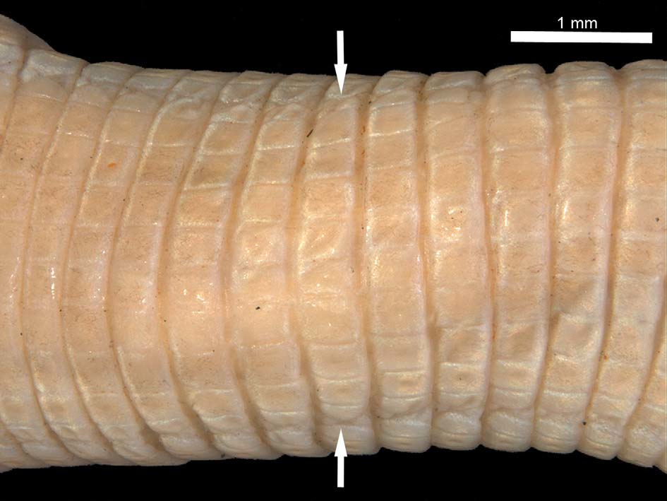
Body annuli distinct. The first body annulus is well-defined only ventrally. Dorsally the first body annulus includes postsupralabial, temporal and the very large postocular dorsally limited by frontal and parietal. There are 306 body annuli. Only one interspersed half-annulus were noted (included in the counts, 303). A midbody annulus contains 10 dorsal segments and 14 ventral segments; the ventral segments, with two central scales rows larger than adjacent scales, only slightly larger than dorsal ones; lateral sulci well marked, separating the dorsal and the ventral segments, starting at the level of 60th body annulus and continued almost to level of vent; no dorsal or ventral sulcus ( Fig. 4 View FIGURE 4 ). No precloacal pores. Cloacal segments regular, with six precloacal and eight postcloacal segments. Three lateral annuli in the vent region.
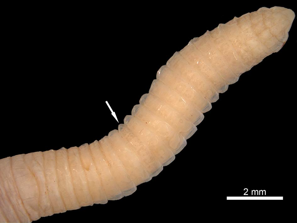
Caudal annuli 21. Tip of tail compressed into a vertical keel ( Fig. 6 View FIGURE 6 ). Amphisbaena maranhensis sp. nov. lacks an externally apparent autotomy constriction, however the eighth tail annulus has more heavily pigmented ventral scales (relative to other tail scales), justifying its definition as an autotomy site [see Gans (1971), Gans & Mathers (1977), and Montero et al. (1997) for similar definition in Mesobaena huebneri Mertens, 1925 , A. medemi Gans & Mathers, 1977 , and A. cegei Montero, 1997 , respectively, that lacked an external autotomy constriction but showed an annulus (autotomy site) slightly more deeply pigmented and otherwise modified] ( Fig. 5 View FIGURE 5 ). Others similar cases are related, e. g., in A. miringoera Vanzolini, 1971 , A. angustifrons Cope, 1861 , and A. caudalis Cochran, 1928 , which were described as lacking externally apparent caudal autotomy. Nevertheless, Mott et al. (2011), Gans & Diefenbach (1972), and Thomas & Hedges (2006) mentioned, respectively for these species, additional specimens that showed a vertebral autotomy plane where the pigmented scales appear. Amphisbaena vanzolinii Gans, 1963 was also described without indication of an externally differentiated autotomy level, being subsequently confirmed an autotomy site more intensely pigmented than the caudal annuli ( Gans, 1963c; Hoogmoed & Mott, 2003).
Coloration in preservative of holotype. Uniform cream ventral and dorsally.
Geographic distribution. The new species is known only from the type locality, municipality of Loreto, Maranhão state, near the boundary with the state of Piauí, northeastern Brazilian Cerrado ( Fig. 1 View FIGURE 1 ). The specimen was collected on the right margin of the Balsas River, affluent of the Parnaiba River, in a region covered with typical Cerrado vegetation. There is no record of sympatric amphisbaenid species with this new species.


| MPEG |
Museu Paraense Emilio Goeldi |
No known copyright restrictions apply. See Agosti, D., Egloff, W., 2009. Taxonomic information exchange and copyright: the Plazi approach. BMC Research Notes 2009, 2:53 for further explanation.
|
Kingdom |
|
|
Phylum |
|
|
Class |
|
|
Order |
|
|
Family |
|
|
Genus |