
Rhinogobius maxillivirgatus Xia, Wu
|
publication ID |
https://doi.org/ 10.11646/zootaxa.4407.4.7 |
|
publication LSID |
lsid:zoobank.org:pub:69D824E3-61C3-43CB-89CE-085C17280210 |
|
DOI |
https://doi.org/10.5281/zenodo.5975947 |
|
persistent identifier |
https://treatment.plazi.org/id/E1414F56-3809-FFDD-6C93-4EFDFCF9FC80 |
|
treatment provided by |
Plazi |
|
scientific name |
Rhinogobius maxillivirgatus Xia, Wu |
| status |
|
Rhinogobius maxillivirgatus Xia, Wu , and Li, sp. n.
( Figure 2 View FIGURE2 )
Holotype. SNHM-F-2017060018, female, 30.1 mm SL; a tributary of Changjiang River , Qimen County, Huangshan City, Anhui Province, China; 4 June 2017, J.- H. Xia.
Paratypes. SNHM-F-2016040025, male, 30.8 mm SL; SNHM-F-2016040026, male, 28.0 mm SL, 28 April 2016; SNHM-F-2016100025, male, 23.0 mm SL; SNHM-F-2016100090, male, 24.3 mm SL, 12 October 2016; SNHM-F-2016040027, female, 31.1 mm SL; SNHM-F-2016040028, female, 29.3 mm SL; SNHM-F-2016040029, female, 31.1 mm SL, 28 April 2016; SNHM-F-2016100032, female, 25.1 mm SL; SNHM-F-2016100091, female, 23.8 mm SL, 12 October 2016. Locality same as holotype, J.-H. Xia & Y.-Q. Wu.
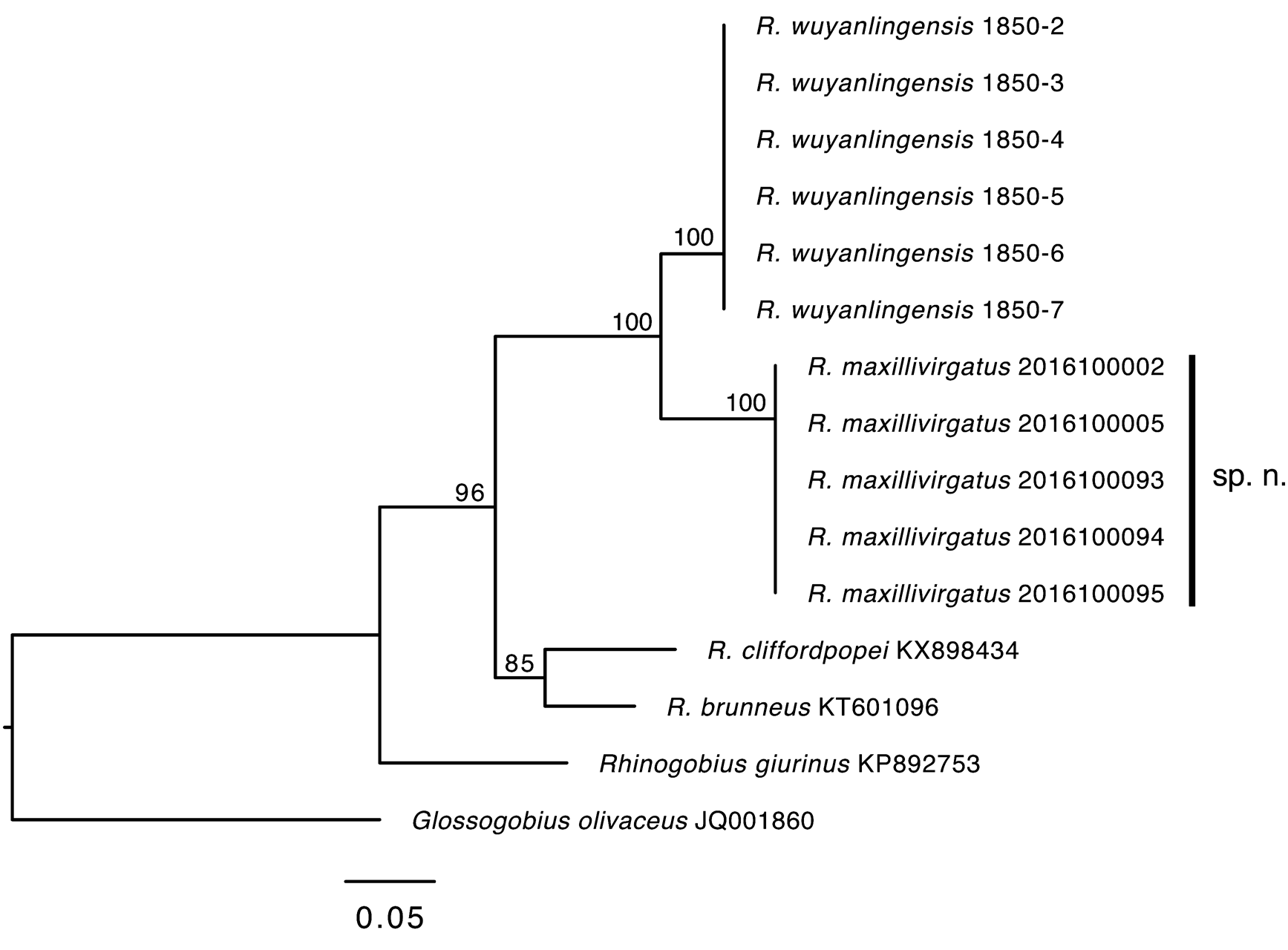
Other material. Five additional specimens were used for DNA barcoding, not measured: 4 females (SNHM- F-2016100002, SNHM-F-2016100005, SNHM-F-2016100093, SNHM-F-2016100095) and 1 male (SNHM-F- 2016100094); 12 October 2016, locality same as holotype, J.-H. Xia & Y.-Q. Wu.
Diagnosis. Rhinogobius maxillivirgatus is distinguishable from the other species in the same genus by the unique combination of the following features: D2 rays modally I/8 (rarely I/9); A rays modally I/7; P rays 13–15; LL 28–30; TR 6–7; predorsal scales 5–9; vertebral count 10+17=27; lateral side with 5–6 longitudinal serrated stripes, ventral ones dark; a whitish band across from snout to dorsal portion of operculum, interrupted by orbit; a black stripe along lower half of upper jaw, followed by a longer parallel stripe across anterior portion of cheek; rear edge of orbit with a black blotch; cheek and operculum spotless, branchiostegal membrane grayish with up to 20 red spots in male; D1 with 3–4 horizontal rows of spots and D2 with 5–6 rows; P base with an arched reddish brown band, membrane with several vertical rows of spots and a bold mark near the origin; and C base with a large dark spot, with vertical rows of spots in membrane and the anterior row enlarged.
Description. Morphometric and meristic data for the holotype and paratypes are presented in Table 1. Body slim, rather cylindrical anteriorly, compressed posteriorly. Eyes dorsolateral, diameter larger than interorbital width. Head moderately conical; snout small, cheeks fleshy. Lips thick. Mouth oblique; lower jaw slightly longer than upper, both with 4–5 rows of conical teeth, outer row enlarged. Isthmus broad, united to branchiostegal membrane. Gill rakers 3+7. Genital papilla flat and triangular in males; column-like with terminal pore in females.
Fins. D1 rays VI (rarely V) with longer third and fourth spinous rays, never extending to base of first branched ray of D2 when adpressed in both sexes. D2 rays modally I/8; A rays modally I/7; P rays 13–15; V rays I/5 + I/5. Rear tips of D2 and A rays not reaching procurrent rays of C when adpressed, A origin inserted below second branched ray of D 2. V resembling disc, with spinous rays with pointed membranous lobe. P and C elliptical, distal margin of C rounded.
Scales. Body with ctenoid scales, mid-trunk scales enlarged. Breast, prepelvic area, and almost whole head naked. PreD cycloid, 5–9; LL 28–30, TR 6–7, SDP 5–7. Anterior part of predorsal area naked. Predorsal squamation with trifurcate anterior edge and anterior boundary of middle series extending just above canal pore Θ, boundary of anterior extension on side of occipital region reaching dorsal origin of pectoral fin.
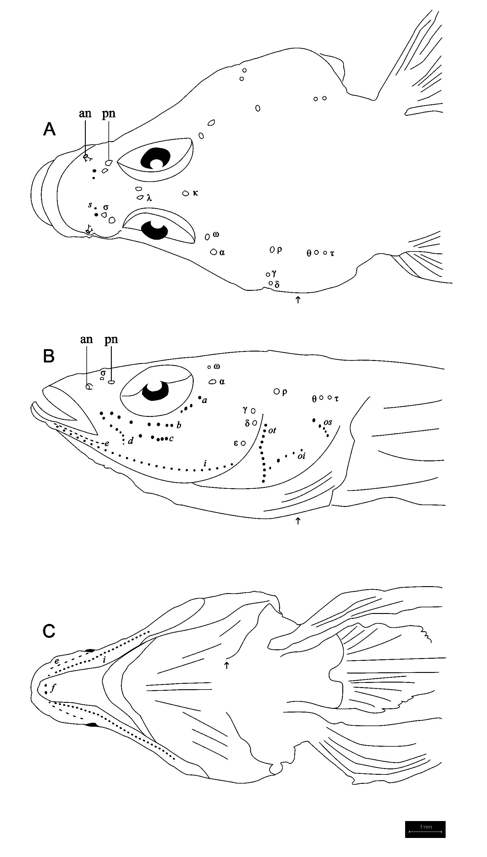
Cephalic canal pores: See Fig. 3 A–B View FIGURE3 . Tubular anterior nares and posterior nares, simple pores without any rim. Canal pore arrangement complete for the genus. Nasal extension of anterior oculoscapular canal with paired terminal pore σ, located anterior to posterior nare, but not reaching anterior nare; anterior interorbital sections separated, with paired pore λ and single pore κ; lateral section over cheek with pore α and terminal pore ρ; pore ω present near posterior border of eye between α and κ. Posterior oculoscapular canal quite short, with two terminal pores Θ and τ. Gap between pores ρ and Θ greater than length of posterior oculoscapular canal. Pores γ, δ, and ε along preopercular canal, distance between γ and δ shorter than that between δ and ε.
Sensory papillae. See Fig. 3 A–C View FIGURE3 . Row a short, extending ventrally along rear edge of orbit, ventral terminal not reaching vertical midline of orbit. Row b extending below orbit, longer than a, but not reaching posteriorly to vertical line through rear margin of orbit. Row c stretching longitudinally on middle cheek, with nearly half length of row b. Row d oblique, along lower half of upper jaw. Operculum with rows ot, oi, and os; row oi attaching ventrally to ventral part of row ot; row os extending ventrally towards posterior, well separated from rows ot and oi. Preopercular-mandibular papillae with rows e, i, and f, paired respectively.
Vertebrae. Shown in Fig. 4A View FIGURE 4 . Total vertebrate 10 + 17 = 27. DP 3/II II I I 0/9
Coloration. See Figs. 2 View FIGURE2 and 4 B–C View FIGURE 4 . Body always with 5–6 longitudinal serrated stripes from dorsum to venter; stripes long and parallel, ventral ones darker. Belly pale. Whitish band running from upper jaw to posterior operculum, interrupted by orbit. Dorsum of snout with U-shaped line united at tip of snout. Black blotch behind orbit. Oblique stripe along lower half of upper jaw, followed by parallel black stripe originating from lower anterior edge of orbit; longer than the previous. Males with red lips in breeding season, faded in fixed material. Branchiostegal membrane pale yellow with up to 20 red spots in males after fresh preservation; spotless in females. Dorsal midline covered with 5 large black blotches.
Fins with rows of black spots alternating with bright rows of white spots in living specimens; whitish rows disappearing when preserved. D1 with 3 longitudinal rows of black spots; D2 with 5 rows. C with 6–7 vertical waved rows of black spots and middle portion of anterior row with a large blotch; base with a large black spot in center, attached to shiny transverse band in life, fading in preservation. P base with deep red mark, followed by bright arched band in live males, red mark vanishing in preservation; 5–6 vertical rows of black spots and anterior row enlarged as a band in both sexes. V grayish in male, whitish and uniform in female.
Distribution. Currently Rhinogobius maxillivirgatus is only known from a particular tributary of the Changjiang Basin in Qimen County, Huangshan City, Anhui Province, China.
Etymology. The name maxillivirgatus is a noun in apposition and derived from the Latin maxilla meaning “upper jaw” and virgas meaning “stripes,” referring to 2 distinctive stripes behind the upper jaw of both sexes.
Biology. This species inhabits a shallow, slow-flowing brook of the upper Changjiang River, with a width of 5–6 meters and cobble substrate. According to three field trips, it was observed that breeding happens from late April to middle June and the number of gravid individuals increased in early June.
Phylogenetic analysis. The best model selected was HKY + G. Rhinogobius maxillivirgatus is a sister species to R. wuyanlingensis in the ML tree rooted with G. olivaceus ( Fig. 5 View FIGURE 5 ). Average genetic p-distance between R. maxillivirgatus and R. wuyanlingensis is 0.081, which is comparable to the genetic distance between R. cliffordpopei and R. brunneus (0.097).
Remarks. Rhinogobius maxillivirgatus is most similar to R. wuyanlingensis , from neighboring Zhejiang Province in Eastern China. Both species have a slim body with about 6 longitudinal lines from the dorsal to ventral region ( Yang et al. 2008). However, R. maxillivirgatus can be distinguished from R. wuyanlingensis by possession of 1) P rays modally 14 (vs. 17–18, modally 18); 2) LL 25–30 (vs. 30–32); 3) TR 5–7 (vs. 9–10); 4) 2 oblique stripes along upper jaws on anterior area of cheek (vs. lacking such pattern of stripes); and 5) branchiostegal membrane with about 20 red big spots in male (vs. 6–7 transverse deep red stripes on branchiostegal membrane in males) (features in parentheses, from Yang et al. [2008]).


| DNA |
Department of Natural Resources, Environment, The Arts and Sport |
No known copyright restrictions apply. See Agosti, D., Egloff, W., 2009. Taxonomic information exchange and copyright: the Plazi approach. BMC Research Notes 2009, 2:53 for further explanation.
|
Kingdom |
|
|
Phylum |
|
|
Class |
|
|
Order |
|
|
Family |
|
|
Genus |