
Marphysa leidii de Quatrefages, 1866
|
publication ID |
https://doi.org/10.11646/zootaxa.4816.1.1 |
|
publication LSID |
lsid:zoobank.org:pub:0475E09C-792F-4F55-9F1F-C85B8A6E44AD |
|
persistent identifier |
https://treatment.plazi.org/id/E3069005-FFB5-FFC4-46D6-FA4B78CFF94D |
|
treatment provided by |
Plazi |
|
scientific name |
Marphysa leidii de Quatrefages, 1866 |
| status |
|
Marphysa leidii de Quatrefages, 1866 View in CoL
Figures 6 View FIGURE 6 , 9F View FIGURE 9 , Table 1
Eunice sanguinea . — Leidy 1855: 147.
Marphysa leidii de Quatrefages, 1866: 22 View in CoL .
Marphysa leidyi . — Verrill 1873: 319, Pt. II, Fig. 64.
Marphysa sanguinea View in CoL . — Webster 1879: 36–40.— Fauvel 1911: 18.— Hartman 1944b: 339.— Hartman 1945: 23–24.— Pettibone 1963: 236–238, Fig. 62 ( non Montagu, 1813).
Material examined. Type material: Neotype USNM 71609 About USNM , EVG 73 123 Little Sheepshead creek by Big stake, Heading North , New Jersey, United State , 3 Oct 1973, coll. Garlo, E. V . Additional material: USMN 4469 ( 2 specimens) , USNM 6194 About USNM ( 1 specimen) Vineyard Sound , Massachusetts, United States, in mud . USNM 3860 About USNM ( 5 specimens) Woods Hole , Massachusetts, United States (4131.1987’N, 7040.9677’W), 5 Sep 2017, 1 m. USNM 61734 About USNM ( 2 specimens), Cape Lookout , North Carolina, United States, 16 Apr 1976 . USNM 61736 About USNM ( 4 specimens) Wrightsville Beach , Banks Channel, North Carolina, United States, 17 Apr 1976 .
Description. Neotype complete, with 175 chaetigers, broken in three parts (first one with 59 chaetigers, second with 31 and third one with 69 chaetigers), with last 24 chaetigers regenerating, L10 = 6.4 mm, W10 = 3.5 mm; LT: 48 mm. Anterior region of the body with dorsum convex and flat ventrum; body depressed from chaetiger 11, widest at chaetiger 33, tapering after chaetiger 81.
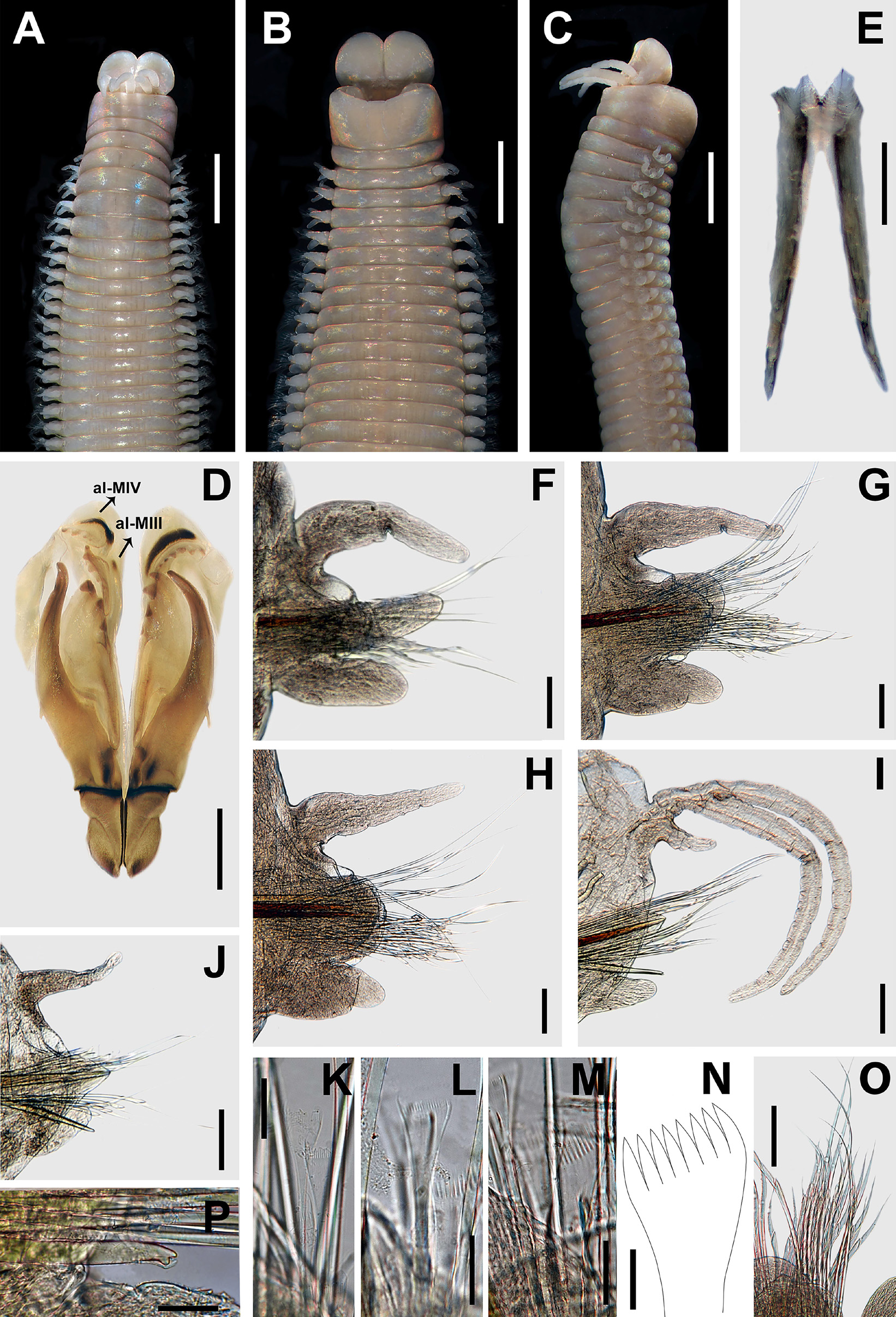
Prostomium bilobed, 1.3 mm long, 2 mm wide; lobes frontally rounded; median sulcus shallow anteriorly and deep ventrally ( Fig. 6 View FIGURE 6 A–B). Prostomial appendages in a semicircle, median antenna isolated by a gap. Palps reaching second peristomial ring; lateral antennae reaching first chaetiger; median antenna reaching the middle of first chaetiger. Palpophores and ceratophores ring-shaped, short, thick; palpostyles and ceratostyles tapering, slender. Eyes brown, reniform, between palps and lateral antennae.
Peristomium ( 1.5 mm long, 3.5 mm wide) wider than prostomium, first ring two times longer than second ring, separation between rings distinct on all sides ( Fig. 6 View FIGURE 6 A–C). Ventral lip with a shallow central anterior depression with a couple of shallow wrinkles ( Fig. 6B View FIGURE 6 ).
Maxillary apparatus with MF = 1 + 1, 5 + 5, 6 + 0, 3 + 7, 1 + 1 ( Fig. 6D View FIGURE 6 ). MI three times longer than length of maxillary carriers. MI forceps-like, MI 4.8 times longer than length of closing system ( Fig. 6 View FIGURE 6 D–E); ligament between MI and MII, sclerotized. MII with recurved triangular teeth; MII three times longer than length of cavity opening ( Fig. 6 View FIGURE 6 D–E); ligament between MII and MIII and right MIV slightly sclerotized. MIII with triangular teeth; with irregular attachment lamella, situated in central of the right edge of the plate, sclerotized ( Fig. 6 View FIGURE 6 D–E). Left MIV with two lateral teeth larger than rest; attachment lamella semicircle, wide, better developed in left side, situated 2/3 of anterior edge of maxilla, sclerotized. Right MIV with four lateral teeth larger than rest; attachment lamella semicircle, wide, better developed in left side, situated along posterior edge of maxilla, sclerotized ( Fig. 6 View FIGURE 6 D–E). MV rectangular, wider than longer, with a short, rounded tooth. Mandibles dark; calcareous cutting plates broken, sclerotized cutting plates brown, with up to 12 growth rings ( Fig. 6F View FIGURE 6 ).
Pectinate branchiae with up to three long filaments, present from chaetigers 20L–21R to 131L–134R ( Fig. 6 View FIGURE 6 J–K). First two and last 21 chaetigers with one filament; reaching the maximum three filaments in chaetigers 33L to 99L ( Fig. 9F View FIGURE 9 ). Branchiae with filaments longer than dorsal cirri except in first and last four branchiae.
First four parapodia smaller, best developed in chaetigers 5–40, following ones becoming gradually smaller. Dorsal cirri conical in all chaetigers; longer than ventral cirri in anterior chaetigers, shorter in median, become similar size in posterior chaetigers; best developed in chaetigers 2–25, following ones gradually smaller ( Fig. 6 View FIGURE 6 G–K). Prechaetal lobes short, as transverse folds in all chaetigers ( Fig. 6 View FIGURE 6 G–K). Chaetal lobes in first 39 chaetigers rounded, shorter than postchaetal lobe, aciculae emerging dorsal to midline; from chaetiger 40 triangular, longer than other lobes, with acicula emerging dorsal to midline ( Fig. 6 View FIGURE 6 G–K). Postchaetal lobes well developed in the first 57 chaetigers; conical in the first six chaetigers, ovoid in chaetigers 7–10, rounded and progressively smaller from chaetiger 11; from chaetigers 58 inconspicuous ( Fig. 6 View FIGURE 6 G–K). Ventral cirri ovoid in the first five chaetigers; in chaetigers 6 to 107 with an oval swollen base and digitiform tip; conical from chaetiger 108 with digital end, gradually reducing in size ( Fig. 6 View FIGURE 6 G–K).
Aciculae blunt, reddish along most of its length, and amber on the distal tip ( Fig. 6 View FIGURE 6 G–K). First four chaetigers with three aciculae; in chaetigers 5–19 with four aciculae; in chaetigers 20–42 with three aciculae; in chaetigers 43–94 with two aciculae; from chaetigers 95 with only one acicula.
Limbate chaetae of two lengths in same chaetiger: long and short, long blade in dorsal position, short blades in ventral position; limbate chaetae reduced in number around chaetiger 18, and then maintained until a similar number until the posterior end. Five types of pectinate chaetae; in anterior chaetigers, thin, isodont narrow, symmetric, with long and slender teeth, 1–2 chaetae with up to 11–12 teeth ( Fig. 6L View FIGURE 6 ); in median chaetigers, thick, isodont wide, symmetric, with short and slender teeth, 1–2 chaetae, up to 17 teeth ( Fig. 6M View FIGURE 6 3 View FIGURE 3 ); in posterior chaetigers, and thick, isodont wide, symmetric, with long and slender teeth, three chaetae, up to 17 teeth ( Fig. 6N View FIGURE 6 1 View FIGURE 1 ), thick, anodont wide, symmetric, with long and thick, 3–4 chaetae, up to seven teeth ( Fig. 6O View FIGURE 6 ), and thick, anodont wide with long and slender, 3–4 chaetae, up to 14 teeth ( Fig. 6N View FIGURE 6 2 View FIGURE 2 ). Compound spinigers present throughout; with blades of two lengths: shorter blades more abundant than longer blades ( Fig. 6P View FIGURE 6 ). Subacicular hook bidentate, translucent; starting from chaetiger 35, one or two per chaetiger, second completely internal within the parapodia and probably a replacement hook, present in all chaetigers subsequently; with blunt teeth, distal tooth smaller than proximal, directed upward, proximal triangular, directed laterally ( Fig. 6Q View FIGURE 6 ).
Pygidium with dorsal pair of anal cirri as long as the last 13 chaetiger; ventral pair short, as long as the last two chaetigers.
Variation. Material examined with L10 = 6.4–17 mm, W10 = 3.5–5.3 mm, TChae = 175–231. Palps reaching middle of first peristomial ring or middle of the second peristomial ring; lateral antennae reaching second peristomial ring or middle of first chaetiger; median antenna reaching first or second chaetiger. The maxillary formula is variable: MII 4–5 + 4–5, MIII 6, MIV 3–4 + 7–9. The proportions of the maxillary apparatus vary as follows: MI are 2.6–3.1 times longer than length of maxillary carriers; MI are 4.5–4.9 times longer than length of closing system; MII are 4.5–5.8 times longer than length of cavity opening. Branchiae from chaetigers 20–27 to 31–41 chaetigers before pygidium. Maximum number of branchial filaments varied from three to five and postchaetal lobes were conspicuous in first 48–70 chaetigers. Ventral cirri with a swollen base from chaetigers 5–7 to 54–68 chaetigers before of pygidium. Start of subacicular hooks in chaetigers 35–75.
Distribution. New Jersey, Rhode Island, Massachusetts, North Carolina, USA.
Habitat. According to Leidy (1855), the specimens were found co-occurring with Lumbrinereis splendida de Blainville, 1828 (b) (= Lumbrineris lumbricalis ( de Blainville, 1825)) in an oyster bed. Verrill (1873) commented that M. leidii lives under stones at low-water mark, but is more common on shelly bottoms in shallow water offshore. In North Carolina, the specimens lives in stiff clay, or in exposed muddy banks at moderately low water, also under boards or large stones ( Hartman 1945). Some specimens studied here were found in mud (USMN 4469, USNM 6194).
Reproduction. According to Pettibone (1963), the eggs are released in firm gelatinous masses between June and July in the Woods Hole region. Hartman (1945) reported that juvenile stages could be found around June in North Carolina.
Remarks. The name Marphysa leidii was assigned by de Quatrefages (1866) to the specimen identified and described by Leidy (1855) as Eunice sanguinea from Great Egg Harbor, New Jersey. de Quatrefages explained the main difference between the species is the beginning of branchiae, starting from chaetiger 60 in M. leidii , whereas in M. sanguinea the branchiae have an earlier beginning. However, Webster (1879) clarified that de Quatrefages misinterpreted the beginning of the branchiae (Leidy described it from chaetiger 16 and not in chaetiger 60). Hence, Webster (1879) considered M. leidii as a small specimen of M. sanguinea with branchiae emerging earlier.
Specimens studied by Leidy are lost; apparently, these materials were not formally deposited in the Academy of Natural Sciences of Philadelphia ( Salazar-Vallejo 2014). However, enough material deposited at the Smithsonian was found; one of the specimens was collected near to the type locality (about 35 km north). Herein, that specimen was assigned as neotype, according to Art. 75 (ICZN 1999), which suggests assigning a neotype to clarify the taxonomic position of a species.
Marphysa leidii is different from M. sanguinea because the former has translucent subacicular hooks and the postchaetal lobe is conical in the first three chaetigers, whereas in latter, the subacicular hook is reddish at the base and translucent distally, and the postchaetal lobe is digitiform in first chaetiger. Moreover, M. leidii has two types of the pectinate anodont, whereas M. sanguinea only has one type of pectinate anodont.
Marphysa leidii resemble M. brasiliensis ( Brazil) , M. bulla (Yellow Sea, China), M. californica (San Diego, California), M. formosa Steiner & Amaral, 2000 ( Sao Paulo, Brazil), M. haemasoma ( South Africa), M. maxidenticulata (Yellow Sea, China), M. parishii ( Brazil) , and M. baileybrockae n. sp. (Hawaii) by having pectinate branchiae with long filaments, the limbate chaetae subacicular absent, the subacicular hook translucent, and the dorsal cirri conical in all chaetigers. However, M. leidii has conical postchaetal lobe in first four chaetigers, whereas in M. brasiliensis , M. bulla , M. californica , M. maxidenticulata , M. parishii , and M. baileybrockae n. sp. the postchaetal lobe is conical with distal edge directed to dorsal side, and in M. formosa and M. haemasoma , the postchaetal is ovoid in the first four chaetigers. Furthermore, M. parishii has five types of pectinate chaetae as in M. leidii , whereas in M. californica , M. haemasoma , and M. maxidenticulata , there are only four types, and in M. brasiliensis , M. formosa , and M. baileybrockae n. sp., there are only three types of pectinate chaetae. Also, M. leidii has eyes, but M. bulla lacks eyes. Finally, M. leidii ( type and additional material, L10 = 10.7–17 mm) has the postchaetal lobe well developed in first 48–70 chaetigers, yet in M. parishii ( type material, L10 = 17.2 mm) the postchaetal is developed in first 292 chaetigers. The comparison of M. leidii with related species is provided in Table 1.


| V |
Royal British Columbia Museum - Herbarium |
No known copyright restrictions apply. See Agosti, D., Egloff, W., 2009. Taxonomic information exchange and copyright: the Plazi approach. BMC Research Notes 2009, 2:53 for further explanation.
|
Kingdom |
|
|
Phylum |
|
|
Class |
|
|
Order |
|
|
Family |
|
|
Genus |
Marphysa leidii de Quatrefages, 1866
| Molina-Acevedo, Isabel C. & Idris, Izwandy 2020 |
Marphysa sanguinea
| Pettibone, M. H. 1963: 236 |
| Hartman, O. 1945: 23 |
| Hartman, O. 1944: 339 |
| Fauvel, P. 1911: 18 |
| Webster, H. E. 1879: 36 |
Marphysa leidyi
| Verrill, A. E. 1873: 319 |
Marphysa leidii
| de Quatrefages, A. 1866: 22 |
Eunice sanguinea
| Leidy, J. 1855: 147 |