
Ogovea cameroonensis, Giribet, Gonzalo & Prieto, Carlos E., 2003
|
publication ID |
https://doi.org/10.5281/zenodo.156214 |
|
DOI |
https://doi.org/10.5281/zenodo.6273703 |
|
persistent identifier |
https://treatment.plazi.org/id/E36E87A7-FFB6-FFF9-FECF-32FFFAECFBF5 |
|
treatment provided by |
Plazi |
|
scientific name |
Ogovea cameroonensis |
| status |
sp. nov. |
Ogovea cameroonensis View in CoL new species
Figs. 1–32 View FIGURES 1 – 2 View FIGURES 3 – 4 View FIGURES 5 – 9 View FIGURES 10 – 15 View FIGURES 16 – 20 View FIGURES 21 – 29 View FIGURES 30 – 32
Type specimens. Male holotype from the forêt d’Ototomo, Région de Yaoundé ( Cameroon), collected 16 December 1968 by J.L. Amiet, deposited at MHNG. One male and three female paratypes, same collection data as holotype, deposited at MHNG. One male and one female paratypes, same collection data as holotype, deposited at MCZ (material used for SEM). Two female paratypes, collected 5 February 1967 by J.L. Amiet, same locality as holotype, deposited at MHNG. Three male and two female paratypes, collected 4–6 April 1969 by J.L. Amiet, same locality as holotype, deposited at MHNG. One female paratype, collected 17 May 1969 by J.L. Amiet, same locality as holotype (with coordinates specified as 3º 40’ N, 11º 18’ E and altitude specified as 700–800 m), deposited at MHNG. One female paratype, collected 30–31 December 1968 by J.L. Amiet, same locality as holotype, deposited at MHNG. One male (penis studied from this specimen) and one female paratypes, collected by J.L. Amiet in Yaoundé, deposited at MRAC 134.818.
3. The type material of O. nasuta is supposed to be deposited at MCSN. Despite several letters and emails to the curators and administrators of the MCSN, no response has been obtained. Fortunately, a male specimen,
supposedly of the type series, was somehow transferred to the ZMUC, making possible the study of this species. 4. Shear (1979) mentioned that one male and one juvenile paratypes were deposited at the MCZ accompany
ing the holotype, however, the tube labeled as “ paratypes ” contains three males and one juvenile.
5. We have not been able to locate the supplement of volume 24 of The Entomologist with the description of
Other material. Male collected 5 February 1967 by J.L. Amiet, same locality as holotype, deposited at MHNG.
Etymology. The species is named after its country of origin.
Diagnosis. This species is the largest in the genus, reaching up to 5 mm in body length. The tricuspidate process of the anterior margin of the scutum is short, with its middle cusp shorter than the lateral ones. The tricuspidate process interlocks with the dorsal crest of the chelicerae; such crest is more conspicuous than in other species of the genus.
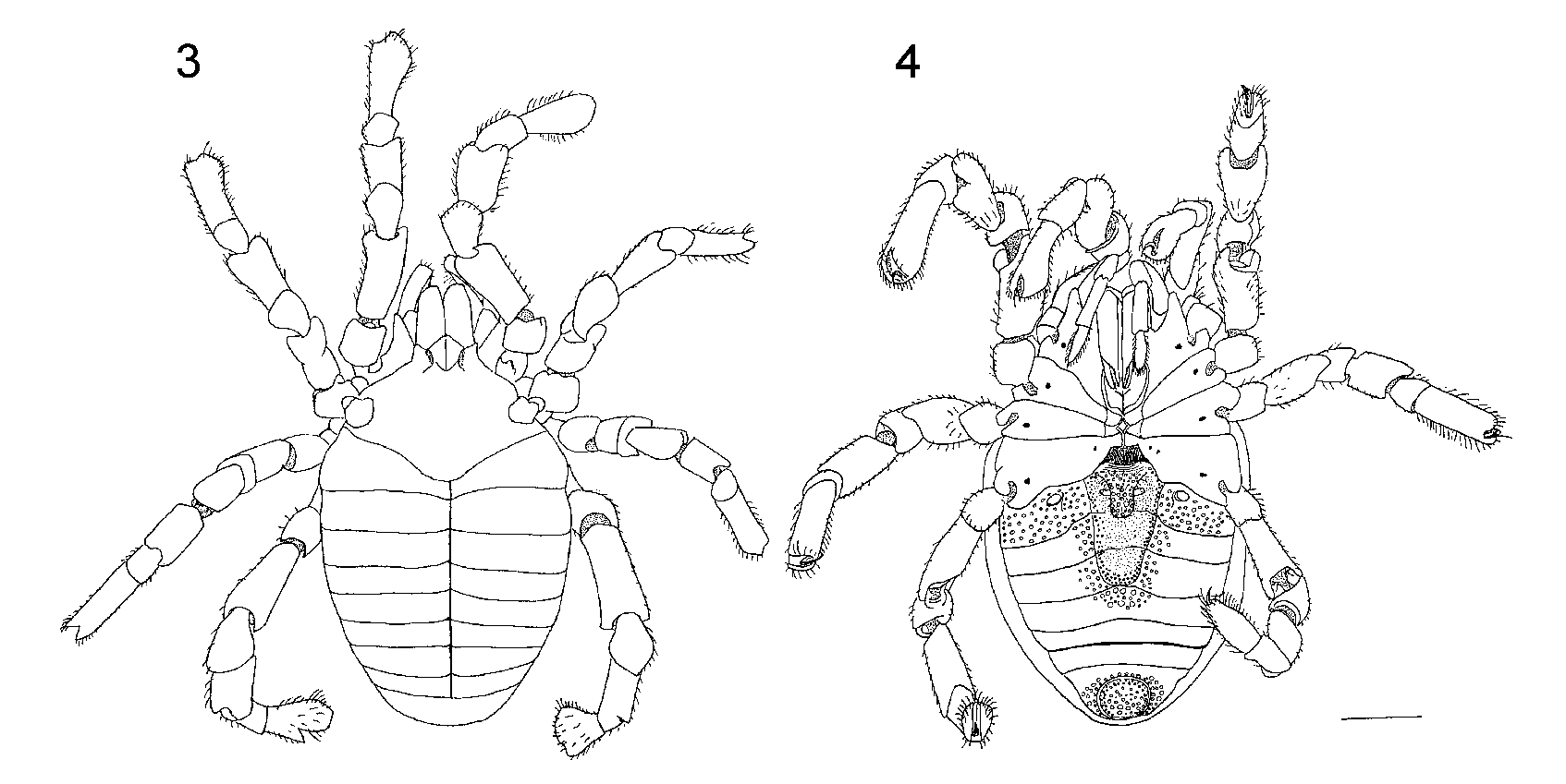
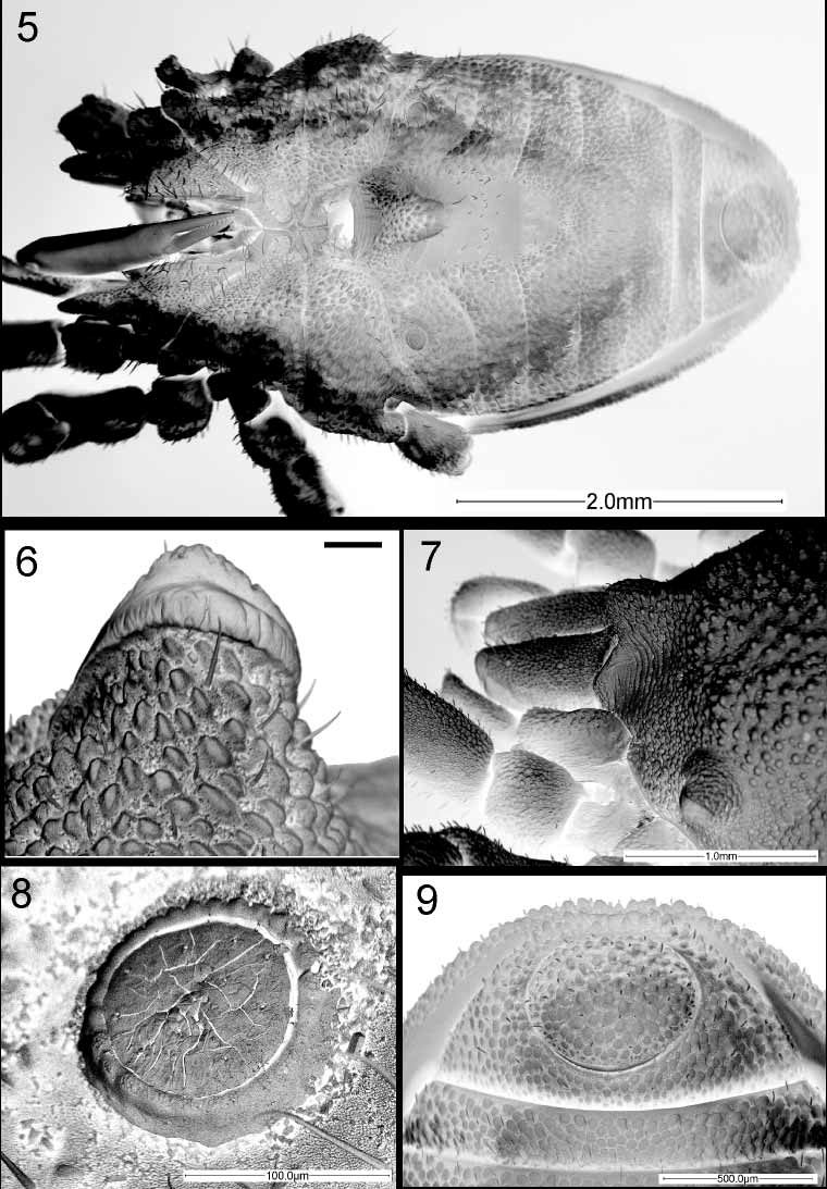
Description. Total length of male holotype ( Figs. 1–4 View FIGURES 1 – 2 View FIGURES 3 – 4 ) (female paratype in parenthesis) 5.0 (5.0), greatest width 2.6 (2.8) at the merging of prosoma and opisthosoma; lengthwidth ratio 1.92 (1.79). Body orange (in alcohol) with most of the dorsal and ventral surfaces and legs showing a dense tuberculategranulate microstructure (nomenclature on ornamentation follows Murphree 1988). Anterior margin of dorsal scutum forming a tricuspidate process (“processus triangulaire prolongeant” of Juberthie 1969), but the median “cusp” (the actual process) barely reaches the other two cusps, allowing the basal cheliceral article to extend horizontally ( Fig. 7 View FIGURES 5 – 9 ). Prosomal region short. Eyes absent. Ozophores conical, raised from the carapace margin, with the aperture resembling closed lips ( Fig. 6 View FIGURES 5 – 9 ). Transverse prosomal sulcus conspicuous ( Figs. 1 View FIGURES 1 – 2 , 3 View FIGURES 3 – 4 ). Transverse opisthosomal sulci equally conspicuous ( Figs. 1 View FIGURES 1 – 2 , 3 View FIGURES 3 – 4 ). Middorsal longitudinal opisthosomal sulcus conspicuous, starting towards the end of tergite I until tergite VIII; tergite IX lacking the middorsal longitudinal sulcus ( Figs. 1 View FIGURES 1 – 2 , 3 View FIGURES 3 – 4 ). Dorsal scutum without special modifications.
Coxae of leg I movable, coxae of the remaining legs fused. Ventral prosomal complex of male with coxae II and IV meeting in the midline, and coxae III barely touching each other ( Figs. 4–5 View FIGURES 3 – 4 View FIGURES 5 – 9 ). Gonostome in the shape of a flattened oval, much wider than long ( Fig. 5 View FIGURES 5 – 9 ). Ventral prosomal complex of females with coxae II, III, and IV not meeting in the midline, and coxae I forming the anterior wall of the gonostome.
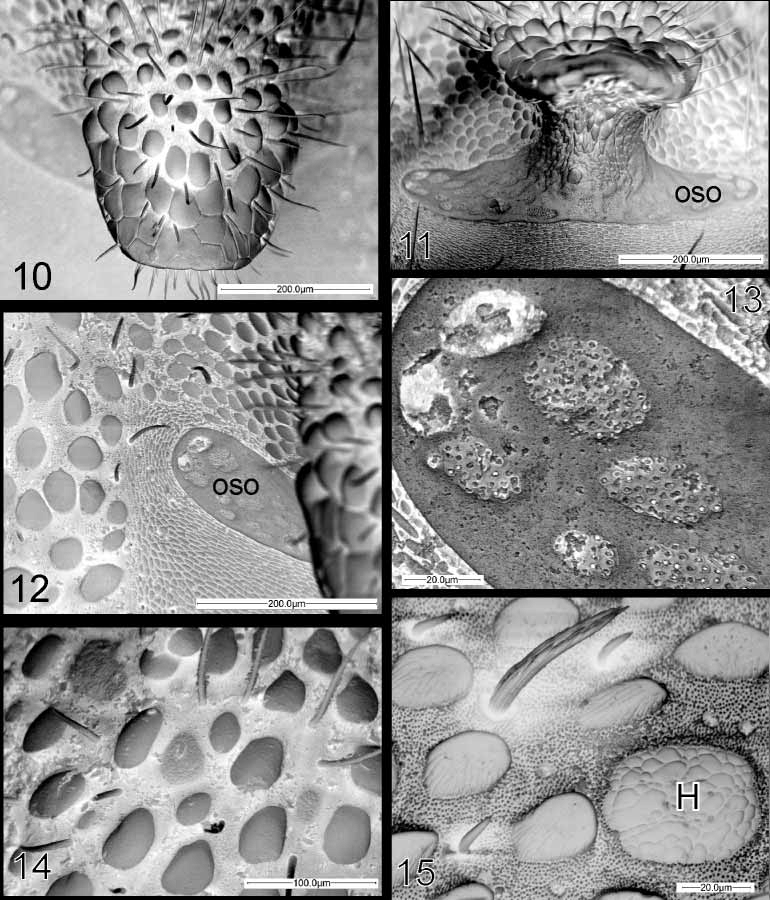
Spiracles small and forming a circle completely closed ( Fig. 8 View FIGURES 5 – 9 ). A large, broad, oblong, subacute process directed downwards and backwards projects behind the gonostome, in the sternal region of the second opisthosomal segment of the male ( Figs. 2 View FIGURES 1 – 2 , 4 View FIGURES 3 – 4 , 5 View FIGURES 5 – 9 , 10–12 View FIGURES 10 – 15 ) (see Hansen 1921; Shear 1980). The process bears numerous long setae ( Figs. 10– 12 View FIGURES 10 – 15 ). The sternal region of segments 2–4 forms a concave depression free of granulation underneath the sternal apophysis, with sternite 5 becoming somehow convex ( Fig. 5 View FIGURES 5 – 9 ). A special structure in the shape of a half moon is present at the base of the apophysis, and is observed at both sides ( Figs. 11–13 View FIGURES 10 – 15 ). Sternites 8 and 9 and tergite IX completely fused forming a corona analis ( Fig. 9 View FIGURES 5 – 9 ). Anal plate without modifications; anal gland pore absent; sexual dimorphism not present ( Fig. 9 View FIGURES 5 – 9 ).
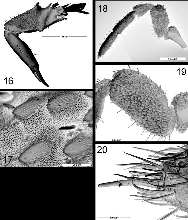
Chelicerae ( Fig. 16 View FIGURES 16 – 20 ) relatively short and strong, with numerous long setae, especially on the second article. Proximal article with ectal surface granulated, a conspicuous dorsal crest, and an inconspicuous ventral process. The dorsal crest collides with the internal surface of the elongated anterior prosomal process, not allowing the extension of the chelicerae forward. Second article fairly robust, lacking granulation. Dentition uniform and similar on both cheliceral fingers. Granulation of the chelicera of special type ( Fig. 17 View FIGURES 16 – 20 ) with short denticlelike setae. Chelicerae lacking Hansen’s organ.
Palps ( Figs. 18–20 View FIGURES 16 – 20 ) similar to those of Ogovea grossa illustrated by Juberthie (1969: Fig. 9 View FIGURES 5 – 9 ), with a trochanter that thins considerably at the base and widens in the trochanterofemoral joint; without a ventral process ( Fig. 18 View FIGURES 16 – 20 ). Femur compressed laterally, suboval in shape ( Fig. 19 View FIGURES 16 – 20 ), and folded slightly over the chelicera, so that the tibiae of both palps enclose the chelicerae ( Fig. 7 View FIGURES 5 – 9 ).
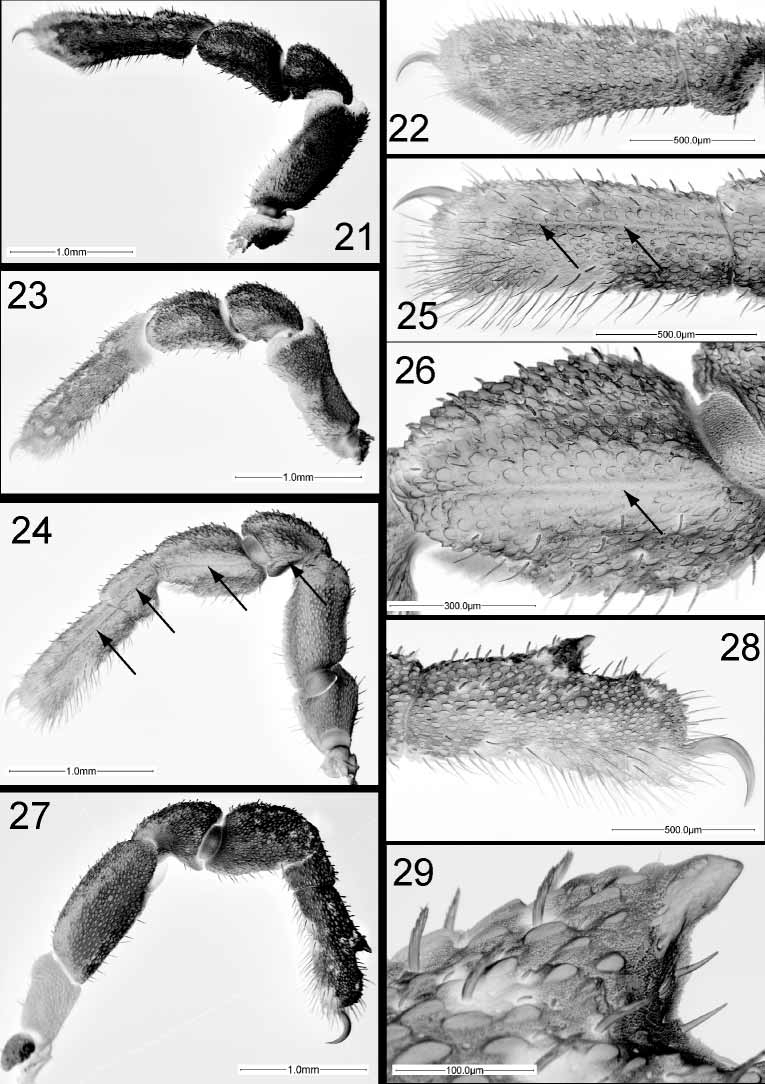
Legs short and robust ( Figs. 21, 23, 24, 27 View FIGURES 21 – 29 ); with all claws smooth, long and hooklike, without dentition or lateral pegs. Surface of all articles, including the base and the dorsal surface of tarsi I and II, clearly and completely ornamented with granules ( Figs. 22, 25, 28 View FIGURES 21 – 29 ). Ventral side of tarsus I with a concentration of short sensory hairs occupying about half of the tarsal length and forming a distinct solea ( Fig. 22 View FIGURES 21 – 29 ). All tarsi with a large dorsal groove for retracting the claws. Patella, tibia, metatarsus and tarsus of leg III with a longitudinal seam in the ectal side ( Figs. 24–26 View FIGURES 21 – 29 ). Tarsus IV of male entire ( Fig. 28 View FIGURES 21 – 29 ), carrying a short thornlike adenostyle that ends abruptly at the beginning of the tarsal groove, past the tarsal half; the adenostyle not ending in a tuft of setae or any bushy formations ( Fig. 29 View FIGURES 21 – 29 ). Tarsus IV of females without modifications.
Penis with numerous setae that make the correct interpretation of all the structures difficult. Ventral plate hypertrophied ( Figs. 30–32 View FIGURES 30 – 32 ) with 4–5 rows of short setae ( Figs. 31–32 View FIGURES 30 – 32 ) in the most distal part of the dorsal side. Ventral setae in a single series. Median plate sclerotized and with 4 apical setae ( Figs. 31–32 View FIGURES 30 – 32 ). Membranous structure around gonopore. Dorsal setae long, forming two groups.
Cuticular structures. The Hansen’s organ ( Figs. 15 View FIGURES 10 – 15 , 28 View FIGURES 21 – 29 ) is found in the surface of all four coxae and legs. On coxae I, II, and III it is found near the socket where the trochanter’s condyle inserts; on coxae IV there is one organ in a similar position, but a much larger one (splitting in two in some cases) occurs near the gonostome opening. Concentrations of these structures occur in most articles of the leg, e.g. up to three per article on tarsus IV.
Distribution. Known only from the type locality.
Remarks. This species is most similar to Ogovea grossa from Gabon, but the new species is much larger, and both species can be differentiated by the anterior process, which appears tricuspidate from above, but the median “cusp” (the actual process) barely reaches the other two cusps in O. cameroonensis , while in O. grossa it is clearly more developed. The chelicerae of O. cameroonensis are longer than those of O. nasuta , and about the same proportional shape than those of O. grossa , but they differ in the dorsal crest, which is much more conspicuous in O. cameroonensis .
A small male, measuring 3.9 mm, was collected from the type locality. The specimen resembles O. cameroonensis in salient morphological features and was collected together with two of the female paratypes. The male specimen has a teratologic right leg IV where the tarsus and tibia are not properly developed and the tarsus lacks an adenostyle. We are uncertain about whether the specimen belongs to a similar species of smaller proportions.


No known copyright restrictions apply. See Agosti, D., Egloff, W., 2009. Taxonomic information exchange and copyright: the Plazi approach. BMC Research Notes 2009, 2:53 for further explanation.