
Spongicola liosomatus, Quintal & Goy, 2019
|
publication ID |
https://doi.org/ 10.11646/zootaxa.4648.2.12 |
|
publication LSID |
lsid:zoobank.org:pub:F6BF068E-606B-471D-B6BD-017394387AA9 |
|
DOI |
https://doi.org/10.5281/zenodo.5945012 |
|
persistent identifier |
https://treatment.plazi.org/id/EC0487C2-0653-FFCD-B0D2-9828FC90F8E8 |
|
treatment provided by |
Plazi |
|
scientific name |
Spongicola liosomatus |
| status |
sp. nov. |
Spongicola liosomatus View in CoL sp. nov.
( Figs. 1–4 View FIGURE 1 View FIGURE 2 View FIGURE 3 View FIGURE 4 )
Material examined. HOLOTYPE: Venezuela. Caribbean Sea, R / V Dr. Fridtof Nansen, stn 477, northern Blanquilla Island , 11°54’N, 64°17’W, 135–160 m, 2.VI.1988, bottom trawling net, male (cl 5.2 mm) inside hexactinellid sponge ( USNM 1573503 About USNM ) GoogleMaps . PARATYPES: Venezuela. Caribbean Sea, R / V Fridtof Nansen, stn 477, northern Blanquilla Island , 11°54’N, 64°17’W, 135–160 m, 2. VI GoogleMaps .1988, bottom trawl net, 2 males (cl 3.8mm; cl 5.4 mm) inside hexactinellid sponge ( USNM 1573504 About USNM ) ; 1 female (cl 4.7 mm) inside hexactinellid sponge (MMM-Crus-0300).
Diagnosis. Small commensal spongicolid shrimp associated with sponges, with slightly depressed body bearing few carapacial spines. Rostrum with dorsal margin slightly upturned toward distal end, bearing 2 to 4 dorsal teeth and small projection at posterior end, ventrally with distal tooth. Carapace without postrostral submedian spine, hepatic spine present in line with antennal spine, 3 or 4 anterolateral spines, 4 to 6 pterygostomian spinules. Abdominal terga entirely smooth, pleura of first to fifth somites broadly rounded without marginal spines. Mandibular palp 2-segmented. First and second pereiopods cutting edges with membranous ridge, palm inflated; chela of third pereiopod with serrated upper margin, lower margin glabrous.
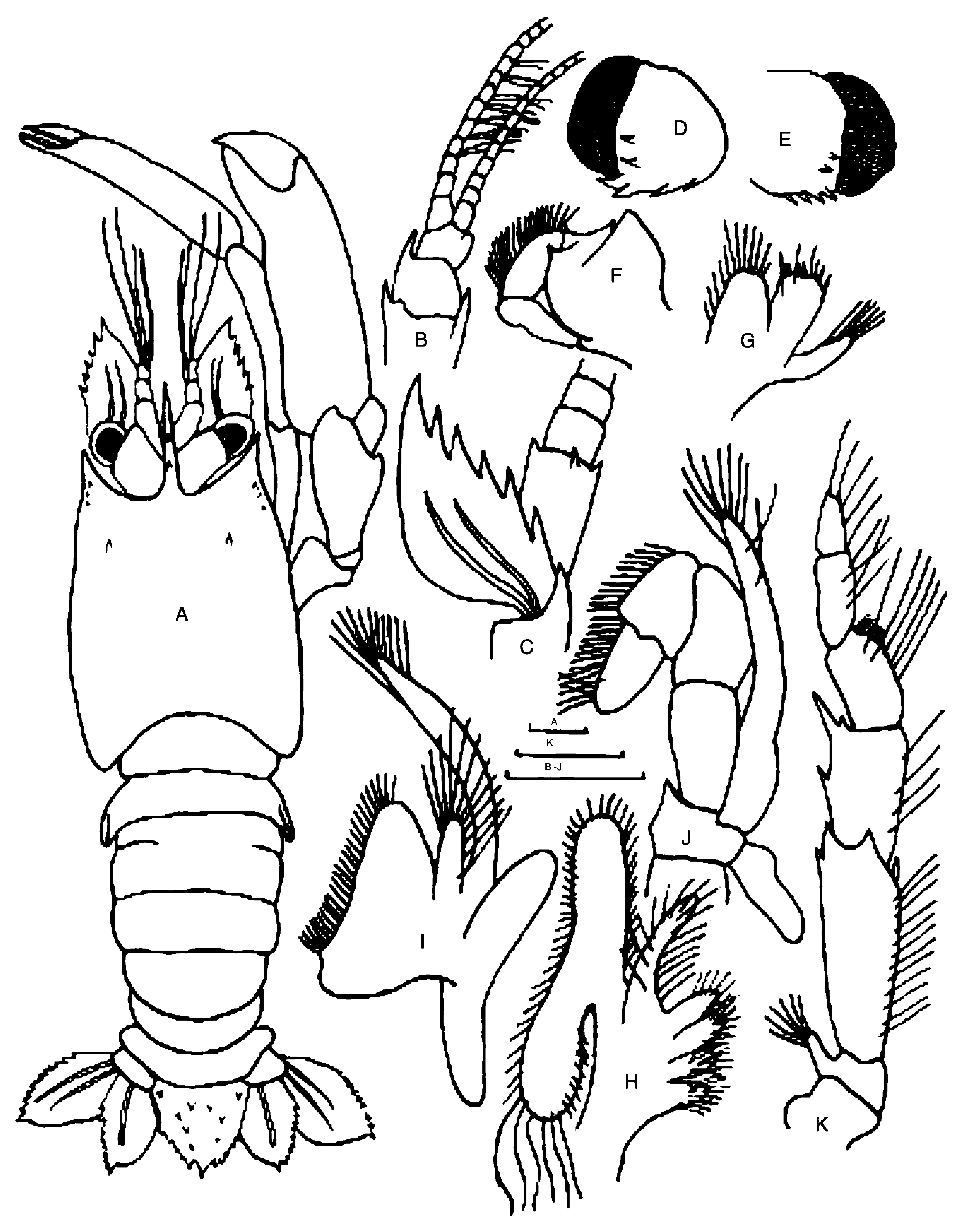
Description. (Holotype, male). Rostrum ( Figs. 1 View FIGURE 1 , 2A View FIGURE 2 ) compressed, triangular at base, reaching slightly past end of second segment of antennular peduncle; dorsally upturned toward distal end, bearing 2 dorsal teeth and small projection at posterior end, ventrally with distal tooth. Carapace ( Figs. 1 View FIGURE 1 , 2A View FIGURE 2 ) subcylindrical, surface glabrous; cervical groove absent; postrostral submedian teeth absent; antennal tooth small, acuminate; hepatic tooth small; 3 anterolateral spines; pterygostomial angle produced, rounded with 6 spinules.
Abdomen ( Figs. 1 View FIGURE 1 , 2A View FIGURE 2 ) smooth; first to sixth terga glabrous; pleura of first to fifth somites broadly rounded without marginal spines; no spiniform process on sixth somite. First somite with distinct transverse carina anteriorly flowing into transverse groove. Second and third somites with faint transverse grooves about midlength. Telson ( Figs 2A View FIGURE 2 , 4A View FIGURE 4 ) lanceolate equally long as broad; with 2 longitudinal carinae on dorsal surface provided with 2 or 3 spines; 2 more spines between bases of carinae; 1 dorsal spine present on each side of base of telson; lateral margin with 3 or 4 spines on each side; posterior margin fringed with long setae (not shown) and provided with 3 spines, 2 subterminal flanking large median terminal.
Eyes ( Figs. 1 View FIGURE 1 , 2A, D, E View FIGURE 2 ) comparatively large, eyestalk covering proximal one-third of antennal scale in dorsal view; with 4 spinules on inner margin, 2 spinules on dorsal surface; cornea darkly pigmented, semi-globular, about same width as eyestalk.
Antennular peduncle ( Figs. 2A, B View FIGURE 2 ) with basal article more than twice as long as second segment, third segment about 0.75 second segment. Stylocerite small, acuminate, reaching near distal margin of third segment, large spine on inner margin reaching middle of second segment. Second segment with distolateral margin bearing long spine. Outer antennular flagellum slightly longer than inner flagellum, proximal portion bearing 7 paired aesthetascs, no setae present.
Antenna ( Fig. 2C View FIGURE 2 ) short. Basicerite stout, bearing distolateral spine, ventrolateral margin unarmed; antennal scale semicircular, about twice as long as broad; outer margin slightly concave, with 6 teeth, terminal one largest, dorsal surface with 2 distinct longitudinal carinae. Antennal flagellum shorter than carapace.
Mandible ( Fig. 2F View FIGURE 2 ) with 2-segmented palp; incisor and molar processes fused bearing few teeth.
Maxillule ( Fig. 2G View FIGURE 2 ) with simple, slender endopod tapering distally; coxal endite suboval, with submarginal row of setae on outer surface; basal endite moderately broad, truncated distally with several setae.
Maxilla ( Fig. 2H View FIGURE 2 ) with curved, slender endopod; coxal and basal endites deeply bilobed; scaphognathite well developed; posterior lobe slightly elongate, suboval terminal margin with very long setae.
First maxilliped ( Fig. 2I View FIGURE 2 ) with endopod unsegmented; basal endite large, subtriangular, with concave mesial margin; exopod well developed; epipod large, distinctly bilobed.
Second maxilliped ( Fig. 2J View FIGURE 2 ) with endopod 7-segmented with coxa and basis fused; ischium short, distal margin sharply pointed; merus and carpus of equal length; propodus cup–shaped; dactylus suboval equal propodal length; podobranch simple; exopod well developed, flagellar.
Third maxilliped ( Fig. 2K View FIGURE 2 ) with 7-segmented endopod; basis short; ischium elongate, distal margin with 3 spines; merus half ischial length, outer margin with small midlength spine, distal end with 1 large, 1 small spine; carpus with well developed distal grooming apparatus, equal propodal length; dactylus tapering distally; exopod rudimentary.
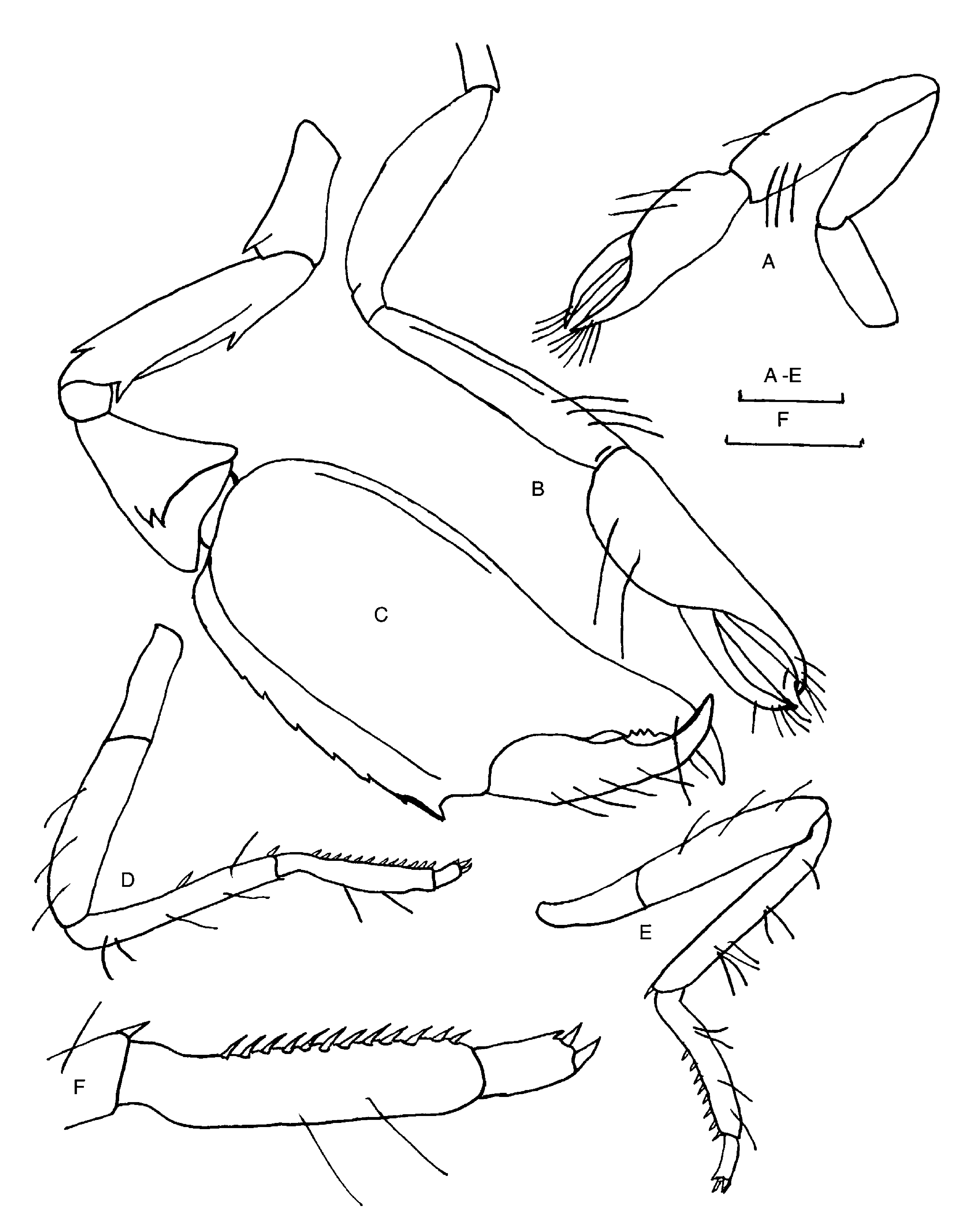
First pereiopod ( Fig.3A View FIGURE 3 ) slender, glabrous, chelate; fingers with membranous ridge on cutting edge. Dactylus 0.33 times length of palm distally ending with setal brush; palm slightly inflated also with setal brush. Carpus longest segment, about 1.5 times as long as chela. Grooming apparatus lacking. Merus about 0.8 times length of carpus. Ischium shorter than merus.
Second pereiopod ( Fig. 3B View FIGURE 3 ) glabrous, slightly longer than third pereiopod, overreaching antennal scale with carpus. Finger cutting edges composed of membranous ridges; dactylus and palm ending in setal brushes; palm inflated, twice dactylar length. Carpus, merus equal in length; ischium 0.5 meral length.
Third pereiopod ( Fig. 3C View FIGURE 3 ) chela large, strongly compressed laterally, almost equal to carapace length; ventral margin glabrous; dorsal margin distally serrate with 7 teeth; fingers distally curving inward, crossing, each ending in sharp point, opposable margin of movable finger with prominent median tooth fitting to large opposing concavity; cutting edge distally armed with 4 small teeth. Carpus triangular, dorsomesial distal angle produced into subtriangular lobe ending in 2 large teeth. Merus 0.7 length of palm, with 1 distal tooth dorsally, ventromedially with large tooth, distal end with large tooth. Ischium 0.5 meral length, strong distodorsal tooth.
Fourth pereiopod ( Figs. 3D, F View FIGURE 3 ) long, slender, sparsely setose; dactylus short, biunguiculate, ventral margin with small accessory tooth at base of ventral unguis; propodus 3.0 times dactylar length, with 12 movable spinules on ventrodistal margin; carpus 1.2 length of propodus, with ventromedial, ventrodistal spinules; merus 0.8 carpal length, glabrous; ischium distinctly shorter than merus.
Fifth pereiopod ( Fig. 3E View FIGURE 3 ) similar to fourth, with only ventrodistal carpal spinule, 8 movable ventrodistal spinules on propodus.
Gill formula typical for genus (cf. Saito & Komai 2008: table 1).
First pleopods unilobed, small, 2-segmented, feebly setose. Second to fifth pleopods bilobed, each fringed with long setae.
Uropods ( Fig. 4A View FIGURE 4 ) with protopod unarmed. Exopod with 8–10 teeth on outer margin; dorsal surface with 2 longitudinal ridges; inner and distal margins with long setae. Endopod with 10 or 11 teeth on outer margin; dorsal surface with longitudinal ridge; inner and distal margins with long setae.
Paratypes similar to holotype. No sexual dimorphism. Rostrum with 2 to 4 dorsal spines; pterygostomian spines vary from 2 to 6 on each side of carapace; telson with 5 or 6 lateral spines. Third pereiopods ( Figs. 4B, C View FIGURE 4 ) with palm bearing 8 or 9 small teeth on dorsal margin, cutting edge lacking small distal teeth; merus with 5 to 7 teeth on ventral margin.
Coloration. Carapace translucent with scarce red spots. Pereiopods translucent with red spots; carpus of fourth and fifth pereiopods with more intense red spots. Antennules with red stripes intercalated with translucent spaces; antennal flagellum similarly banded but starting 1/3 from the base.
Distribution. Known only from the type locality, off northern Blanquilla Island, Venezuela, Caribbean Sea, 135– 160 m.
Host. No information on the host was available.
Etymology. From the Greek “liosomatos” (= smooth-bodied), in reference to the reduced armature on the carapace and abdomen of the new species.
Remarks. The present new species is referred to the genus Spongicola because of the rudimentary exopod on the third maxilliped and the compressed chelae of the third pereiopods. The presence or absence of an exopod on the third maxillipeds has been considered important in separation of genera within the Spongicoloidae (de Saint Laurent & Cleva 1981; Holthuis 1993; Saito & Takeda 2003; Saito & Komai 2008; Goy 2010). However, the development of the exopod of the third maxilliped has been shown to be variable among species of Microprosthema ( Saito & Anker, 2014) . Moreover, species of Spongiocaris and Spongicoloides show some variation in the number of branchial exites (de Saint Laurent & Cleva 1981; Garcia Raso 1996) where certain exites can be missing, rudimentary, or well developed in specimens of the same species collected at one location. Differences are also evident in relation in the size and degree of development in some of the spongicoloids. These facts, plus the fragility of the gills and the relatively small number of known specimens, makes the usefulness of branchial formulae in separating species of spongicoloids suspect. A reappraisal of the characters used to separate the genera within Spongicoloidae is necessary ( Saito & Takeda 2003; Chen et al. 2016).
Spongicola liosomatus View in CoL sp. nov. appears closest to S. levigatus View in CoL , widely distributed in the western Pacific ( Saito & Komai 2008). These two species lack a postrostral submedian spine on the carapace; have smooth, rounded abdominal pleura; and have an unsegmented endopod on the first maxilliped. The carapacial and abdominal spination shows similarity to S. teres ( Komai, 2015) View in CoL from French Polynesia and the 2-segmented mandibular palp has only been found in Microprosthems looensis from the Florida Keys ( Goy & Felder 1988). The new species has inflated palms of the first and second pereiopods as seen in S. inflatus (de Saint Laurent & Cleva 1981) View in CoL . However, S. liosomatus View in CoL sp. nov. differs from these species and all other members of Spongicola View in CoL by the unserrated ventral margins of the third pereiopod chela and the membranous ridges present on the cutting edges of the first and second pereiopods. With the transfer of S. cubanicus to the genus Spongiocaris (Saito 2008) View in CoL , S. liosomatus View in CoL sp. nov. is the first member of Spongicola View in CoL sensu stricto to be found in the Atlantic Ocean.


| VI |
Mykotektet, National Veterinary Institute |
No known copyright restrictions apply. See Agosti, D., Egloff, W., 2009. Taxonomic information exchange and copyright: the Plazi approach. BMC Research Notes 2009, 2:53 for further explanation.
|
Kingdom |
|
|
Phylum |
|
|
Class |
|
|
Order |
|
|
Family |
|
|
Genus |
Spongicola liosomatus
| Quintal, Bladimir Rodríguez & Goy, Joseph W. 2019 |
Spongicola liosomatus
| Quintal & Goy 2019 |
S. liosomatus
| Quintal & Goy 2019 |
S. liosomatus
| Quintal & Goy 2019 |
Spongicola
| Quintal & Goy 2019 |
S. teres (
| Komai 2015 |
Spongiocaris
| Saito 2008 |
S. cubanicus
| Ortiz, Gomez & Lalana 1994 |
S. levigatus
| Hayashi & Ogawa 1987 |
S. inflatus
| de Saint Laurent & Cleva 1981 |
Spongicola
| de Haan 1844 |