
Schizochilus banesensis, Diez & Reygel & Artois, 2019
|
publication ID |
https://doi.org/10.11646/zootaxa.4646.1.1 |
|
publication LSID |
lsid:zoobank.org:pub:2A39D2E1-262F-423F-9B7F-89C376912FFC |
|
persistent identifier |
https://treatment.plazi.org/id/ED01879A-FFFD-FF94-FF68-F8BAFB720885 |
|
treatment provided by |
Plazi |
|
scientific name |
Schizochilus banesensis |
| status |
sp. nov. |
Schizochilus banesensis sp. n.
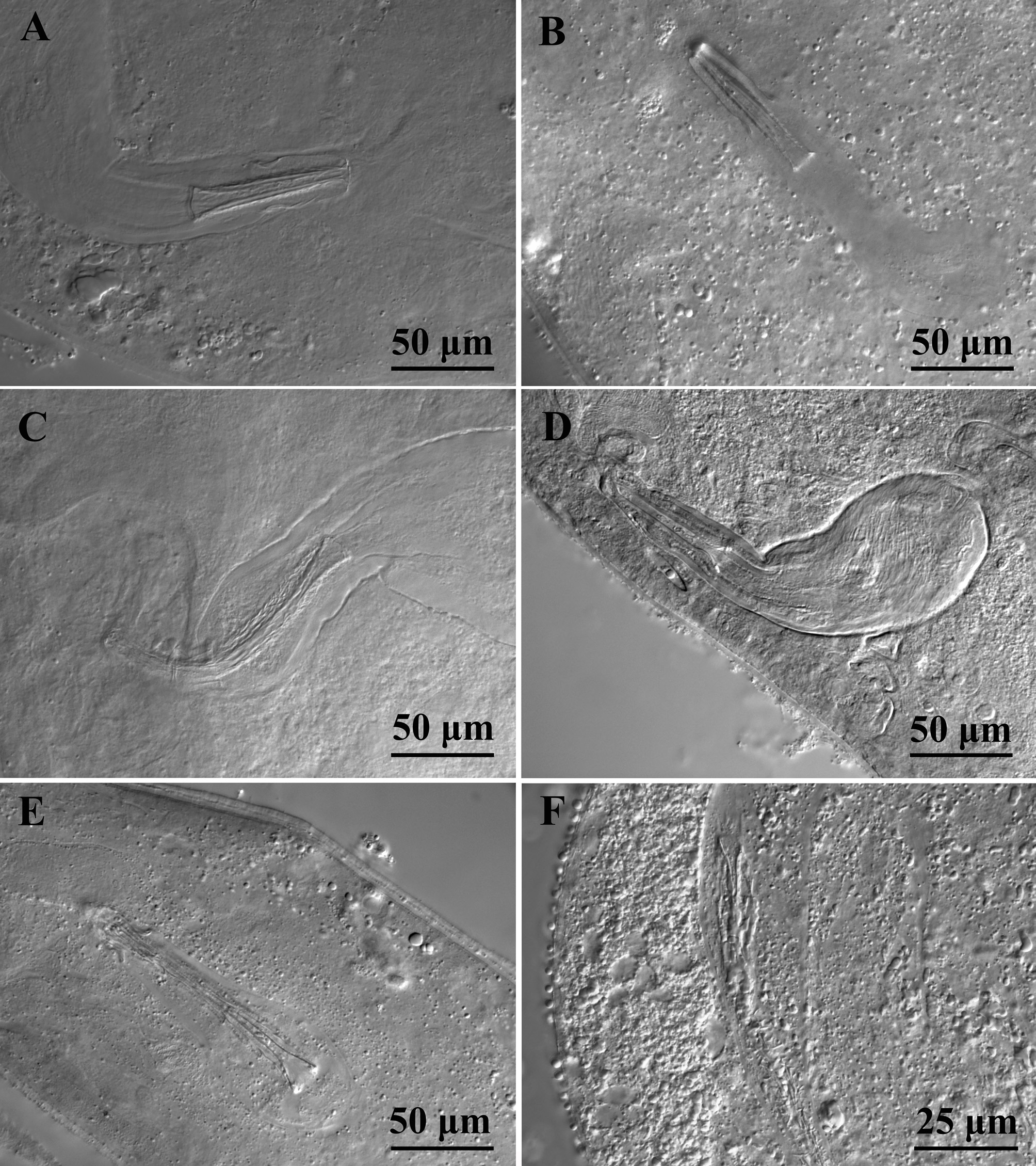
( Fig. 12G View FIGURE 12 , 15C View FIGURE 15 )
http://zoobank.org/ urn:lsid:zoobank.org:act:
Material. Observations on one live animal, whole mounted afterwards, designated holotype ( FMNH https:// id.luomus.fi/KV.619), collected in Playa Morales (type locality) ( January 4, 2017), intertidal, upper 10 cm of finegrained sand, salinity 35 ‰.
Etymology. The epithet refers to its occurrence in Banes, area of the type locality.
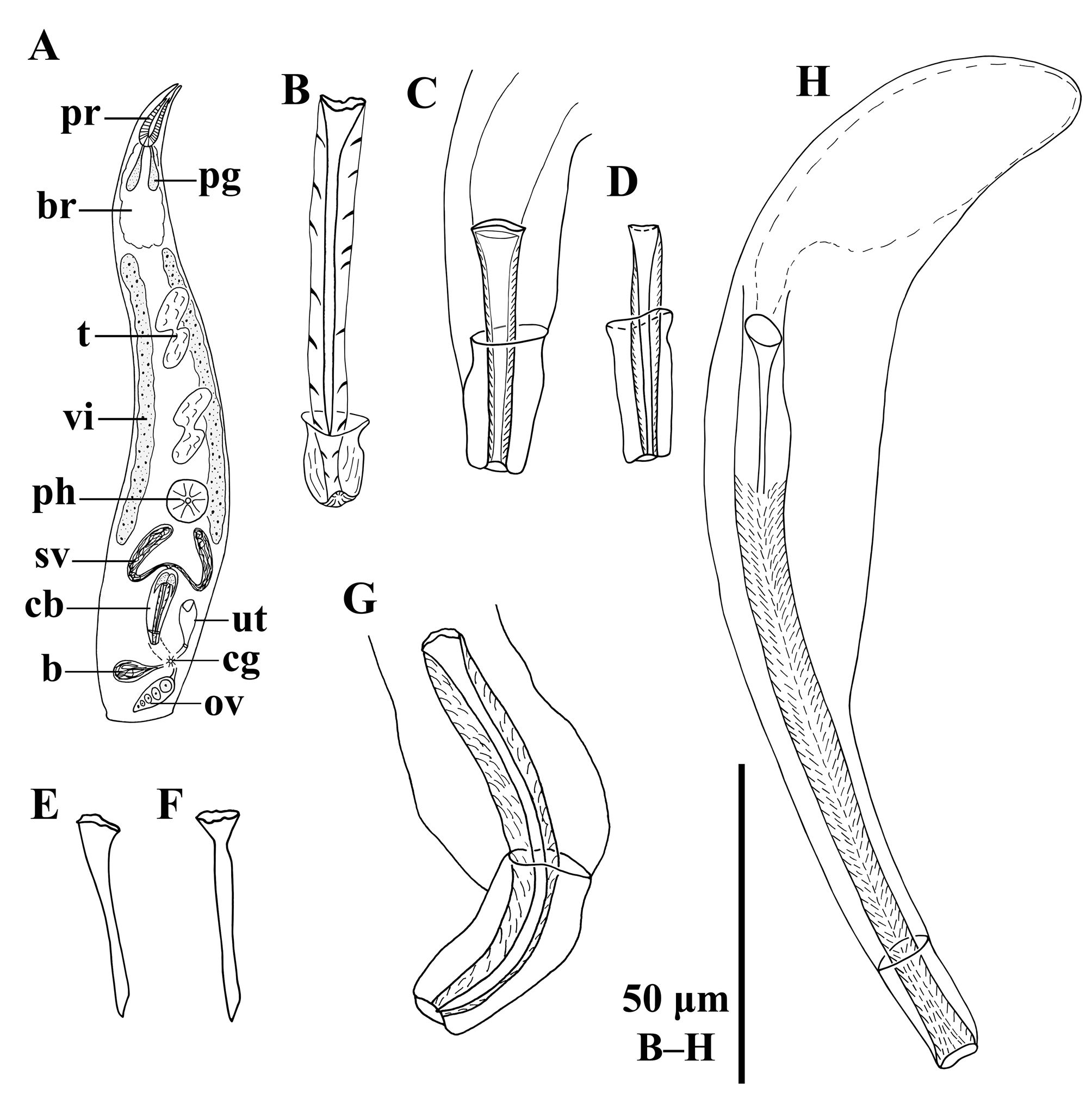
Diagnosis. Species of Schizochilus with a spiny cirrus, covered with fine spines of ±2 μm long.A needle-shaped, 70-μm-long stylet runs centrally through the cirrus. Copulatory bulb with a 32-µm-long sclerotized distal cap.
Description. Live animal about 1.5 mm long, translucent, without eyes. Habitus and internal organisation as in S. espinosai sp. n.. Proboscis lips 279 μm long (n = 2). Pharynx 226 μm in diameter, located at 75%.
Two vitellaria extend between the brain and the copulatory bulb, one at each side of the body. The rest of the genital organs are situated in the caudal fourth of the body, with the ovary and the bursa near to the caudal body end. Two long testes are located laterally between the brain and the pharynx, one on each body side. Seminal vesicles open proximally into the copulatory bulb. Copulatory bulb 144 μm long, comprising the prostate vesicle and the cir- rus. Cirrus ( Fig. 12G View FIGURE 12 , 15C View FIGURE 15 ) 70 μm long and ±8 μm wide, armed with fine spines that are 1.7–2.3 μm long ( x = 2 μm; n = 9). A stylet ( Fig. 12G View FIGURE 12 , 15C View FIGURE 15 ) lies centrally in the cirrus. It is slightly curved in the live animal; more curved in the whole mount (probably because of the squeezing). It is 70 μm long and 8 μm wide proximally, needle shaped, taper- ing to a distal sharp tip. The distal walls of the copulatory bulb are sclerotized, forming a 32-μm-long cap, which is 13 μm wide at the widest point. As such, it surrounds the cirrus for 46% of the latter’s length.
| FMNH |
Field Museum of Natural History |
No known copyright restrictions apply. See Agosti, D., Egloff, W., 2009. Taxonomic information exchange and copyright: the Plazi approach. BMC Research Notes 2009, 2:53 for further explanation.
|
Kingdom |
|
|
Phylum |
|
|
Class |
|
|
Order |
|
|
Family |
|
|
Genus |