
Schizochilus atlanticus, Diez & Reygel & Artois, 2019
|
publication ID |
https://doi.org/10.11646/zootaxa.4646.1.1 |
|
publication LSID |
lsid:zoobank.org:pub:2A39D2E1-262F-423F-9B7F-89C376912FFC |
|
persistent identifier |
https://treatment.plazi.org/id/ED01879A-FFFD-FF97-FF68-FC26FBFA0D56 |
|
treatment provided by |
Plazi |
|
scientific name |
Schizochilus atlanticus |
| status |
sp. nov. |
Schizochilus atlanticus sp. n.
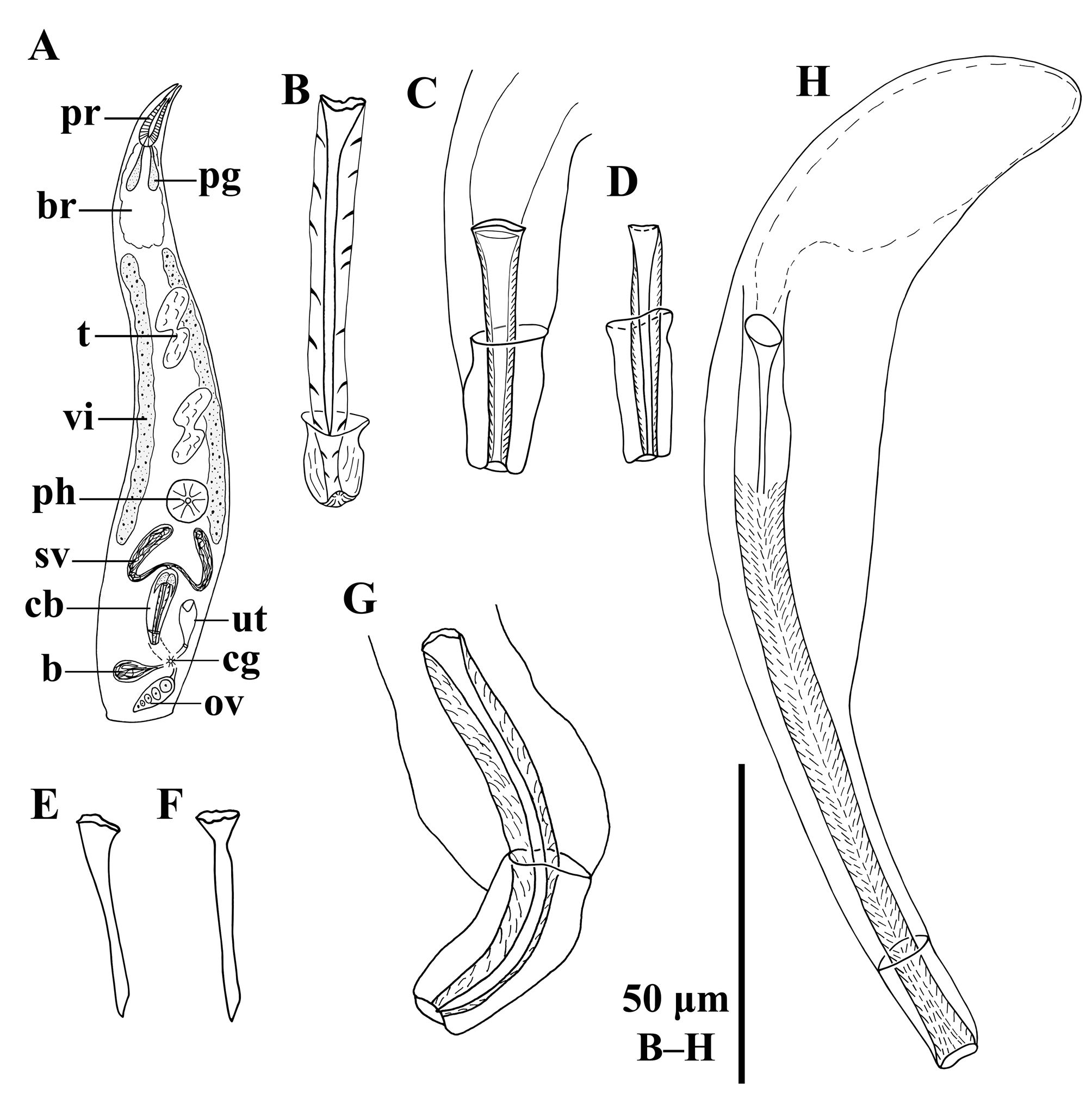
( Fig. 12 View FIGURE 12 C–D, 15A–B)
http://zoobank.org/ urn:lsid:zoobank.org:act:
Material. Observations on live animals, whole mounted afterwards. Four whole mounts from Macabí (type locality) ( April 23, 2017), one of which is designated holotype ( FMNH https://id.luomus.fi/KV.618), the other material in HU (X.1.19– X.1.21), intertidal, upper 10 cm of fine-grained sand, salinity 34 ‰. Two whole mounts from Siboney ( April 4, 2017) (HU X.1.22– X.1.23), intertidal, upper 10 cm of fine-grained sand, salinity 34 ‰.
Etymology. The epithet refers to the fact that the species was found at the Atlantic coast of Cuba.
Diagnosis. Species of Schizochilus with a spiny cirrus; fine spines about 1.5 μm long. A straight, tubular, ±39- µm-long stylet is present in the cirrus. Copulatory bulb with a distal cap of ±19 μm long.
Description. Live animals about 1–1.5 mm long, translucent, without eyes. Habitus and internal organisation as in S. espinosai sp. n.. Proboscis lips 162–184 μm long ( x = 176 μm; n = 12), with a pair of gland sacs that enter the proboscis at its proximal end. Pharynx 141–172 μm in diameter ( x = 162 μm; n = 5), located at 70%.
Four (in the specimens from Macabí) to five or six (in the specimens from Siboney) oval- to kidney-shaped testes lie in a single row rostrally from the pharynx. Two vitellaria extend between the brain and the copulatory bulb, one at each side of the body. The rest of the genital organs are situated in the caudal fourth of the body, with the ovary and the bursa near to the caudal body end. Copulatory bulb 81–131 μm long ( x = 107 μm; n = 6), com- prising the prostate vesicle and a spiny cirrus. Cirrus ( Fig. 12 View FIGURE 12 C–D, 15A–B) ornamented with 0.5–1.8-μm-long fine spines ( x = 1.2 μm; n = 30). A tubular stylet ( Fig. 12 View FIGURE 12 C–D, 15A–B) lies centrally in the cirrus. Cirrus and stylet are 34–43 μm long ( x = 39 μm; n = 6) and proximally 5–7 μm wide ( x = 6 μm; n = 6). The distal walls of the copula- tory bulb are sclerotized, forming a 13–24-μm-long cap ( x = 19 μm; n = 6) that is 9–14 μm at the widest point ( x = 11 μm; n = 6). As such it surrounds the cirrus for 38–56% of the latter’s length.
| FMNH |
Field Museum of Natural History |
No known copyright restrictions apply. See Agosti, D., Egloff, W., 2009. Taxonomic information exchange and copyright: the Plazi approach. BMC Research Notes 2009, 2:53 for further explanation.
|
Kingdom |
|
|
Phylum |
|
|
Class |
|
|
Order |
|
|
Family |
|
|
Genus |