
Schizochilus favus, Diez & Reygel & Artois, 2019
|
publication ID |
https://doi.org/10.11646/zootaxa.4646.1.1 |
|
publication LSID |
lsid:zoobank.org:pub:2A39D2E1-262F-423F-9B7F-89C376912FFC |
|
persistent identifier |
https://treatment.plazi.org/id/23613044-29FD-45CF-A694-B984B0164D12 |
|
taxon LSID |
lsid:zoobank.org:act:23613044-29FD-45CF-A694-B984B0164D12 |
|
treatment provided by |
Plazi |
|
scientific name |
Schizochilus favus |
| status |
sp. nov. |
Schizochilus favus sp. n.
( Fig. 13 View FIGURE 13 , 14 View FIGURE 14 A–B)
http://zoobank.org/ urn:lsid:zoobank.org:act:
Material. Observations on live animals, whole mounted afterwards. Six whole mounts from Siboney (type locality) ( February 7, 2016), one of which is designated holotype ( FMNH https://id.luomus.fi/KV.621), the others in HU (X.1.27– X.1.31), intertidal, upper 20 cm of the fine-grained sand, salinity 35 ‰.
Etymology. The epithet reflects the fact that the bases of the cirrus spines together have the appearance of a honeycomb. Lat. favus : honeycomb.
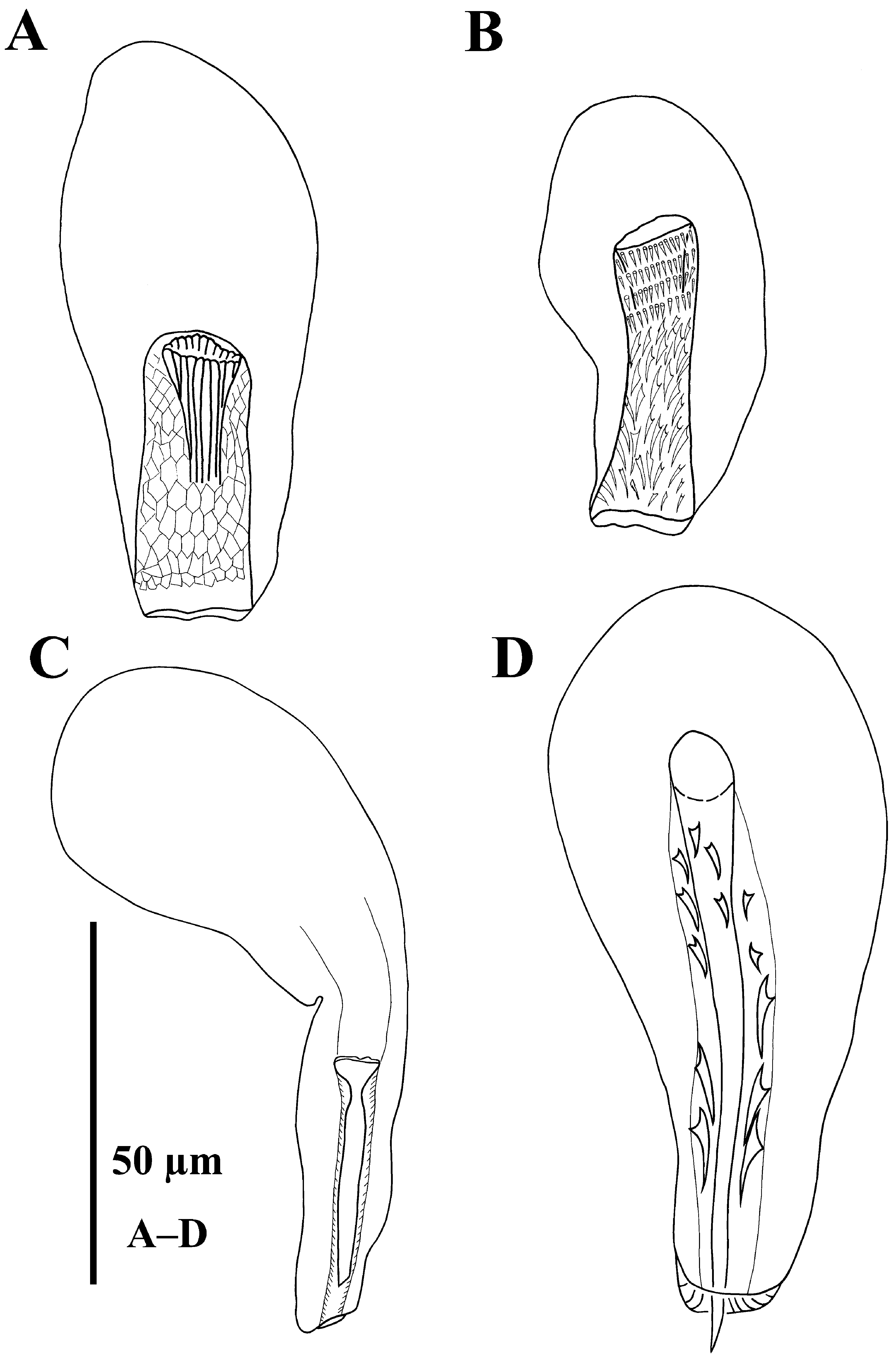
Diagnosis. Species of Schizochilus with a single large testis rostrally from the pharynx. Spiny cirrus 37 μm long; spines about 2 μm long in the proximal half of the cirrus and 7 μm long in the distal half. The bases of the spines are arranged in such a way that in the live animal they have the appearance of a honeycomb. Inside the cirrus a ±36-μm-long stylet is present. Copulatory organ without a distal cap.
Description. Live animals about 1–1.5 mm long, 0.6–1.4 mm long in the whole mounts ( x = 1 mm; n = 5), translucent, without eyes. Habitus and internal organisation as in S. espinosai sp. n.. Proboscis ( Fig. 13A View FIGURE 13 : pr) 84– 88 μm long ( x = 86 μm; n = 6), with a pair of gland sacs ( Fig. 13A View FIGURE 13 : pg) that enter the proboscis at its proximal end. Rostrally from the proboscis, in the proximal tip of the body, a pair of brownish-pink glands ( Fig. 13A View FIGURE 13 : gl) can be found, one on each body side. The brain ( Fig. 13A View FIGURE 13 : br) is located caudally from the proboscis. Pharynx ( Fig. 13 View FIGURE 13 A–B: ph) has a diameter of 93–98 μm ( x = 96 μm; n = 4), and is located at 70%.
A large testis, up to 272 μm long in one whole mount ( Fig. 13 View FIGURE 13 A–B: t), is located rostrally from the pharynx. Two vitellaria ( Fig. 13 View FIGURE 13 A–B: vi) extend between the brain and the copulatory bulb, one at each side of the body. The rest of the genital organs are situated in the caudal fourth of the body, with the ovary and the bursa near to the caudal body end. Copulatory bulb ( Fig. 13A View FIGURE 13 : cb, 14A–B) comprising the prostate vesicle and a spiny cirrus. Spiny cirrus ( Fig. 13B View FIGURE 13 : ci, 13C–F, 14A–B) 34–38 μm long ( x = 37 μm; n = 5) and 9–11 μm wide ( x = 10 μm; n = 5). In the proximal half of the cirrus, the spines are about 2 μm long (n = 18); in the distal half they are 5–12 μm long ( x = 7 μm; n = 12). The spines are arranged in such a way, that their bases together form a honeycomb-like struc- ture (see Fig. 13C View FIGURE 13 ), mainly in the distal half of the cirrus. Inside the cirrus lies a tubular stylet, 35–37 μm long ( x = 36 μm; n = 2) and 8–12 μm wide proximally ( x = 10 μm; n = 4). This stylet bears longitudinal ridges (see Fig. 13D View FIGURE 13 , 14A View FIGURE 14 ). In general the stylet is not well visible because the spines hamper observation. Often only the proximal part is visible, as shown in Fig. 14A View FIGURE 14 . The copulatory bulb does not form a sclerotized cap surrounding its distal tip. The common gonopore opens at 90%.

| FMNH |
Field Museum of Natural History |
No known copyright restrictions apply. See Agosti, D., Egloff, W., 2009. Taxonomic information exchange and copyright: the Plazi approach. BMC Research Notes 2009, 2:53 for further explanation.
|
Kingdom |
|
|
Phylum |
|
|
Class |
|
|
Order |
|
|
Family |
|
|
Genus |