
Sphecozone rostrata Millidge, 1991
|
publication ID |
https://doi.org/ 10.5281/zenodo.208645 |
|
DOI |
https://doi.org/10.5281/zenodo.6175348 |
|
persistent identifier |
https://treatment.plazi.org/id/EE318B5F-FF8A-FFB0-FF41-FAF83F7BF83F |
|
treatment provided by |
Plazi |
|
scientific name |
Sphecozone rostrata Millidge, 1991 |
| status |
|
Sphecozone rostrata Millidge, 1991 View in CoL
( Figs. 1–5 View FIGURES 1 – 10 , 20 View FIGURES 20, 21 )
Sphecozone rostrata Millidge, 1991:175 View in CoL , figs. 743–746 (Holotype 3, Encruzilhada, Bahia, Brazil, XI.1973, M. Alvarenga col., in AMNH, not examined); Rodrigues, 2005: 105; Platnick, 2011.
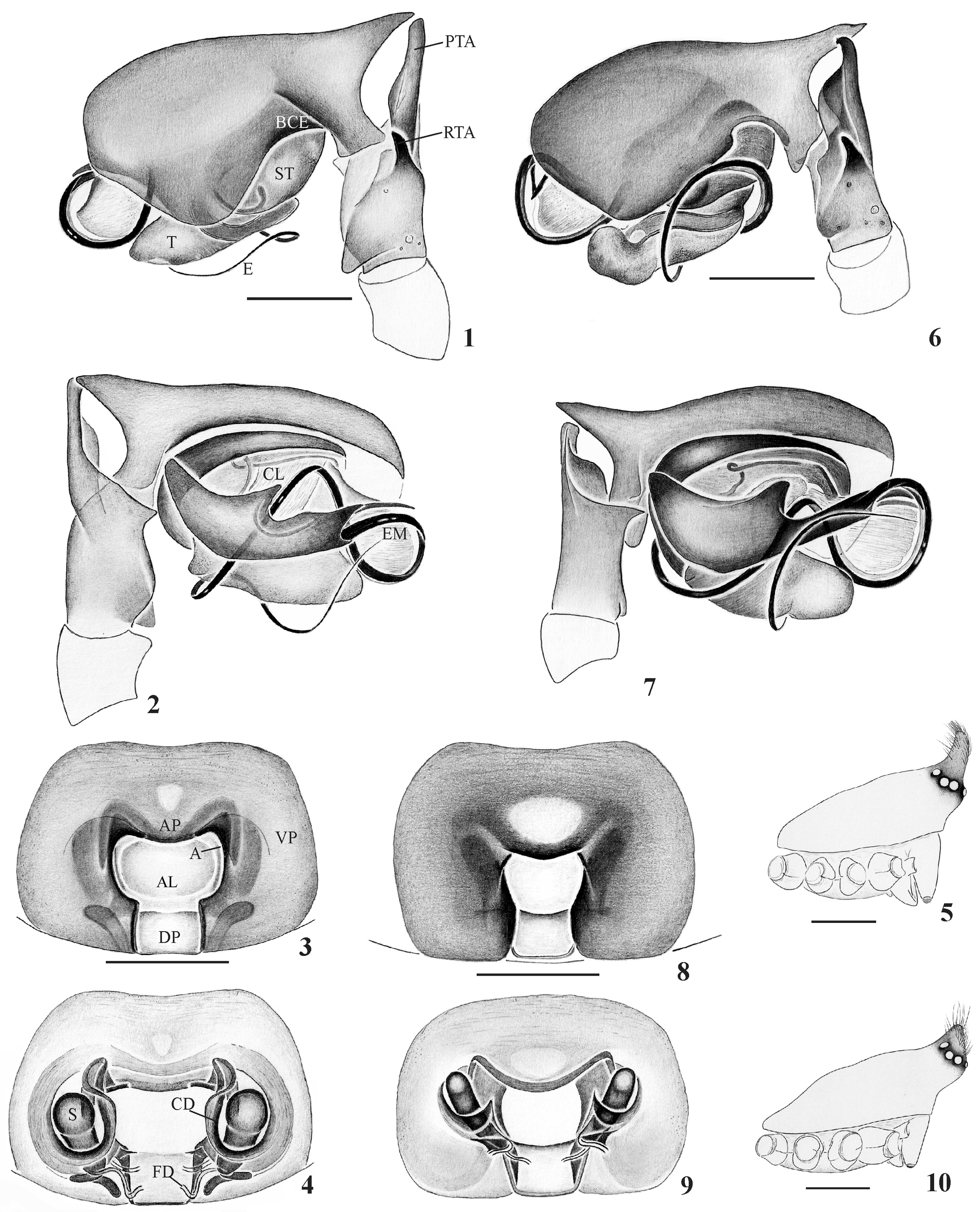
Diagnosis. The male of Sphecozone rostrata resembles that of S. personata by the dorsally projected cymbium, the long prolateral tibial apophysis ( Figs. 1, 2 View FIGURES 1 – 10 ) and the cephalic projection ( Fig. 5 View FIGURES 1 – 10 ). The males of Sphecozone rostrata differ from those of S. personata by having the palp tibiae with a more developed tubercle near the retrolateral margin ( Fig. 1 View FIGURES 1 – 10 ), by the embolus without a conspicuous loop at the retrolateral portion of tegulum and subtegulum ( Fig. 1 View FIGURES 1 – 10 ) and by the more slender and pronounced cephalic projection ( Fig. 5 View FIGURES 1 – 10 ). The female of S. rostrata is close to S. personata by the presence of a larger anterior process on the ventral epigynal plate ( Fig. 3 View FIGURES 1 – 10 ); differs from it by a wider anterior lobe of the dorsal epigynal plate ( Fig. 3 View FIGURES 1 – 10 ), larger spermathecae and fertilization ducts closer to the edge of the dorsal plate ( Fig. 4 View FIGURES 1 – 10 ).
Description. Female (MCN 47515): Total length 1.55. Carapace length 0.62, width 0.55, height 0.35. Clypeus height 0.10. Sternum length 0.37, width 0.37. Abdomen length 1.0 5, width 0.85, height 0.87. Leg formula IV/I/II/III; lengths (I/II/III/IV): femora 0.50/0.52/0.42/0.57; patellae 0.17/0.17/0.15/0.15; tibiae 0.42/0.37/0.30/0.47; metatarsi 0.35/ 0.35/0.32/0.47; tarsi 0.25/0.25/0.25/0.25; total 1.69/1.66/1.44/1.91. Coxae III slightly smaller, coxae IV separated by more than their width. TmI 0.40. Metatarsal trichobothria I–III present, IV absent. Tibial spine formula: 1-1-1-1. Eye diameters: AME 0.0 5, ALE, PME and PLE 0.0 6. Eyes with dark borders. Ocular area dark-brown. Clypeus glabrous.
Carapace not projected, pale orange, setae present at ocular area and one median row with 3–4 setae. Chelicerae and endites pale orange, promargin with five very small teeth and retromargin with four. Labium brownish. Sternum pale orange with reddish-brown borders, posteriorly truncated. Coxa yellowish. Femura and patellae pale orange, tibiae, metatarsi and tarsi brown. Abdomen brownish, longer than wide, with longitudinal stripe. Spinnerets brownish. Colulus well developed. Epigynum with short fertilization ducts, near the base of the dorsal plate; copulatory ducts forming a loop around the spermathecae and ventral plate wider than long ( Figs. 3, 4 View FIGURES 1 – 10 ).
Male (MCN 47515; as female except noted): Total length 1.55. Carapace length 0.75, width 0.55, height 0.30. Clypeus height 0.15. Sternum length 0.40, width 0.37. Abdomen length 0.87, width 0.67, height 0.65. Leg formula IV/I/ II/III; lengths (I/II/III/IV): femora 0.57/0.50/0.40/0.62; patellae 0.17/0.17/0.15/0.12; tibiae 0.44/0.50/0.32/0.50; metatarsi 0.37/0.37/0.37/0.50; tarsi 0.25/0.25/0.25/0.25; total 1.80/1.79/1.49/1.99. TmI 0.39. Metatarsi I–III with trichobothrium, IV absent. Eye diameters: AME 0.0 3, ALE, PME and PLE 0.0 5. Carapace red-brown, projected and pubescent in cephalic area. Chelicerae and endites orange-brown. Palpal tibiae with one retrolateral trichobothrium. Retrolateral tibial apophysis short and curved. Prolateral tibial apophysis long, pointed dorsally. Presence of membranous column and embolic membrane ( Fig. 2 View FIGURES 1 – 10 ). Cymbium projected with apex directed to the tip of the prolateral tibial apophysis. Embolus long and filiform ( Figs. 1, 2 View FIGURES 1 – 10 ).
Material examined. Brazil, Rio Grande do Sul: Maquiné (riparian forest, Maquiné river), 193, 22Ƥ, 18.IX.2007 - 07.VI.2009 (MCN 47499, 47500, 47515-47517, 47519, 47520); Parobé (riparian forest, Sinos river), 1Ƥ, 07.II.2009 (MCN 47520); Cristal (riparian forest, Camaquã river), 13, 1Ƥ, 19.XII.2007, (MCN 47518), all collected with a beating tray by E.N.L. Rodrigues.
Distribution. Brazil (Bahia and Rio Grande do Sul).
Ecology. Linyphiidae were the sixth most abundant spider family in the samples from riparian forests of southern Brazil, recording 45 species. Sphecozone rostrata was the second most abundant linyphiid with 100 specimens. More females (N=60) than males (40), were captured (sex ratio 1.5:1).
This spider was more abundant in Maquiné river (61%), followed by Sinos river (37%) and less abundant in Camaquã river (2%). Their abundance was greater in the forest interior (72%) than in forest-grassland edges (28%) and not recorded in the forest-river edge.
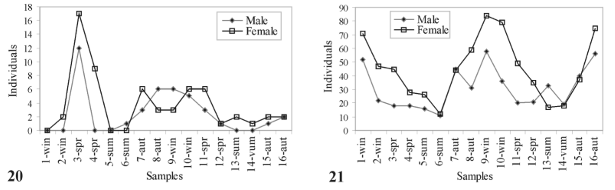
This species was recorded from almost every sample taken in the two years ( Fig. 20 View FIGURES 20, 21 ). The highest abundances were registered in the spring of the first year and in the winter of the second year. Males and females have very similar abundance peaks with lower abundances during summer ( Fig. 20 View FIGURES 20, 21 ).
No known copyright restrictions apply. See Agosti, D., Egloff, W., 2009. Taxonomic information exchange and copyright: the Plazi approach. BMC Research Notes 2009, 2:53 for further explanation.
|
Kingdom |
|
|
Phylum |
|
|
Class |
|
|
Order |
|
|
Family |
|
|
Genus |
Sphecozone rostrata Millidge, 1991
| Rodrigues, Everton Nei Lopes, Ott, Ricardo & Mendonça, Milton De S. 2012 |
Sphecozone rostrata
| Rodrigues 2005: 105 |
| Millidge 1991: 175 |