
Chaetonotus rhombosquamatus, Kolicka, Małgorzata, Kisielewski, Jacek, Nesteruk, Teresa & Zawierucha, Krzysztof, 2013
|
publication ID |
https://doi.org/ 10.11646/zootaxa.3717.2.7 |
|
publication LSID |
lsid:zoobank.org:pub:27BD65FD-18CF-4E9D-AE77-C7C0137CF1DC |
|
DOI |
https://doi.org/10.5281/zenodo.6164622 |
|
persistent identifier |
https://treatment.plazi.org/id/EF4A2F6C-6D5A-FFC9-FF52-F9FBFEFBF961 |
|
treatment provided by |
Plazi |
|
scientific name |
Chaetonotus rhombosquamatus |
| status |
sp. nov. |
Chaetonotus View in CoL (Tristratachaetus subgen. nov.) rhombosquamatus sp. nov. ( Figs 4–12, Table 5 View TABLE 5 )
Locality: Site 1; session II.
Material: 1 sample, 9 specimens (all adults), all photographed. The photographs of holotype and four paratype are available in Natural History Collection at Adam Mickiewicz University in Poznań under accesion number NHC-GCTR- 1-1-19 /h (holotype) and NHC-GCTR- 1-20-69 /p (paratypes). Additional all photographs are available at Adam Mickiewicz University in Poznań, in the collection of the first author.
Etymology: From Latin “rhombus”—rhomb, and Latin “squama”—scale, referring to the specific shape of the body scales.
Diagnosis: As for the subgenus.
Description: Chaetonotus (Tristratachaetus subgen. nov.) rhombosquamatus sp. nov. is a species with relatively large and massive body. The head is of a rounded shape with five lobes, i.e. the cephalion (U1–U3) and two pairs of pleures (U2–U8). The cephalion, epipleuria and hypopleuria are visible in the outline of the body ( Fig. 11 View FIGURE 11 ). The indentations between the head plates are shallow. The hypopleuria are only slightly larger than the epipleuria, and are located on U4–U8. On the head there are two pairs of ciliary tufts. The tufts of the first pair (three cilia per tuft) are shorter than the posterior pair (four cilia per tuft). Large globular cuticle formations located dorsally on the head (at U4–U5) strongly reflect light and are probably a kind of ocellar granules ( Fig. 10 A). The mouth is situated subterminally at U2–U4. In the mouth ring, leaf–like cuticular reinforcements typical of the genus Chaetonotus and short cuticular hairs occur. A large pentagonal–shaped hypostomium is located at U4–U8. Near the hypostomium anterior edges, there are short transverse cuticular bars. The neck widens gradually, turning smoothly into the trunk (U25–U93). The trunk widens gradually to ca. U58 and subsequently gradually narrows from U76 up to the furca base. The furca begins at U93. The furca branches point slightly outwards. The short and straight adhesive tubes are gradually tapering distally (U97–U100). They are very short in comparison with the body length.
The head, neck and trunk on the dorsal, lateral and ventrolateral parts are covered with convex, conical scales with four keels (two pairs, perpendicular to one another) ( Fig. 6 View FIGURE 6 ). On the head, neck and trunk the scales are rhomboidal in shape and have weakly rounded edges, without posterior notches ( Fig. 9 View FIGURE 9 ). The scales are arranged in 38–46 longitudinal alternating rows, 41–49 scales in each row. Scale edges contact neighbouring scales; the longitudinal rows start at U6 and extend to the furca base. The length and width of scales gradually increase from the head, through the neck to the widest trunk part (U58–U76), and subsequently gradually decrease to the furca base. Spines arise perpendicularly from the scale centre at the confluence of two pairs of keels, and are subsequently strongly curved. The spines narrow and taper explicitly and uniformly distally so that the ends are very thin. Lateral denticles are absent in all the body spines. The length of the spines gradually increases from anterior to posterior end of body. From U83 there is a strong elongation of the spines. The posteriormost lateral spines are always considerably longer and thicker than the other body spines. Dorsal sensory bristles are absent.
A—Dorsal view, B—Inside view, C—Ventral view. All measurements in micrometers.
On the ventral body surface longitudinal ciliary bands start at U5 and end at U91. The interciliary field (U6– U94) is covered with numerous one–lobed scales without keels, and the spines are arranged alternately. In the pharynx section of the ventral interciliary field, directly under and around the hypostomium, minor round scales are present. In the intestinal section of the ventral interciliary field scales are of an elongated, rounded rectangular shape. The terminal scales of the ventral interciliary field are arranged in three pairs and are localised at U90–U94. They have an elongated, rounded rectangular shape, are void of keels and possess straight spines reaching beyond the furca indentation. These scales are considerably larger than the other ventral scales. The central pair is larger than the lateral ones. The most important feature of this species is thick, three–layer cuticle ( Fig. 8 View FIGURE 8 ). The outer layer of the cuticle has a granulated irregular structure (of varying thickness and texture in particular individuals) and covers the dorsal, ventrolateral and ventral scales as well as the scaleless part of body ( Fig. 12 View FIGURE 12 ). The middle layer of cuticle consist scales with spines, the inner cuticle layer is base of cuticle. The entire cuticle (including scales) is of a distinct orange–brown (“russet”) colour.
The pharynx (U3–U25) possesses distinct anterior (U3–U7) and posterior (U20–U24) dilatation (swellings) typical of the genus Chaetonotus . The posterior dilatation is wider than the anterior one. The intestine has well– developed enzymatic section (U26–U31).
Taxonomic remarks: The scales in C. (T.) rhombosquamatus sp. nov. correspond in the shape to those of Neogossea antennigera Gosse, 1851 belonging to the family Neogosseidae Remane, 1927 . This species has a very similar rhomboidal shape and the same type of scale arrangement as in the new Chaetonotus species. However, this similarity probably reflects convergent evolution and in N. antennigera the spines arise near the posterior edges of the scales, unlike in C. (T.) rhombosquamatus sp. nov. Previous genetic studies took no account of the family Neogosseidae ; therefore, the relation to the various groups of the non–monophyletic genus Chaetonotus and its representatives remains unknown (Kånneby et al. 2013).
A—Dorsal view, B—Ventral view of distribution and shape of interciliary field scales, C—Dorsal view of scales in trunk region. All measurements in micrometers.
A—Head with visible globular cuticle formations; B—Furca. All measurements in micrometers.
Next specific feature in the new species is the absence of dorsal sensory bristles. In Chaetonotus (Chaetonotus) sp. 1 (see below) the dorsal sensory bristles are also absent. In the other known species one or two pairs of sensory bristles are present. Perhaps the specific construction of the thick cuticle (three–layer system) is the reason of the reduction observed in dorsal sensory organs.
Differential diagnosis: Chaetonotus (Tristratachaetus subgen. nov.) rhombosquamatus sp. nov. is not similar to any other known species of Chaetonotidae . Of all the described species, it seems to share a few morphological features with Chaetonotus (Chaetonotus) hirsutus Marcolongo, 1910 , Chaetonotus (Chaetonotus) linguaeformis Voigt, 1902 and Chaetonotus (Chaetonotus) maximus Ehrenberg, 1830 . All three compared species are entirely different in the shape, number and distribution of scales from C. (T.) rhombosquamatus sp. nov. Additionally, the spines themselves are differently shaped. In the new species the spines arise from the scale centre, not from the vicinity of the posterior edge. Also, none of the compared species have ocellar granules or ocellar–like formations similar to those found in the new species. The structure of the cuticle in the compared species is also entirely different.
A—Ventral view, B—View of internal morphology, C—Dorsal view. All measurements in micrometers.
C. (C.) hirsutus Marcolongo, 1910 (150–230 Μm in length) is smaller than C. (T.) rhombosquamatus sp. nov. Additionally, the spines are longer than in C. (T.) rhombosquamatus sp. nov. and their length (neck: 12–15 Μm; trunk: 12–22 Μm) gradually increases from the anterior to ca. the middle of the trunk (not to the body posterior end). In the compared species the length of the rearmost pair of lateral spines (25–52 Μm) is definitely longer than that of the other body spines. This is also the case in the new species, but their spines are generally shorter. Furthermore, the furca length is marginally shorter (21 Μm) than in C. (T.) rhombosquamatus sp. nov.
C. (C.) linguaeformis Voigt, 1902 (180–370 Μm in length) has a significantly thinner body than C. (T.) rhombosquamatus sp. nov. The head of the compared species is of a three–lobed shape with one pair of cephailic cilia. The trunk spines in C. (C.) linguaeformis are relatively long (15–20 Μm) and do not extend gradually towards the end of the body. The rearmost pair of lateral spines in this species is slightly longer (21 Μm) than the trunk spines as opposed to the newly described species. Moreover, the furca of C. (C.) linguaeformis is significantly longer (30–33 Μm).
C. (C.) maximus Ehrenberg, 1830 (112–330 Μm in length) has a pronounced and deep indentation between the head plates, in contrast to the shallow indentation in the newly described species. The spines’ length (neck: 6–15 Μm; trunk: 8–12 Μm) gradually increases from the anterior to the posterior body end and is similar to the length range of the spines of the new species. However, significantly different is the length of the last pair of dorsal spines (12–14 Μm). In this particular species it does not differ from the other dorsal spines, but in C. (T.) rhombosquamatus sp. nov. the pair of the last lateral spines is definitely longer and thicker than the rest of the body spines. Additionally, C. (C.) maximus has adhesive tubes that are significantly longer (16–18 Μm) than those of the new species.


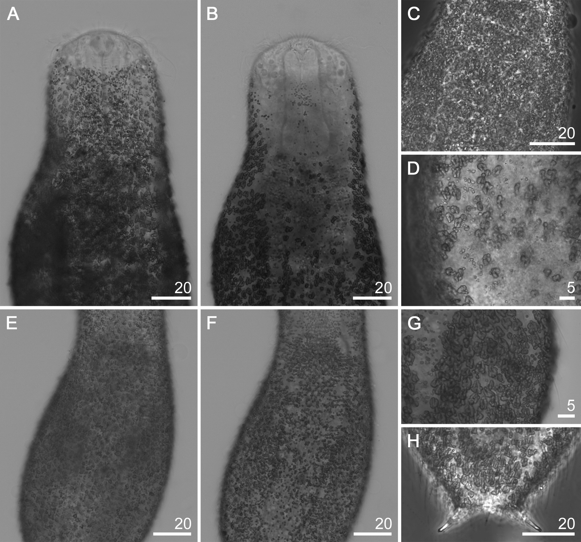
TABLE 5. Morphometric parameters for Chaetonotus (Tristratachaetus subgen. nov.) rhombosquamatus sp. nov.; Nnumber of specimens or structures analysed, Range—the smallest and the largest structure found among all specimens measured, SD—standard deviation.
| Characters N | Holotype | Range | SD |
|---|---|---|---|
| Body length 7 | 238.5 | 221.8–239.9 | 6.89 |
| Pharynx length 5 | 50.7 | 50.7–52.3 | 0.66 |
| Width of anterior pharynx thickening (a) 5 | 16.0 | 15.6–17.3 | 0.68 |
| Width of pharynx narrowing that follows 5 anterior thickening (n) Width of pharynx at its middle length (m) 6 | 13.5 14.3 | 12.3–14.3 13.8–14.9 | 0.44 0.83 |
| Width of posterior pharynx thickening (p) 5 | 20.4 | 20.4–21.9 | 0.76 |
| Length of cephalic cilia 7 | 18.5–32.7 | (18.3–23.6)–(32.7–39.2) | 2.09–2.36 |
| Hypostomium length 5 | 9.0 | 7.9–9.0 | 0.46 |
| Cephalion length 5 | 11.2 | 9.3–11.9 | 1.02 |
| Cephalion width 6 | 23.3 | 19.1–23.3 | 1.48 |
| Diameter of mouth ring 7 | 7.8 | 7.7–8.7 | 0.36 |
| Furca length 9 | 22.2 | 21.5–22.8 | 0.45 |
| Length of adhesive tube 9 | 9.6 | 8.9–11.2 | 0.67 |
| Neck spine length 6 | 10.6–13.4 | (9.0–10.8)–(12.4–13.6) | 0.71–0.49 |
| Trunk spines length 6 | 13.5–14.0 | (12.5–13.7)–(14.0–15.3) | 0.46–0.48 |
| Length of extending spine of the posterior body part 6 | 14.3–22.4 | (14.3–15.7)–(20.0–23.4) | 0.21–1.23 |
| Length of posteriormost pair 8 of lateral spine Neck scale length 5 | 23.2 2.7–4.8 | 21.3–26.2 (2.2–2.8)–(4.5–5.3) | 1.57 0.25–0.31 |
| Neck scale width 5 | 1.6–2,8 | (1.5–2.1)–(2.2–3.2) | 0.31–0.38 |
| Trunk scale length 6 | 4.9–7.6 | (4.7–5.4)–(7.2–7.6) | 0.32–0.16 |
| Trunk scale width 6 | 2.9–4.8 | (2.3–3.3)–(4.8–5.7) | 0.35–0.32 |
| Number of cephalic cilia in one tuft 9 Number of separated cephalic tufts 9 | 3 (anterior); 4 (posterior) 4 | 3 (anterior); 4 (posterior) 4 | 0.00 0.00 |
| Number of scales in single longitudinal row 9 | 43 | 41–49 | 3.18 |
| Total number of longitudinal alternating rows of scales 9 | 42 | 38–46 | 2.45 |
| Pharynx formula a 5 | 31.6 | 30.4–33.1 | 0.01 |
| Pharynx formula n 5 | 26.6 | 24.1–28.1 | 0.02 |
| Pharynx formula m 5 | 28.2 | 27.0–29.1 | 0.01 |
| Pharynx formula p 5 | 40.2 | 40.2–42.4 | 0.01 |
| Ratio of scale distribution 9 | 97.7 | 89.8–107.3 | 0.05 |
| Length ratio of terminal spines 7 | 9.7 | 9.7–10.9 | 0.00 |
No known copyright restrictions apply. See Agosti, D., Egloff, W., 2009. Taxonomic information exchange and copyright: the Plazi approach. BMC Research Notes 2009, 2:53 for further explanation.
|
Kingdom |
|
|
Phylum |
|
|
Order |
|
|
SubOrder |
Paucitubulatina |
|
Family |
|
|
SubFamily |
Chaetonotinae |
|
Genus |