
Parastenocaris distincta, Cottarelli, Vezio, Bruno, Maria Cristina & Berera, Raffaella, 2006
|
publication ID |
https://doi.org/10.5281/zenodo.174828 |
|
DOI |
https://doi.org/10.5281/zenodo.6256827 |
|
persistent identifier |
https://treatment.plazi.org/id/F15F87A8-6872-FFA9-5618-FA11FDE5FB7F |
|
treatment provided by |
Plazi |
|
scientific name |
Parastenocaris distincta |
| status |
sp. nov. |
Parastenocaris distincta sp. nov.
Material examined—Holotype: male, dissected and mounted on slide labelled: Parastenocaris distincta holotype male ( MCSNG 53622a). Paratype (i.e. allotype): female, dissected and mounted on slide labelled: Parastenocaris distincta allotype female ( MCSNG 53622b). Paratypes: two male, each dissected and mounted on slide labelled: Parastenocaris distincta paratype male no. 1, 2 respectively ( DSAUT); two males, each mounted on slide, labelled: Parastenocaris distincta paratype male no. 3, 4 respectively ( DSAUT). One female, mounted on slide labelled: Parastenocaris distincta paratype female no. 1 ( DSAUT); 2 female, dissected and mounted on slide labeled: Parastenocaris distincta paratype female no. 2, 3 respectively ( DSAUT). Two female copepodites, stage V, each mounted on slide labeled: Parastenocaris distincta , copepodite female no. 1, 2, respectively ( DSAUT). All material collected by V. Cottarelli on 02-08-05 (adults) and 02- 23-04 (copepodites).
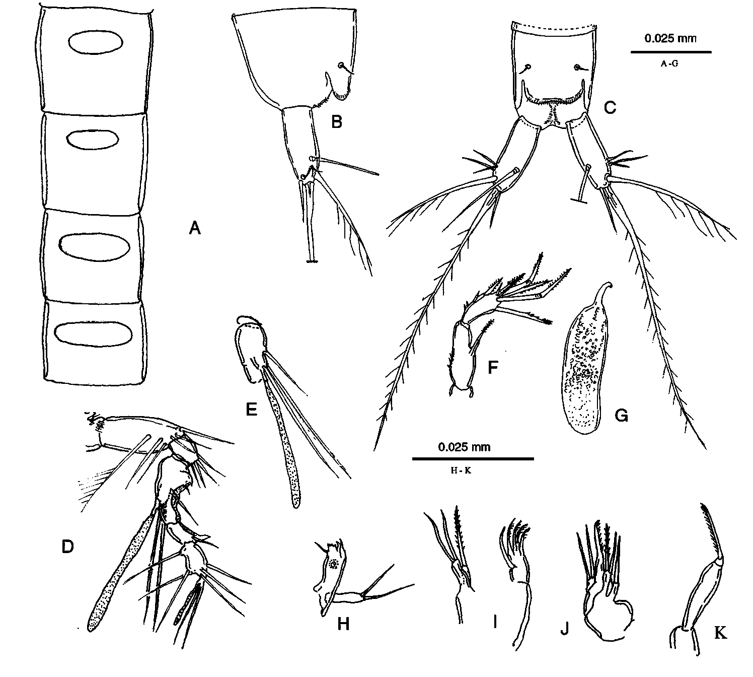
Description of male—Body vermiform, slender, unpigmented, eyeless. Length, measured from rostrum to apex of caudal rami: 0.389 mm. Hyaline frills of somites smooth. Genital somite and first 3 abdominal somites with oval dorsal integumental windows, the one on the first abdominal somite smallest ( Fig. 1 View FIGURE 1 A). Anal somite ( Fig. 1 View FIGURE 1 B–C) with paired sensilla on dorsal side, and 2 medial rows of small longitudinal spinules near the anus. Anal operculum ( Fig. 1 View FIGURE 1 B–C) with slightly concave, smooth distal margin, a row of thin distal spinules visible under transparent operculum. Caudal rami ( Figs 1 View FIGURE 1 B–1C) shorter than last abdominal somite, length to width ratio 2.5; anterolateral accessory seta (I) and seta II short, subequal; seta III slightly longer; outer terminal seta (IV) long (length seta/length caudal ramus: 1.7), pinnate; inner terminal seta (V) without breaking plane; terminal accessory seta (VI) smooth, short (length seta/length caudal ramus: 0.6); dorsal seta (VII) articulate (length seta/length caudal ramus: 1.2). All caudal setae inserted on the distal third of the caudal ramus. Spermatophore as in Fig. 1 View FIGURE 1 G.
Rostrum ( Fig. 1 View FIGURE 1 D) small, reaching half of first segment of antennule; with 2 apical sensilla.
Antennule ( Fig. 1 View FIGURE 1 D–E) geniculate, 8-segmented; first segment bare with transverse row of distal spinules; second segment with 6 setae, 1 one-side plumose; segment 3 with 4 distal setae; segment 4 represented by U-shaped, bare sclerite. Segment 5 enlarged ( Fig. 1 View FIGURE 1 D–E), on the ventral side a distal tubercle carrying 2 setae of different lengths and 1 long apical aesthetasc, which reaches past end of antennule; 1 subapical, short seta at the base of the tubercle. The medial side of the segment has a proximal expansion curved inwards, with 1 small seta and 1 small pointed tip, and a distal hyaline membrane. Segment 6 bare, partially fused to segment 5. Segment 7 short, slightly curved inwards, with medial hyaline membrane, slender seta on the medial distal corner, and medio-distal process with a single pointed tip. Segment 8 with 6 setae and apical trithek consisting of 2 subequal setae and 1 slender, short aesthetasc.
Antenna ( Fig. 1 View FIGURE 1 F), coxa unarmed; allobasis with 1 transversal row of spinules on medial margin; exopod 1-segmented, not-defined at base, with 1 short, pinnate apical spine; endopod bearing 2 geniculate and 1 transformed setae and 2 spines ( Fig. 1 View FIGURE 1 F), 2 spinules along medio-distal margin, 2 spines and some spinules along lateral margin.
Mandible ( Fig. 1 View FIGURE 1 H), cutting edge of coxal gnathobase with 2 strong teeth and 1 row of smaller teeth; palp 1-segmented, with 2 distal setae.
Maxillule ( Fig. 1 View FIGURE 1 I), praecoxal arthrite with 4 claw-like spines and 1 subapical, curved seta; coxa with pinnate distal seta; basis with 2 apical setae.
Maxilla ( Fig. 1 View FIGURE 1 J), syncoxa with 2 endites, proximal one with 1 seta, distal one with 2 setae, 1 pinnate; allobasis with apical pinnate claw; endopod with 2 setae.
Maxilliped ( Fig. 1 View FIGURE 1 K), prehensile; syncoxa small, unarmed; basis slim, elongate, unarmed; endopod represented by distally unipinnate claw.
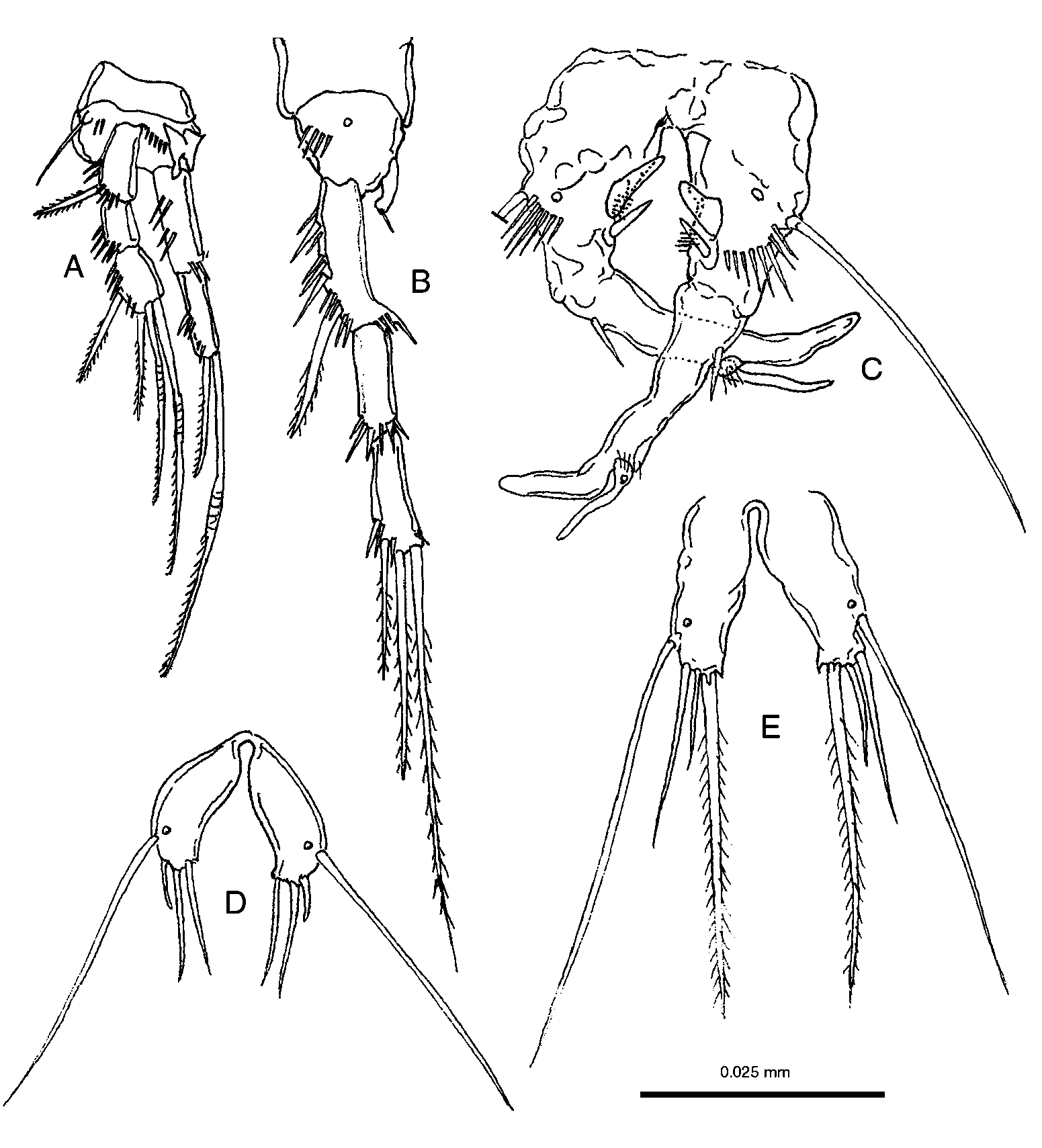
P1 ( Fig. 2 View FIGURE 2 A), basis with thin lateral seta, rows of transverse spinules and 1 enlarged, chitinous protrusion, with rounded bifid tip near endopod insertion; exopod 3-segmented, shorter than endopod; third segment with 1 pinnate and 2 geniculate apical setae, and 1 subapical pinnate seta. Endopod 2-segmented; enp-2 with a long, geniculate, pinnate seta and 1 short, pinnate seta on apex.
P2 ( Fig. 2 View FIGURE 2 B), basis with 1 pore and 1 row of spinules, without lateral seta; exopod 3- segmented, with fringed extension on medial distal corner of segment 1, armature shown in figure; endopod very small, reaching 1/4 of exp-1, represented by a small, outwardly curved cylindrical segment, with 1 apical short seta.
P3 ( Fig. 2 View FIGURE 2 C), elongated, basis with long, lateral seta, a pore and 1 transverse row of spinules; endopod represented by a thin, pointed segment, on the medial margin proximal to the endopod one strong, triangular laminar process; longitudinal row of long spinules ( Fig. 2 View FIGURE 2 C) and row of thin setae posterior to the endopod and process. Exp-1 fused with exp-2, slender, smooth, with spine inserted at about 1/5 of the lateral margin. Exp-2 ending in a long, inwardly curved apophysis with blunt tip. Outer spine of Exp-1 represented by a leaf-like, pointed thumb, shorter than apophysis, with proximal pore, and 3 short spinules at its insertion.
P4 ( Fig. 3 View FIGURE 3 A), basis with long, lateral seta; exopod long, 3-segmented, chaetotaxy shown in figure. Exp-1 and exp-3 with fringed extensions on medial distal corner. Endopod represented by a cylindrical segment with a round protrusion at midlength, followed by bifid blunt tip, reaching to 1/2 length of exp-1; 2 long spines, 1 hooked, near the endopod insertion.
P5 ( Fig. 2 View FIGURE 2 E), without intercoxal plate, represented by 2 almost rectangular, elongated plates fused at the base. A medial subapical tooth-like, small curved expansion; 3 apical setae, medial one longest, pinnate, the middle one is shortest; 1 very long, lateral, subapical seta with a pore near its insertion.
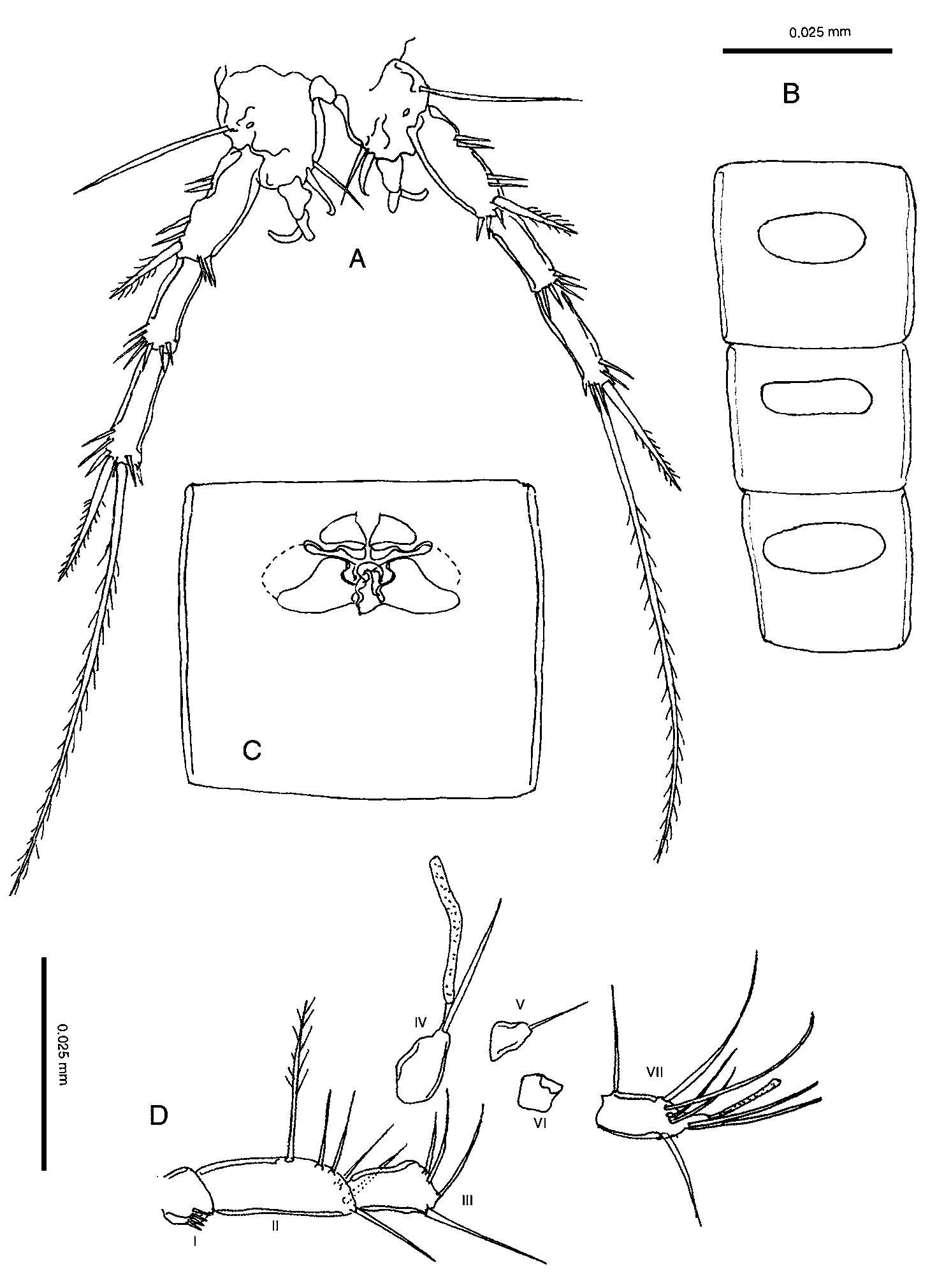
Description of female—Length, measured from rostrum to apex of caudal rami: 0.410 mm. Genital somite, and abdominal somites 2–3 with oval dorsal integumental windows larger than in male ( Fig. 3 View FIGURE 3 B). Genital somite and first abdominal somite fused, forming genital double-somite ( Fig. 3 View FIGURE 3 C). Genital field located in the proximal 1/2 of the genital double-somite ( Fig. 3 View FIGURE 3 C). Anal somite, anal operculum, rostrum, antenna, oral appendages, maxilliped, P1 and P2, as in the male. Caudal rami ( Fig. 4 View FIGURE 4 F) similar to those of the male, outer terminal seta relatively shorter than in male.
Antennule ( Fig. 3 View FIGURE 3 D), 7-segmented, with aesthetasc on segment 4 reaching past end of segment 7. Segment 1 with row of short spinules. Setal formula (from proximal segment): 0, 6, 4, 1 + aesthetasc, 1, 0, 6 + trithek. Apical trithek represented by 2 setae of same lengths and 1 slender, short aesthetasc.
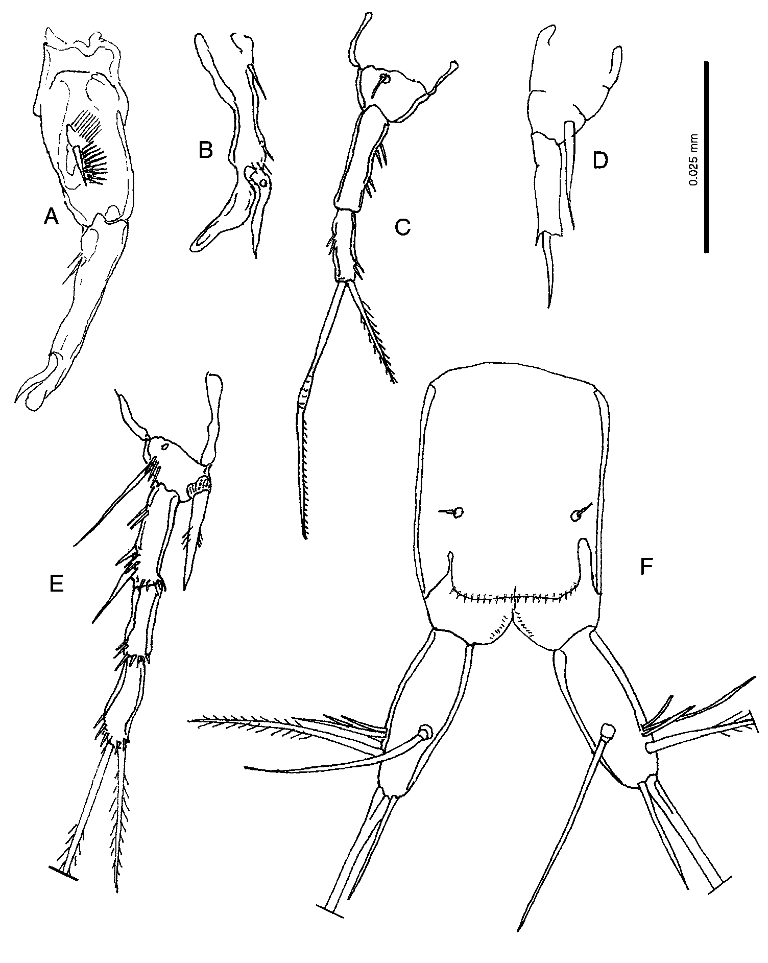
P1 basis ( Fig. 4 View FIGURE 4 C), lateral ornamentation as in the male, the medial margin with 1 slender, short seta.
P3, exopod 2-segmented as normal in the genus; endopod ( Fig. 4 View FIGURE 4 D) represented by a thin, pointed segment, reaching past the end of exp-1.
P4 ( Fig. 4 View FIGURE 4 E), basis with lateral seta, a row of spinules and 1 pore; exopod similar to that of male, with fringed extension on medial distal corner of each segment; endopod inserted on a small chitinous plate, represented by a small cylindrical, pointed segment as long as exp-1, with few longitudinal spinules.
P5 ( Fig. 2 View FIGURE 2 D), without intercoxal plate, fused at the base. P5 similar to that of the male, but less elongate, lacking medial subapical tooth-like expansion; all setae are relatively shorter than in male, the lateralmost apical seta shortest.
Variability—All morphological characters described above appear to be constant, except the P3 of one male paratype which has two spinules on the exp-1 lateral margin at about 1/5 of the length ( Fig. 4 View FIGURE 4 A); the P3 of another male paratype has one spinule on the exp-1 lateral margin at about 1/5 of the length, and a second spinule on the lateral margin at about 3/5 of the length ( Fig. 4 View FIGURE 4 B).
Etymology—The specific name from the Latin adjective “ distinctus ” meaning “different”, it refers to the peculiar shape of the male P1 basis and P3. The epitheton is an adjective in feminine singular.
No known copyright restrictions apply. See Agosti, D., Egloff, W., 2009. Taxonomic information exchange and copyright: the Plazi approach. BMC Research Notes 2009, 2:53 for further explanation.
|
Kingdom |
|
|
Phylum |
|
|
Class |
|
|
Order |
|
|
Family |
|
|
Genus |