
Phareicranaus rohei, Colmenares, Pío A. & Tourinho, Ana Lúcia, 2014
|
publication ID |
https://doi.org/10.11646/zootaxa.3768.1.4 |
|
publication LSID |
lsid:zoobank.org:pub:702D1957-4C7A-4CC8-BE71-7EF45FCDE609 |
|
DOI |
https://doi.org/10.5281/zenodo.6126965 |
|
persistent identifier |
https://treatment.plazi.org/id/F74B87B2-FFB8-FF8D-B5CA-FF64240D48BA |
|
treatment provided by |
Plazi |
|
scientific name |
Phareicranaus rohei |
| status |
sp. nov. |
Phareicranaus rohei View in CoL sp. nov.
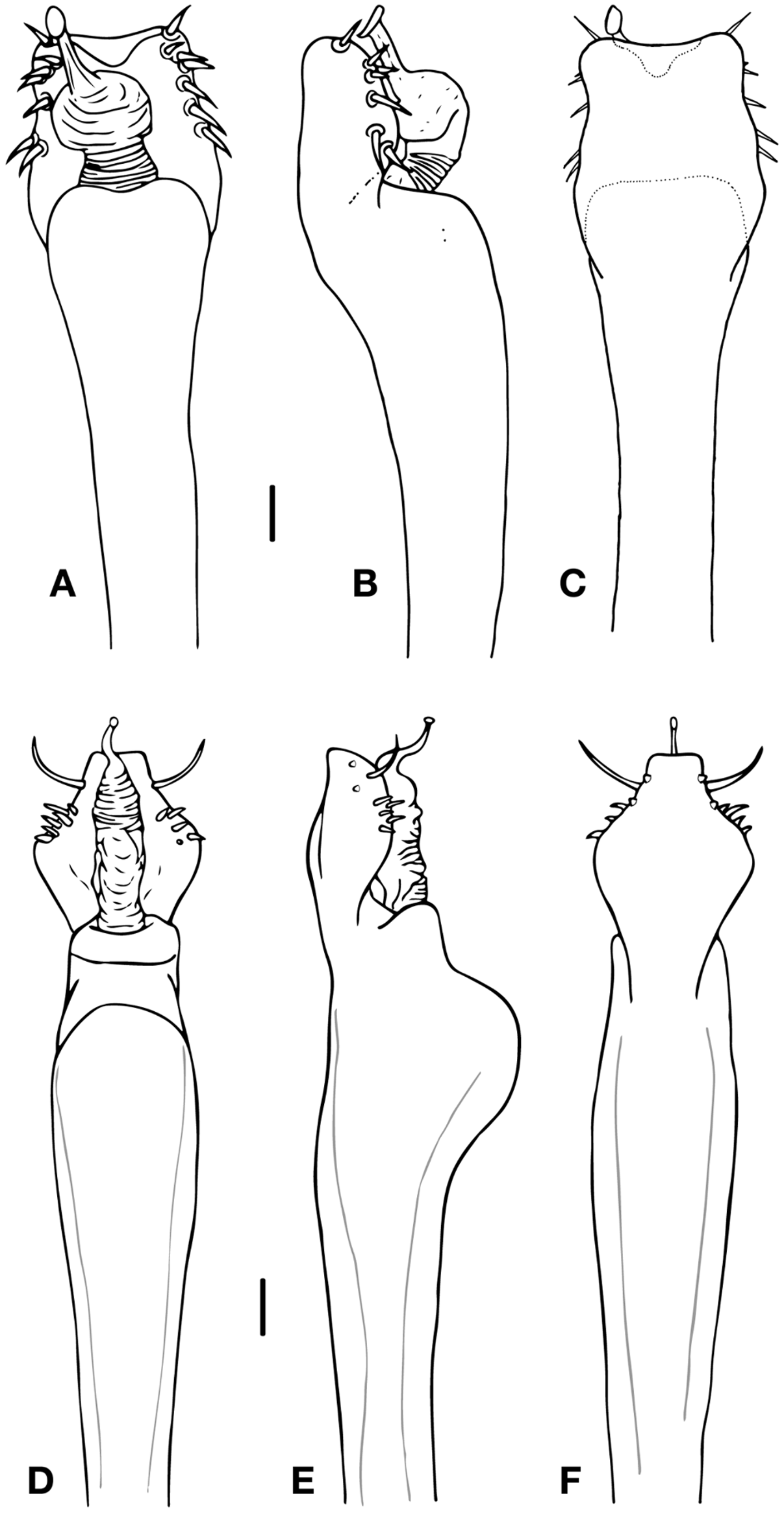
( Figs. 1 View FIGURE 1 A–D; 3 A–C; 6)
Type material. Male holotype: (INPA-OP-2073), Brazil, Amazonas state, RDS Ucari, right border of “Médio Rio Juruá”, Bauana Community, near Rio Bauana, Fabio Röhe leg. Paratypes: 1 male and 1 female (INPA-OP-2074), same data as holotype.
Etymology. The name is a patronymic in honor of Fabio Röhe, a Brazilian mammalogist who has contributed largely to the studies of Amazonian harvestmen by collecting specimens of this species and several other important samples of both species and genera of harvestmen from many localities in the Amazon basin.
Diagnosis. Is related to the other species possessing white circles on the dorsal scutum [ Phareicranaus angelicus ( Roewer, 1963) , Phareicranaus divisor Pinto-da-Rocha & Bonaldo, 2011, Phareicranaus gracilis (Pintoda-Rocha & Kury, 2003) , Phareicranaus hermosa (Pinto-da-Rocha & Kury, 2003), Phareicranaus ortizi ( Roewer, 1952) and Phareicranaus singularis ( Soares, 1970) ]. It can be distinguished from P. angelicus and P. ortizi by the absence of black areas on the scutal scutum; from P. gracilis , by the absence of white circles in lateral areas; from P. divisor , P. hermosa and P. singularis by number of white circles: prosoma with 14, area I with 13, area II with 9, area III with 14 and posterior margin with 18.
Distribution. Known only from the type locality ( Fig. 6 View FIGURE 6 ).
Description. Male Holotype. Measurements: Dorsal scutum length 7.58; width 7.5; prosomal length 3.5; width 5.41; pedipalpal femur 4.16; femur IV 16; leg I 23.08; II 50.66; III 35.75; IV 48.83.
Dorsal scutum ( Fig. 1 View FIGURE 1 A, B). Anterior border with a median projection between the chelicerae, two paramedian and a lateral row of 2–3 small tubercles on each side. Eye mound with two spiniform tubercles, five tubercles behind them and two lateral. Area I with an anterior row of 2–3 small tubercles and a posterior row of four tubercles, II with a posterior row of 4–5 small tubercles, III with two sharp, divergent and high spiniform paramedian tubercles, two small tubercles behind them and two lateral rows (anterior and posterior) of three small tubercles. Posterior border with 18 tubercles. Free tergite I with a pair of paramedian tubercles and a smaller tubercle on each side, II–III with a pair of larger paramedian spiniform tubercles and a smaller tubercle each side. Anal operculum with three rows of 4–5 of small tubercles.
Venter. Coxa I with median row of 4–5 tubercles, three anterior, five posterior and three apical (the anterior one larger); II with a median row of 8–9 small tubercles, three anterior, three posterior and five apical; III with a median row of 7–8 median tubercles, 3–4 anterior, 8–9 posterior and five apical; IV with a median row of 7–8 small tubercles, 6–7 anterior, 8–9 posterior and a pair large apophyses close to the spiracles. Free sternites II–III with two well defined tubercles each side; IV–V with one well defined tubercle each side.
Chelicerae. Basichelicerite with six tubercles on lateral of bulla; hand with several small frontal tubercles; fixed finger with four teeth; movable finger with four teeth.
Pedipalps ( Fig. 1 View FIGURE 1 C). Coxa with 2–3 small tubercles. Trochanter with two dorsal and three ventral tubercles. Femur with a row of 6–7 ventral strong tubercles (three basal and 3–4 median-apical), a retrolateral row of 7–9 tubercles, a dorsal row of 6–7 tubercles (apical larger and sharp). Patella granular, with 12–14 dorsal tubercles unequally distributed and one prolateral apical tubercle. Tibia ventrally with four ectal and four mesal spines (IiIi). Tarsus dorsally granular, ventrally with four ectal (IiIi) and 3–4 mesal spines (IiIi on the left pedipalp and IiI on the right pedipalp).
Legs ( Fig. 1 View FIGURE 1 D). Coxa: I and II with an anterior dorsal tubercle; III smooth; IV with 4–5 latero-dorsal tubercles and an apical spiniform tubercle. Trochanter: I with one dorsal and three ventral tubercles; II with one dorsal median tubercle, two retrolateral and three ventro-apical tubercles; III dorsally with one apical tubercle, three prolateral, four retrolateral and three ventral tubercles; IV with one apical anterior dorsal tubercle, five prolateral, three retrolateral and 4–5 small ventral tubercles. Femora: I–IV straight, with rows of small tubercules; III with one basal retrolateral tubercle and with two dorso-apical sharp tubercles; IV with two ventral rows of more larger tubercles in the first half, one curved retrolateral spiniform tubercle in the second half and two dorso-apical tubercles. Patella I–IV granular. Tibia: I–III granular; IV with two ventral basal tubercles (the basal larger than the other). Tarsal formula: 8(3)/13–14(3)/8/9.
Penis ( Fig. 3 View FIGURE 3 D–F). Ventral plate not very cleft in the distal border, distal corners with flange forming two subequal apical lobes. With 7 setae not easily distinguishable in groups along the lateral borders; the distal pair on the corners of the ventral plate. Gland without dorsal process, with a membranous sac. Stylus smooth, slightly curved and arising straight from glans. Apex bent at an obtuse angle, not swollen.
Color (in alcohol). Body and legs dark brown, except in trochanter, which are more clear. Eye mound, anterior border, quelicerae and pedipalps with a darker reticule. Chelicerae fingers reddish. Tubercles of the dorsal scutum and free tergite I with white tip, and circled by a greenish area that finishes in white rings. Spiniform tubercles of the eye mound and free tergites II–III yellowish. Tarsus clear brown.
Female paratype. Anterior margin with two paramedian and two tubercles on each side. Eye mound with only 4–5 tubercles behind the spiniform tubercles. Pedipalp with tubercles slightly smaller than the male; Tarsus with four mesal spines in both sides (IiIi). Body generally darker than in the male. Tarsal formula: 8(3)/12–13(3)/8/9.


No known copyright restrictions apply. See Agosti, D., Egloff, W., 2009. Taxonomic information exchange and copyright: the Plazi approach. BMC Research Notes 2009, 2:53 for further explanation.
|
Kingdom |
|
|
Phylum |
|
|
Class |
|
|
Order |
|
|
SubOrder |
Laniatores |
|
Family |
|
|
Genus |