
Neodolodus colombianus Hoffstetter & Soria, 1986
|
publication ID |
https://doi.org/10.5252/geodiversitas2023v45a15 |
|
DOI |
https://doi.org/10.5281/zenodo.8319192 |
|
persistent identifier |
https://treatment.plazi.org/id/F7748797-067D-FFF4-FF49-FB876A029C74 |
|
treatment provided by |
Felipe |
|
scientific name |
Neodolodus colombianus Hoffstetter & Soria, 1986 |
| status |
|
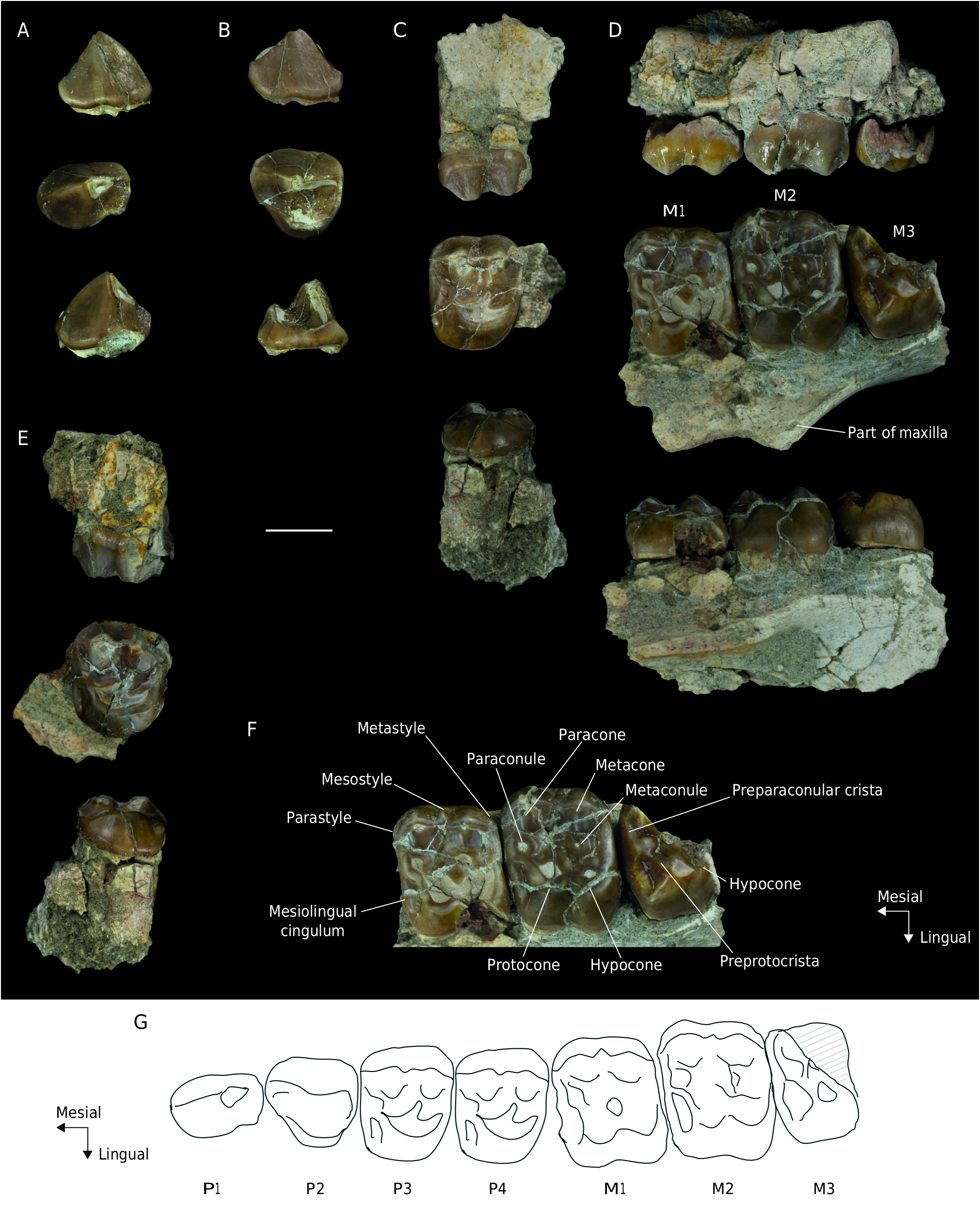
Neodolodus colombianus Hoffstetter & Soria, 1986 ( Figs 1 View FIG ; 5-8 View FIG View FIG View FIG View FIG )
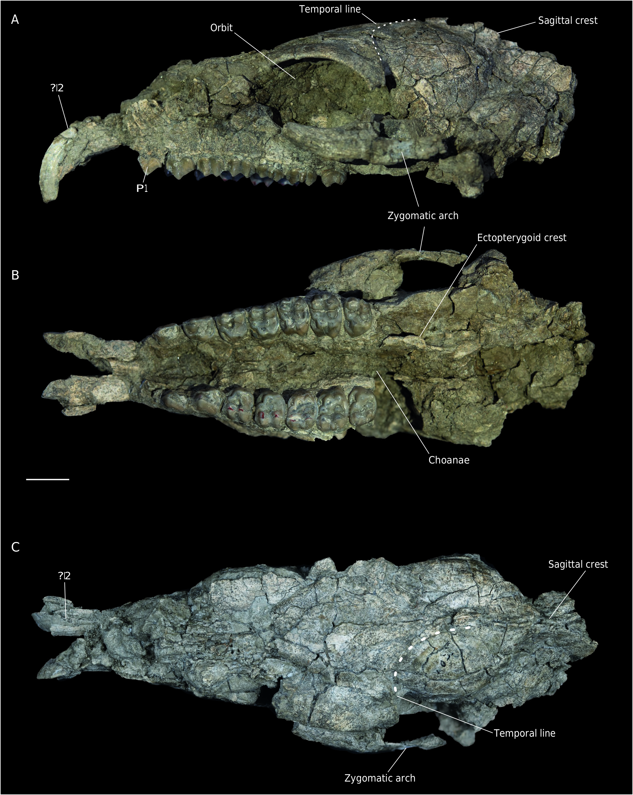
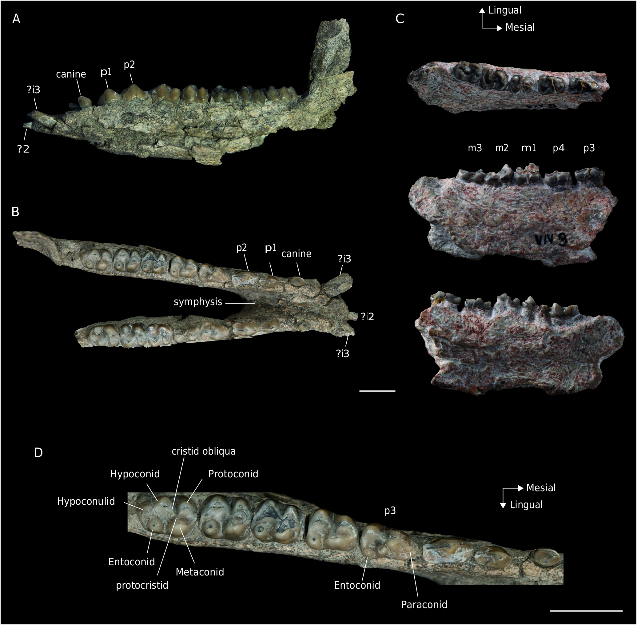
HOLOTYPE. — Specimen MNHN.F.VIV9, partial right mandible bearing p3-m3 ( Fig. 7C View FIG ).
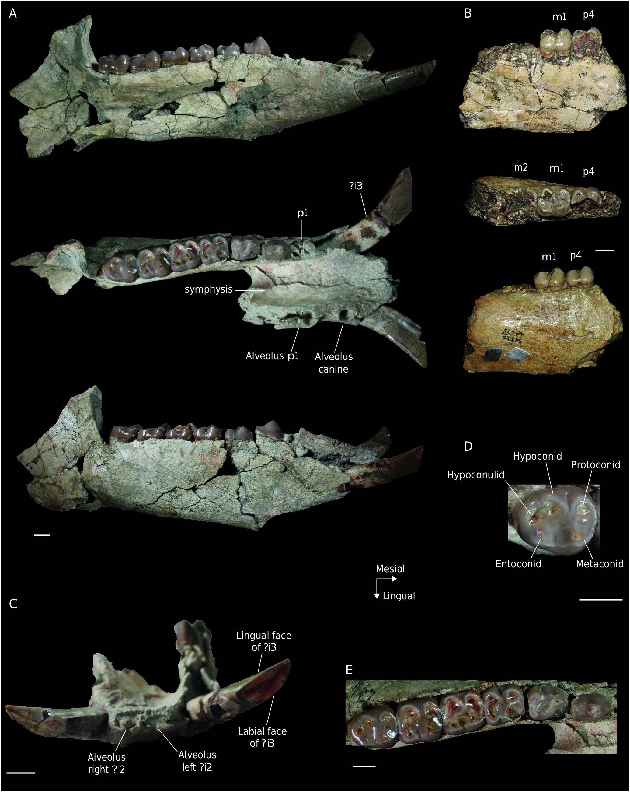
REFERRED MATERIAL. — VPPLT 183, isolated right M1 ( Fig. 6B View FIG ). VPPLT 1696, almost complete skull with left and right?I2 and P1-M3 ( Figs 5-6A View FIG View FIG ). Partial mandible with complete right and left dentition except for left?i2 ( Fig. 7 View FIG A-B, D). Partial right scapula ( Fig. 8A View FIG ), partial right tibia ( Fig. 8B View FIG ), left metacarpal III ( Fig. 8C View FIG ), right metatarsal III ( Fig. 8D View FIG ), and fragments of undetermined (possibly for metapodials III) phalanx.
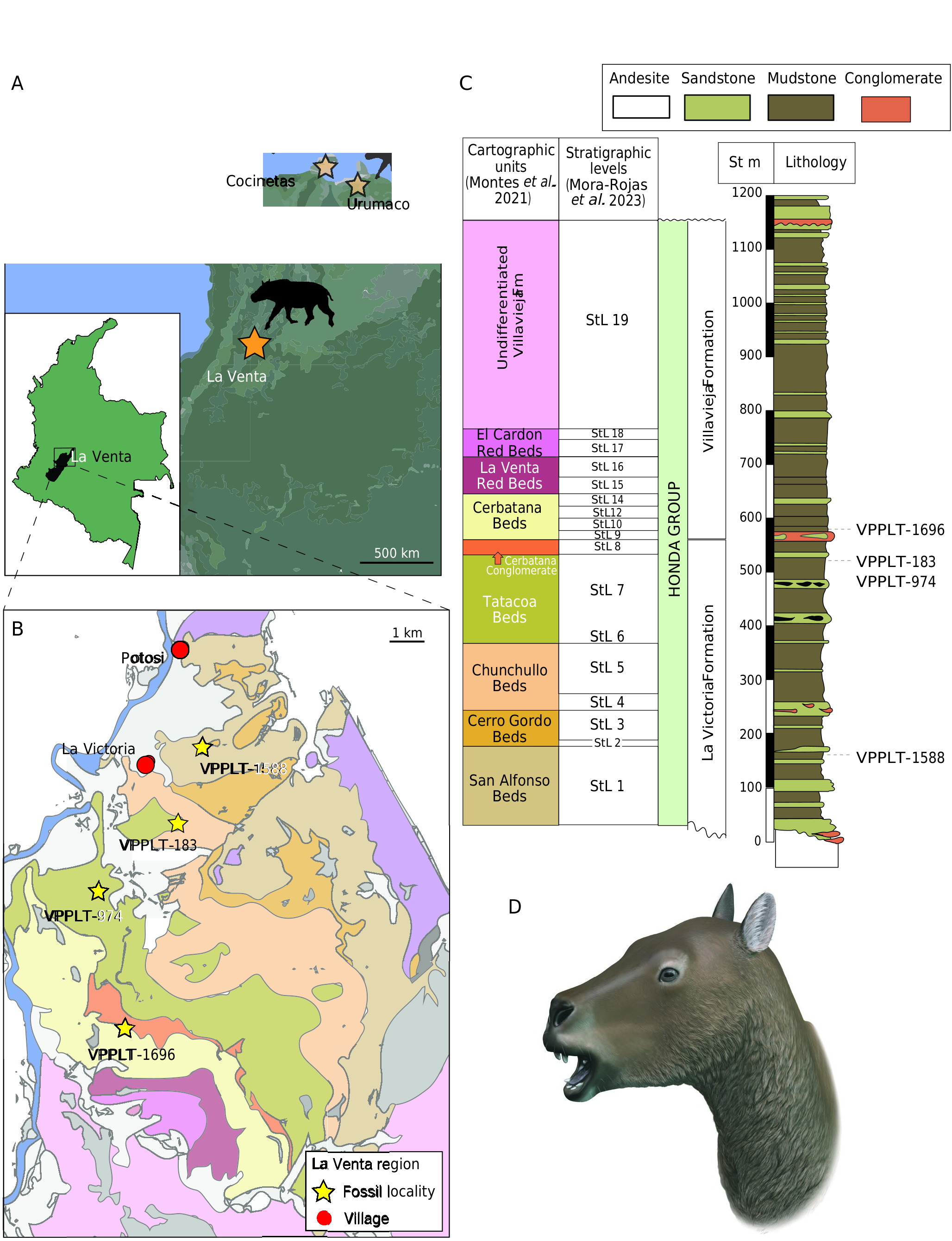
TYPE LOCALITY AND HORIZONS. — VPPLT 183 comes from Kilómetro 121 ( 3°19’29”N, 75°10’55”W), La Victoria Formation in the bed set below the Cerbatana conglomerate (StL 7; Mora-Rojas et al. 2023). VPPLT 1696 comes from Piedra Gorda, San Nicolás ( 3°16’45”N, 75°11’46”W), Villavieja Formation, Monkey beds (StL 9; Mora-Rojas et al. 2023) ( Fig. 1 View FIG ).
DIAGNOSIS. — Same as for the genus due to monotypy.
REMARKS
Previously known craniodental and postcranial material referred to Neodolodus colombianus included partial mandibles that preserved p2, p3, p4, m1, m2 and m3. Partial premaxilla with?I2, and maxillae that preserved P2, P3, P4, M1, M2 and M3 ( Table 3 View TABLE ). The known postcranial elements include partial humerus, radii, metacarpal III and II or IV, manual phalanx, partial pelvis, femur, tibia, astragalus, calcaneus, metatarsal III and pedal phalange ( Cifelli & Guerrero Díaz 1989).
Previously known specimens referred to Neodolodus did not preserve the rostral portion of the cranium and mandible, and the new material clarifies the number and morphology of the upper and lower incisors, canines and anterior premolars ( Table 3 View TABLE ).
DESCRIPTION
VPPLT 1696 preserves most of the cranium although many parts are damaged with cracks and crushed fragments of bone. Parts of the maxilla, palatines, the left zygomatic arch, the nasal, frontal, squamosal, parietal, alisphenoid and occipital bones, and most of the mandible are preserved but, except for teeth, the bad state of preservation does not permit to describe subtle anatomical details ( Figs 5 View FIG ; 6A View FIG ; 7A, B, D View FIG ). VPPLT 1696 is referred to Neodolodus based on its small size ( Table 5 View TABLE ), brachydont and bunodont dentition, the absence of a mesostyle on P3-4 ( Figs 5B View FIG ; 6A View FIG ), a small paraconid present on p3 but absent on p4-m3, and the isolated entoconid on m1-3 ( Fig. 7B, D View FIG ) ( Hoffstetter & Soria 1986; Cifelli & Guerrero Díaz 1989; McGrath et al. 2020a).
The cranium measures c. 10.5 cm from the most caudal point of the sagittal crest to the most rostral point of the snout between the incisors. It is damaged, lacking portions of the prexamilla and maxilla. Due to the preservation, no sutures or synchondrosis are clearly visible. In ventral view, the palate looks narrow, but it is unclear whether this is due to a slight post-mortem mediolateral compression of the cranium or represents the unaltered morphology. The choanae open directly posteromedially to the distal edges of the M3, with a rounded rostral edge. From there, the basipharyngeal canal is bordered laterally by the sagittally-oriented ectopterygoid crests, up to the auditory region caudally. In lateral view, the rostrum is low and elongated ( Fig. 5A View FIG ). The orbit is large, with a large frontal postorbital process of the frontal posteriorly. The zygomatic arch does not extend much laterally but this might be due in part to postmortem deformation. In dorsal view, the temporal lines are concave caudally, extending from the caudal margin of the orbit and meeting on the roof of the cranium at the level of the rostral edge of the posterior root of the zygomatic arch ( Fig. 5A View FIG ). The union of these temporal lines forms a well-defined sagittal crest caudally.
The mesial-most pair of the upper teeth (?I2) are developed into tusks, which are triangular in cross-section and curved ventrally ( Fig. 5A View FIG ). The most apical portion of the tusks is not preserved, and is not possible to assess if the upper tusks touched with the lower ones, as it appears to be the case in Megadolodus ( Fig. 3C View FIG ). In Neodolodus , the crown of P1 is simple with a single centrally-located cusp with a crest extending mesially, as in Megadolodus . The distal portion of the crown has an oblique wear facet oriented distolingually ( Figs 5B View FIG ; 6A View FIG ). The P2 also has one cusp, located on the labial side and positioned mesial of the midline of the tooth. The tooth exhibits a lingual basin which is stretched distomesially and bordered by a distolingual cingulum ( Figs 5B View FIG ; 6A View FIG ). A small cusp was also likely present on the distolingual edge of this basin, but the wear stage of the tooth precludes a definite statement on this morphology. On the distolabial border of the crown there is a metastyle.
The upper premolars and molars of VPPLT 1696 increase in size from P3 to M2 ( Table 5 View TABLE ), and the M3 is similar in size to P4, as in Megadolodus ( Cifelli & Villarroel 1997) , Anisoplophus, Diadiaphorus , Megadolodus and Villarroelia , and unlike Tetramerorhinus where the M3 is larger or of similar size to M1-2. The P3-4 are partly molariform, they have a well-developed protocone, paracone and metacone, but they lack a fully individualized hypocone (an incipient cingulum-derived hypocone is visible on P4) ( Figs 5B View FIG ; 6A View FIG ). As in Megadolodus ( Fig. 2C, E View FIG ), there is a parastyle and metastyle on the ectoloph of the P3 and P4 of Neodolodus , but no mesostyle ( Fig. 6A View FIG ). Two faint labial ridges are visible on the labial wall of the paracone and metacone ( Figs 5B View FIG ; 6A View FIG ). As seen in other specimens of Neodolodus ( Cifelli & Guerrero Díaz 1989), the posterior premolars of VPPLT 1696 have a thick mesiolingual cingulum that defines a small basin directed labiolingually. The paraconule and metaconule are distinct, the former being connected to the protocone by the preprotocrista, whereas the latter is isolated ( Fig. 6A View FIG ).
The upper molars exhibit a labially protruding mesostyle, especially on M1-2. The parastyle and metastyle are less developed than the mesostyle ( Fig. 6A View FIG ). A cingulum of varying thickness is present at both labial corners at the base of the crown, as described in other specimens of Neodolodus ( Cifelli & Guerrero Díaz 1989). All the upper molars have a postcingulum-derived hypocone ( Figs 5B View FIG ; 6A View FIG ). The paracone and metacone are well-defined and not connected to the paraconule or metaconule. As in Megadolodus , there are two ridges on the labial flank of the paracone and metacone, although in Neodolodus these ridges are more developed ( Figs 2F View FIG ; 6A View FIG ). The upper molars of Neodolodus also exhibit a thick mesiolingual cingulum that forms a basin just like on P3-4 ( Figs 5B View FIG ; 6A View FIG ) ( Cifelli & Guerrero Díaz 1989). The paraconule is connected by the preprotocrista to the protocone, as in Megadolodus, ( Cifelli & Villarroel 1997) . The metaconule is isolated from other cusps in all the molars. The hypocone is small and connected to the protocone on M3 by a small lingual crista ( Fig. 6A View FIG ), as in other specimens of Neodolodus ( McGrath et al. 2020a) .
The mandible of VPPLT 1696 preserves most of the right and left horizontal rami and part of the left ascending ramus ( Fig. 7A View FIG ). The mandibular symphysis extends caudally until the level of the p2 ( Fig. 7B View FIG ). Of the two mesial-most incisors, only the crown of the right?i2 is preserved. It is spatulate with flat lingual and labial faces. The base of the crown of the right and left?i3 are preserved ( Fig. 7A, B View FIG ). The?i3 have flat lingual and convex labial faces, separated by medial and labial ridges. The?i3 are considerably larger than the?i2. As in Megadolodus , there are two short diastemata ( Fig. 7B View FIG ), one between the?i3 and the canine (5.0 and 4.0 mm in right and left side respectively), and one between the canine and the p1 (4.0 and 3.0 mm in right and left side respectively). The canine is small and somewhat spatulate, with a flat lingual face and a convex labial one ( Fig. 7A, B View FIG ).
The p1-2 are simple with a single cuspid located slightly mesial to the centre of the tooth, which results in a nearly triangular shape in labial view. On both teeth, crests extend downward mesially and distally from the main cuspid ( Fig. 7B View FIG ). On p2, the distal crest gently curves lingually and upwards at its distal end which resembles an incipient talonid ( Cifelli & Guerrero Díaz 1989). The size of the premolars increases from p1 to p4. The p3 is bicrescentic, with the trigonid being narrower than the talonid ( Fig. 7D View FIG ). A low cuspid is present at the mesial edge of p3, which may well represent an incipient paraconid; this cuspid is absent on p4 and lower molars, as already described for this species ( Hoffstetter & Soria 1986; Cifelli & Guerrero Díaz 1989). The p4 is molariform, without paraconid ( Fig. 7D View FIG ). The lingual portion of the crown of the right p4 of VPPLT 1696 is broken, but the left p4 is complete. In Neodolodus , the hypoconulid is absent on p3-4 ( Fig. 7D View FIG ). There is a low metaconid on p3, and the same cuspid is more developed on p4. The metaconid is separated from the protoconid by a sulcus on its mesial edge. The entoconid is isolated from other cuspids on both teeth ( Fig. 7D View FIG ).
The lower molars of Neodolodus are bicrescentic and increase in length from m1 to m3 ( Cifelli & Guerrero Díaz 1989). VPPLT 1696 has high wear on m1 and moderate wear on m2, whereas the m3 is only slightly worn ( Fig. 7B, D View FIG ). The less worn state of m3 allows the preservation of the protocristid, which connects the protoconid and the metaconid along their distal edges ( Fig. 7D View FIG ). There is a mesial crest extending from the protoconid. The absence of paraconid results in an ovate outline of the trigonid. The hypoconid is the largest cusp on the molar talonid. It is connected to the base of the metaconid and trigonid through a low cristid obliqua and to the hypoconulid through a low distal cristid (hypocristid). The entoconid is isolated from other cusps on all the lower molars ( Hoffstetter & Soria 1986) and located in a more lingual position than the hypoconulid ( Cifelli & Guerrero 1997; McGrath et al. 2020a). The hypoconulid is smaller than the entoconid on m1-2 ( Fig. 7D View FIG ). The hypoconulid on m3 is located distally from the hypoconid and entoconid and is connected to a cristid extending lingually from it, without reaching the entoconid ( Fig. 7D View FIG ).
MNHN. Duke-ING UCMP Duke-ING IGN UCMP IGN VPPLT 1696 VPPLT F.VIV9* 86-093** 86-274** 86-288** 37691** 88-332** 182578** 38910** 182568** left right 183
?i2 Antero-posterior – – – – – – – – – – – – length
Transverse length – – – – – – – – – – – –
?i3 Antero-posterior – – – – – – – – – 4.1 4.2 – length
Transverse length – – – – – – – – – 2.6 2.6 –
Associated postcranial elements of VPPLT 1696 include a partial scapula and limb bones. The right scapula preserves the glenoid fossa, part of the spine and the blade ( Fig. 8A View FIG ). The glenoid fossa is semi-circular, being rounded on the lateral edge and straighter in the dorsal edge. The neck is short and wide. The spine is broken, it is narrow and located in the middle of the blade. Most of the blade is not preserved and the relative size and shape of the supraspinous and infraspinous fossae cannot be assessed. The right tibia preserves the shaft and the distal epiphysis ( Fig. 8B View FIG ). The medial malleolus extends further distally than the lateral malleolus, as in Diadiaphorus majusculus ( Schmidt et al. 2019b) . On the lateroposterior part of the distal end of the tibia, there is a facet for the articulation with the fibula ( Fig. 8B View FIG ). The metapodials III ( Fig. 8 View FIG C-D) have a straight and long diaphyses, and the distal a well-defined keels, as seen in other proterotheriids ( Carrillo et al. 2018; Schmidt et al. 2019b). By comparison with the known metapodials III of Megadolodus ( Cifelli & Villarroel 1997) , we tentatively identify one element as left metacarpal III ( Fig. 8C View FIG ) because it is shorter and more robust than the other element, which we identify as the right metatarsal III ( Fig. 8D View FIG ). The metacarpal III has a small facet for the articulation with the metacarpal II on the medial side, and the proximal epiphysis is not fused ( Fig. 8C View FIG ).


| MNHN |
Museum National d'Histoire Naturelle |
No known copyright restrictions apply. See Agosti, D., Egloff, W., 2009. Taxonomic information exchange and copyright: the Plazi approach. BMC Research Notes 2009, 2:53 for further explanation.
|
Kingdom |
|
|
Phylum |
|
|
Class |
|
|
Order |
|
|
Family |
|
|
SubFamily |
Megadolodinae |
|
Genus |