
Aolacoccus angophorae, Gullan, Penny J. & Williams, Douglas J., 2016
|
publication ID |
https://doi.org/10.11646/zootaxa.4117.1.4 |
|
publication LSID |
lsid:zoobank.org:pub:5C240849-6842-44B0-AD9F-DFB25038B675 |
|
DOI |
https://doi.org/10.5281/zenodo.6070256 |
|
persistent identifier |
https://treatment.plazi.org/id/85E4AA88-B7D1-44A9-9ECF-6A929A208A2D |
|
taxon LSID |
lsid:zoobank.org:act:85E4AA88-B7D1-44A9-9ECF-6A929A208A2D |
|
treatment provided by |
Plazi |
|
scientific name |
Aolacoccus angophorae |
| status |
sp. nov. |
Aolacoccus angophorae sp. nov.
urn:lsid:zoobank.org:act:85E4AA88-B7D1-44A9-9ECF-6A929A208A2D
Etymology. The specific name is based on the genus name of the host plant, is in the Latin genitive singular and means "of Angophora ".
Type material examined. Holotype: Adult female (body 740 µm long), on slide with its second-instar exuviae. AUSTRALIA, New South Wales: Sydney, Cremorne Point, under bark of Angophora , 15.x.1980, A.M. Richards, A12605 View Materials , B3 ( ANIC).
Paratypes: 5 slides, each with 1 adult female and 4 also with female's second-instar exuviae, 1 slide with 1 second-instar female exuviae and 5 embryos, 3 slides with 13, 10 or 2 first-instar nymphs respectively, 1 slide with 1 second-instar male and its first-instar exuviae, all with same data as holotype (8 slides in ANIC; 2 slides each with 1 adult female and its second-instar exuviae in ASCU); 4 slides, each with 1 adult female and its second-instar exuviae and 1 of these slides also with 1 first-instar nymph, 1 slide with 2 adult females and 3 second-instar exuviae, 1 slide with 16 embryos, all with same data as holotype except "B1" instead of "B3" (4 slides in ANIC; 2 slides each with 1 adult female and its second-instar exuviae in BMNH).
Other material examined: AUSTRALIA, New South Wales: 2 slides each with 1 male pupa (with pharate adult male in one pupa but broken in half), Bungwahl (misspelled on label as "Bungwalil") [N of Newcastle], ex Angophora , 16.v.1984, C. Ballard ( ANIC); 1 slide with 5 second-instar males, Cowan [N of Sydney], under bark of Angophora sp., 2.x.1980, A.M. Richards, A12605 View Materials , B2 ( ANIC); 1 slide of adult male [poorly cleared], Hornsby Heights [N of Sydney], 29.v.1984, A.M. Richards ( ANIC); 1 slide with 2 adult females, 1 slide with 1 adult female and its second-instar exuviae and 12 first-instar nymphs, 2 slides each with 1 adult female and its second-instar exuviae, 1 slide with 8 second-instar exuviae (4 with adult female inside), and 1 slide with 1 second-instar male, Sydney, Cronulla, Gunnamatta Park, roadside, ex trunk of Angophora costata , under small pieces of loose bark, 13.ii.2015, P.J. Gullan, P.S. Cranston & T. Kondo ( ANIC).
Note: The second-instar male from the type collection was compared with those collected at other localities and they were identical. The pharate adult male from Bungwahl and the adult male from Hornsby Heights also appeared identical and, although no associated female specimens are available for slide mounting, A.M. Richards said that females were present and she was certain that they were conspecific with specimens from Cremorne Point (pers. comm. to DJW).
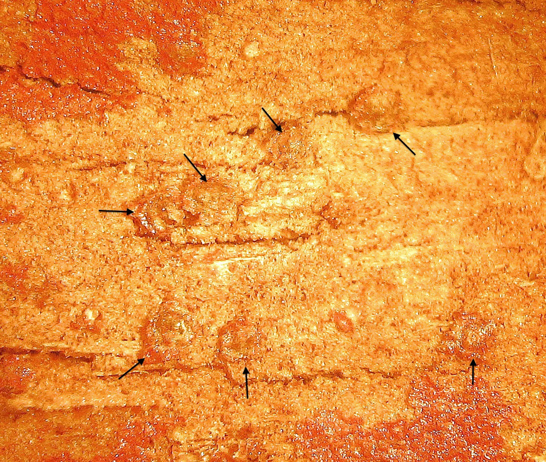
Adult female ( Figs 1 View FIGURE 1 , 2 View FIGURE 2 ) Unmounted material. On trunk and branches of host trees, usually beneath bark; each adult female hidden beneath a thin layer of bark cells ( Fig. 1 View FIGURE 1 ) and pharate within sclerotised cuticle of penultimate instar.
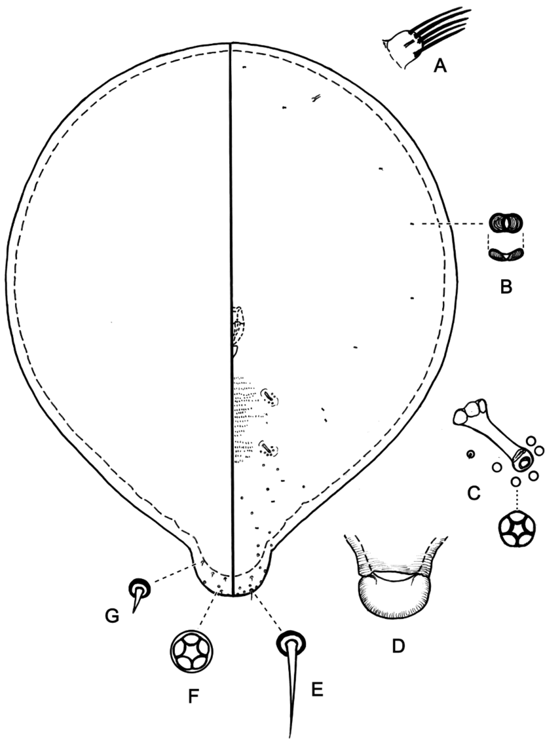
Mounted material (description based on 12 females). Body 400–760 µm long, 400–750 µm wide, rounded, almost circular when mature, with posterior of abdomen tapering to a rounded end with light sclerotisation for posteriormost 55–100 µm. Margin not defined, lacking setae. Derm membranous, except for posterior of abdomen and rows of minute microtrichia ventromedially between spiracles. Segmentation indistinct to absent.
Dorsum. Most setae ( Fig. 2 View FIGURE 2 F) minute and sparsely distributed, each about 2 µm long and 2.0–2.5 µm across socket, except 8–15 longer setae, each 5–10 µm long, on posterior of abdomen. Macrotubular and microtubular ducts absent. Loculate pores absent. Anal lobes absent.
Venter. Setae ( Fig. 2 View FIGURE 2 C) mostly minute and sparsely distributed as on dorsum, with 7–12 longer setae each 7–20 µm long on posterior of abdomen, and several setae each 5–10 µm long just posterior to vulva. Macrotubular and microtubular ducts absent. Loculate pores ( Fig. 2 View FIGURE 2 D) all quinquelocular (5 loculi) (except for a single 9-locular pore detected on one female), restricted to 2 places: (i) 3–8 pores, each 3–4 µm in diameter, around atrial opening of each spiracle, and (ii) a dense cluster of 110–150 pores, each 4–5 µm in diameter, surrounding vulva. Small disclike structures ( Fig. 2 View FIGURE 2 E), each 2.5–5.0 µm in diameter, perhaps cicatrices, in cluster of about 90–110 posterior to vulva, partly intermixed with quinquelocular pores. Eyespots absent. Antennae ( Fig. 2 View FIGURE 2 A) each a small unsegmented tubercle 12–23 µm long and 10–17 µm wide, bearing 4 or 5 fleshy setae, each seta 13–30 µm long. Frontal lobes and antennal tubercles absent. Clypeolabral shield 120–160 µm long, 90–120 µm wide. Labium 50– 75 µm long, 45–60 µm wide, segmentation poorly defined, probably with 3 segments and 4 pairs of setae. Stylets 2000–2200 µm long (based on 2 females with intact stylets), coiled into about 4 loops when retracted. Spiracles ( Fig. 2 View FIGURE 2 B) well developed, subequal in size, each with muscle plate expanded medially; length including muscle plate 25–40 µm; width across peritreme at atrial opening 10–15 µm and across widest part of muscle plate 16–26 µm; each peritreme surrounded by a group of quinquelocular pores (see above). Legs absent. Vulva well developed, its position marked by radiating wrinkles. Anal ring apparently ventral and represented by a small, partially sclerotised ring, lacking pores and setae.
Second-instar female ( Fig. 3 View FIGURE 3 ). Mounted material (description based on 8 females). Body 550–1000 µm long, 500–900 µm wide, rounded, with posterior of abdomen tapering to a sclerotised and rounded knob, 65–90 µm long, that splits to become a hinged flap or operculum ( Fig. 3 View FIGURE 3 D) to allow emergence of crawlers. Anal lobes absent and anus not detected. Margin not defined, lacking setae. Derm membranous when young, becoming sclerotised with age, and with 12–17 rows of minute microtrichia ventromedially between spiracles. Segmentation not discerned.
Dorsum. Setae absent on dorsal derm except for 8–12 short setae ( Fig. 3 View FIGURE 3 G) on knob of posterior of abdomen, each seta 2–4 µm long and ~2 µm across socket. Macrotubular and microtubular ducts absent. Loculate pores absent except for several on posterior apex of abdomen on operculum (see below for venter).
Venter. Setae few and each mostly 1–2 µm long and 2 mm across socket, 1–3 pairs near spiracles, a few on posterior of abdomen and a pair of longer setae ( Fig. 3 View FIGURE 3 E), each 10–13 µm long and 3 µm across socket, on posterior margin of abdomen (the latter may be apical setae). Macrotubular ducts and typical eriococcid microtubular ducts absent, but a sclerotised pore-like structure ( Fig. 3 View FIGURE 3 B), 2 µm across widest dimension, possibly a bilocular microduct, sparsely distributed on abdomen. Loculate pores ( Fig. 3 View FIGURE 3 F) all quinquelocular (5 loculi), each 2.5–3.0 µm in diameter, restricted to 2 places: (i) 2–6 pores around atrial opening of each spiracle, and (ii) a scattering of pores on abdomen posterior to spiracles with a denser grouping of 20–30 on posterior apex of abdomen, of which some may be dorsal. Eyespots absent. Antennae ( Fig. 3 View FIGURE 3 A) each a small unsegmented tubercle 7.5–8.0 µm long and 8–10 µm wide, bearing 4–5 fleshy setae, each 7–16 µm long. Frontal lobes and antennal tubercles absent. Clypeolabral shield 75–100 µm long, 50–60 µm wide. Labium 35–45 µm long, 30–35 µm wide, segmentation poorly defined, probably with 3 segments and at least 3 pairs of setae. Stylets at least 1200 µm long (broken or missing on all but one specimen). Spiracles ( Fig. 3 View FIGURE 3 C) small but well developed, subequal in size, each with muscle plate slightly expanded medially; length including muscle plate 1 8–24 µm; width across peritreme at atrial opening 5–7 µm and across widest part of muscle plate 8–10 µm; each peritreme surrounded by a small group of quinquelocular pores (see above). Legs absent.
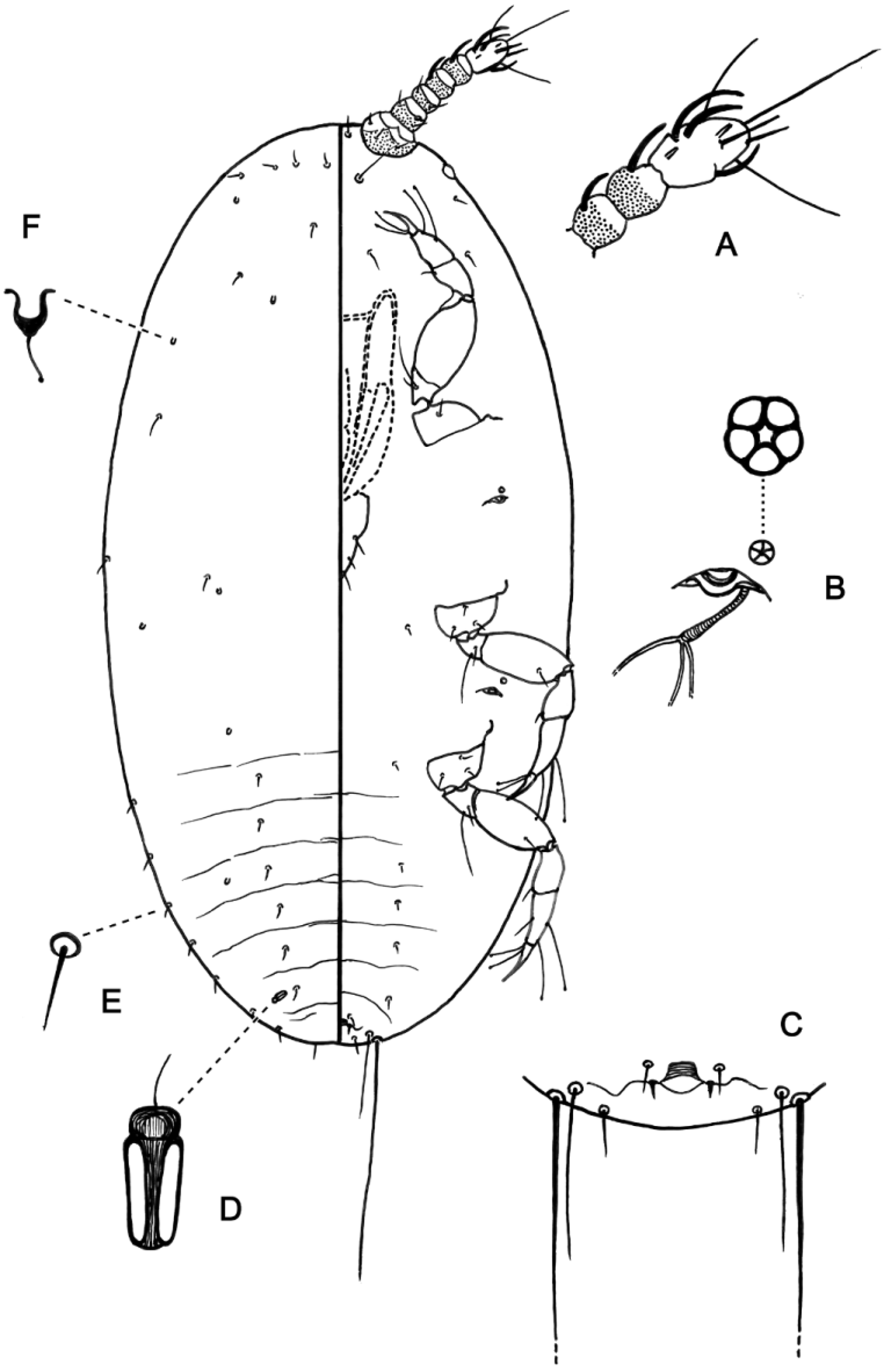
First-instar nymph ( Fig. 4 View FIGURE 4 ). Mounted material (sex not determined; description based on 8 nymphs). Body 230– 255 µm long, 110–135 µm wide, ovoid, with posterior of abdomen ( Fig. 4 View FIGURE 4 C) rounded, lacking distinct anal lobes, anus located ventrally. Margin not defined by differentiated setae. Derm membranous; segmentation discernible only on abdomen.
Dorsum. Setae ( Fig. 4 View FIGURE 4 E) minute hair-like, each 3–5 µm long, in marginal and submedial longitudinal rows on each side of abdomen, rare on thorax and scattered mostly anteriorly on head. Macrotubular ducts and loculate pores absent. Microtubular ducts of 2 kinds: (i) a single robust, sclerotised duct ( Fig. 4 View FIGURE 4 D), 5.0–5.5 µm long and 2– 3 µm wide, submarginally on each side of posterior of abdomen, and (ii) shallow, sclerotised ducts ( Fig. 4 View FIGURE 4 F), 1.5– 2.0 µm deep, mostly 1.5 µm wide, scattered, few in number (10–12 total).
Venter. Setae hair-like, each 2–4 µm long on abdomen and thorax, 5–12 µm long on head with longest situated submedially posterior to antennae; 1 pair of long flagellate apical setae, each 70–75 µm long, with a shorter apical seta, each 22–24 µm long, medial to each apical seta, and a pair of shorter setae, each 5–6 µm long, between 2 pairs of apical setae ( Fig. 4 View FIGURE 4 C). Macrotubular and microtubular ducts absent. Loculate pores quinquelocular, each 2.0–2.5 µm in diameter, with 1 pore anterior to opening of each spiracle. Eyespot 8–10 µm in greatest dimension, situated on margin posterior to base of antenna. Antennae ( Fig. 4 View FIGURE 4 A) each 43–53 µm long with 6 segments; segment I mostly lightly sclerotised and basal part of each of 5 distal segments with light sclerotisation; apical segment 13–15 µm long and 7.0–7.5 µm wide, bearing 3 curved fleshy setae each 8–11 µm long, 1 slender straight fleshy seta 6–9 µm long, 3 long hair-like setae each 16–26 µm long (apical one longest), 2 slender setae each 6–7 µm long, and 2 coeloconic sensilla; other antennal segments as follows: segment I with 3 or 4 hair-like setae; II with 2 hair-like setae and a campaniform sensillum; III with 2 or 3 hair-like setae; IV with 1 fleshy seta 5–6 µm long and no other setae; V with 1 fleshy seta 6–7 µm long and 2 or 3 hair-like setae. Frontal lobes and antennal tubercles absent. Clypeolabral shield 55–62 µm long, 30–35 µm wide. Labium 20–27 µm long, 18–20 µm wide, segmentation poorly defined, probably with 3 segments and 3 pairs of setae, with apical setae spike-like. Stylets 5 00–800 µm long, coiled into almost 2 loops when retracted. Spiracles ( Fig. 4 View FIGURE 4 B) subequal in size, each with elongate sclerotised peritreme of length 7–8 µm, maximum width 2.0–2.5 µm; each spiracle associated with a quinquelocular pore (see above). Legs well developed; segment lengths (metathoracic leg (µm)): coxa 9–10; trochanter + femur 30–34; tibia 13–17; tarsus 12–15; claw 11–13; long trochanteral seta 14–17 µm long; a campaniform sensillum present at base of each tarsus (difficult to see); claw with a subapical denticle; both tarsal digitules on all legs capitate, with one digitule distal and other much more proximal, distal digitule 12–15 µm long, other digitule 14–19 µm long, with longest digitules on metathoracic legs; claw digitules alike, each 9–10 µm long, with small capitate apex. Anus represented by a sclerotised arc 2–3 µm across that may represent a partial anal ring; flanked by 2 pairs of setae, more distal pair spine-like and each 2–3 µm long, other pair hair-like and each ~4 µm long.
Second-instar male ( Fig. 5 View FIGURE 5 ). Unmounted material (only one live nymph seen). On bark of host plant; in white waxy test of typical eriococcid type.
Mounted material (description based on 7 nymphs). Body 500–780 µm long, 300–455 µm wide, ovoid, with posterior of abdomen rounded, lacking distinct anal lobes and anus located ventrally. Margin not defined by differentiated setae. Derm membranous but with rows of minute microtrichia ventromedially between thoracic legs. Segmentation distinct dorsally, weakly indicated on ventral abdomen.
Dorsum. Setae ( Fig. 5 View FIGURE 5 D) robust hair-like, each 10–17 µm long in a sparse row on each thoracic and abdominal segment; longer and sparsely scattered on head, each 25–30 µm. Macrotubular ducts ( Fig. 5 View FIGURE 5 E) each with main duct and outer opening membranous (difficult to discern), up to 25 µm deep, with inner cup about 3.5 µm across and a membranous inner ductule 5–6 µm long; ducts sparsely distributed across all segments and with 1 duct opening on lateral margin of each side of each abdominal and thoracic segment and several opening on margin around head. Microtubular ducts absent. Loculate pores ( Fig. 5 View FIGURE 5 B) all quinquelocular, each about 3 µm in diameter, sparsely scattered on every body segment.
Venter. Setae robust hair-like, each 10–30 µm long on abdomen, 15–38 µm long on thorax and laterally on head; but 6 or 7 (usually 7) setae, each 30–70 µm long, situated medially to submedially on head; one pair of long flagellate apical setae, each 85–90 µm long, with a pair of shorter setae, each 17–20 µm long, present between apical setae. Macrotubular and microtubular ducts absent. Loculate pores ( Fig. 5 View FIGURE 5 B) all quinquelocular, each about 3 µm in diameter, sparsely scattered on every body segment but with a loose aggregation around each posterior spiracle and 2 or 3 pores anterolateral of atrial opening of each spiracle. Eyespots each 15–19 µm in greatest dimension, situated on margin posterior to base of antenna. Antennae ( Fig. 5 View FIGURE 5 A) each 37–50 µm long with 6 segments; base of each of 5 distal segments with light sclerotisation but segment I entirely membranous; apical segment 10–15 µm long and 10–13 µm wide, bearing 3 subequal, curved fleshy setae, each 15–24 µm long, 6 hairlike setae, each 10–25 µm long, and a coeloconic sensillum; other antennal segments as follows: segment I with 3 or 4 hair-like setae; II with 2 hair-like setae and a campaniform sensillum; III with 2 or 3 hair-like setae; IV with 1 fleshy seta 12–15 µm long and no other setae; V with 1 fleshy seta 15–21 µm long and 4 hair-like setae. Frontal lobes and antennal tubercles absent. Clypeolabral shield 88–100 µm long, 70–80 µm wide. Labium 40–50 µm long, 30–40 µm wide, segmentation poorly defined, probably with 3 segments and 4 pairs of setae. Stylets 1000– 1400 µm long. Spiracles elongate, subequal in size, each with muscle plate slightly expanded medially; length including muscle plate 20–26 µm; width across peritreme at atrial opening 4–5 µm and across widest part of muscle plate 7–10 µm; always associated with a few quinquelocular pores (see above). Legs well developed; segment lengths (metathoracic leg (µm)): coxa 40–50; trochanter + femur 58–70; tibia 35–42; tarsus 27–40; claw 12–15; long trochanteral seta 30–50 µm long; a campaniform sensillum present at base of each tarsus; claw with a subapical denticle; both tarsal digitules on all legs capitate and equal in size, each 17–20 µm long; claw digitules alike, each 10–12 µm long, with small capitate apex. Anus ( Fig. 5 View FIGURE 5 C) represented by a sclerotised arc 5–7 µm across that may represent a partial anal ring; flanked on each side by a tiny sclerotised patch bearing a seta 7–8 µm long and with a slightly more anterolateral seta 10–12 µm long.
Adult male ( Fig. 6 View FIGURE 6 ). Mounted material (notes based on 2 adult males, one poorly cleared, other pharate in pupal cuticle and mostly poorly visible). Body ~800 µm long with abdomen ~430 µm long, with middle segments partially telescoped into each other. Head with 2 pairs of simple eyes and antennae each with 8 segments. Wings and hamulohalteres absent. Body with hair-like and peg-like setae; pores absent. Glandular pouches absent. Genital capsule only slightly longer than wide, tapered to a bluntly rounded apex.
Head. Broad conical with truncate anterior margin; head width across ocular sclerites ~120 µm; head ridges and most sclerites not apparent; width across each ocular sclerite 15–17 µm. Head setae: ventral surface with ~24 peg-like setae, each 3–4 µm long, and ~14 hair-like setae each 12–15 µm long; dorsal surface with ~6 peg-like setae, each 2–3 µm long, and ~12 hair-like setae each ~10 µm long. Antennae ( Fig. 6 View FIGURE 6 A) each ~160 µm long, approximately twice length of head; scape and pedicel each about as long (~25 µm) as wide (25–30 µm); segment III pedicellate, 30–32 µm long, 20–23 µm wide at distal margin; distal 4 segments (V–VIII) forming an elongate club of maximum width 30 µm; apical segment rounded, 19 µm long, 20–22 µm wide, with 3 apical capitate hairlike setae each 23–25 µm long, 1 long antennal bristle 15 µm long, 2 basiconic sensilla each ≤3 µm long, and 4 fleshy setae each 12–16 µm long. Other segments of antennal club with several fleshy setae, each 6–7 µm long, and hair-like setae, each 15–23 µm long; more proximal antennal segments only with hair-like setae, each 12–20 µm long.
Thorax. Ridges and sclerites apparently degenerate although thoracic detail obscured by body contents; setae (as far as can be discerned): peg-like setae, each 4–5 µm long, on ventral and dorsal thoracic margins; dorsal hairlike setae in sparse transverse row across each thoracic segment (and abdominal segment I), each seta 7–9 µm long; ventral hair-like setae apparently represented by 1 pair between each pair of legs, each seta 13–15 µm long. Mesothoracic and metathoracic spiracles subequal in size, length including muscle plate 23–25 µm, width across peritreme 11–12 µm. Legs subequal in size; metathoracic legs with following segment lengths (in µm); coxa: 43– 45; trochanter + femur: 100–103; tibia: 83–85; tarsus: 40; claw: 15–17, with subapical denticle; paired tarsal digitules capitate, 22–23 µm long; claw digitules capitate, longer than claw; long trochanteral seta ~25 µm long.
Abdomen. Segments I and II reasonably normal, but segments III–VII long and narrow with telescoping confounding estimate of their length relative to total body length; each of segments V and VI with fold where anterior part of segment telescopes into posterior part of preceding segment, or into anterior part of same segment for segment V. Tergites and sternites of segments I–VII considered absent. Abdominal segment VIII 43 –46 µm long, with a pair of internal rod-like structures about the same length as segment. Caudal extensions, glandular pouches and glandular pouch setae absent. Abdominal setae of 2 types: hair-like, each 3–8 µm long, and peg-like, each 4–6 µm long. Genital segment with genital capsule (penial sheath) lightly sclerotised, 27–28 µm long, 18–22 µm wide at base; aedeagus 18–20 µm long, 4–5 µm wide at apex, parallel-sided; genital sensilla few: 3 pairs of stout fleshy setae (or bristles), one pair dorsal and 2 pairs marginal, each seta 6–8 µm long, plus one pair of ventral hair-like setae each 3–4 µm long, and a pair of subapical campaniform sensilla each 3 µm across.
Comments. The adult male of A. angophorae has the general body and antennal form of the apterous adult male of Pseudochermes fraxini , as illustrated by Afifi (1968), except that the posterior of the abdomen of A. angophorae is more elongate and is telescopic, whereas the legs are more typical of alate adult male eriococcids than those of P. fraxini . Presumably the resemblance of males of A. angophorae and P. f r ax i n i is convergence due to the adult males of both species being very small and wingless. The second-instar and adult females of the two taxa are extremely different in appearance (the first-instar nymph and the two female instars of Ps. fraxini are described and illustrated by Williams 1985b). We note that Pseudochermes Nitsche and Cryptococcus Douglas were placed in the family Cryptococcidae by Kozár et al. (2013) but current phylogenetic evidence ( Gwiazdowski et al. 2006; Nan et al. 2013) does not support the recognition of a family just for these two genera of eriococcids s.l.
We also compared the adult male of A. angophorae with the adult males of Apiomorpha and Opisthoscelis ( Theron 1968) , Callococcus Ferris ( Afifi & Kosztarab 1967 [therein as " Sphaerococcus tomentosus Fuller ?"]; Coles et al. 1988), Cystococcus Fuller ( Semple et al. 2015) and Lobimargo Hardy & Beardsley (formerly part of Lachnodius ) ( Hardy et al. 2011), which are all members of the eriococcid Myrtaceae-feeding clade of Cook & Gullan (2004). Only the adult males of Cystococcus species bore any resemblance to the male of Aolacoccus , as discussed above under "Species concepts and relationships".
Biology. Little is known of the biology of A. angophorae , although Dr A.M. Richards observed that the species was common on all Angophora trees around Sydney and she noticed two species of lady beetle ( Coleoptera : Coccinellidae ) present among infestations of the scale (pers. comm. to DJW). It appears that A. angophorae does not produce honeydew. Dr Richards never observed ant attendance even though ants were numerous and, in one case, were attending aphids on a nearby tree (pers. comm. to DJW). Each of the feeding stages (first instar, second instar of both sexes and adult female) has a highly reduced anus lacking a typical eriococcid anal ring and without wax-exuding pores (anus not definitely detected in the second-instar female), suggesting that no honeydew is produced. Similarly, in adult females of Cystococcus , the gut is blind-ended, the anus apparently non-functional and the short mouthpart stylets suggest that they do not access phloem tissue ( Semple et al. 2015). Armoured scale insects, the diaspidids, produce no honeydew and have an unusual digestive system, but many are thought to imbibe sap from phloem sieve tubes close to the cambium ( Banks 1990). The tiny insects of A. angophorae must feed by accessing plant cells in the stems or trunk of their Angophora hosts and yet the body length of all life stages never exceeds 1 mm and for most it is much less. The fully extended feeding stylets of each instar are longer than the insect's body (e.g., about four times as long for some adult females) but even the adult female's 2 mm long stylets might not be able to reach the phloem on a main branch or trunk of an Angophora tree. The cortex lies below the epidermis on the stem or trunk of Angophora ( Chattaway 1953; Bryant & Trueman 2015) and probably these insects feed from cortical parenchyma tissue.
The elongate abdomen of the adult male of A. angophorae presumably allows copulation with an adult female protected within its second-instar exuviae under the host-plant bark ( Fig. 1 View FIGURE 1 ). Similarly, the elongate abdomens and/ or genitalia of the adult males of a number of gall-inducing eriococcid genera allow access to females hidden in galls, as discussed by Gullan et al. (2005).


No known copyright restrictions apply. See Agosti, D., Egloff, W., 2009. Taxonomic information exchange and copyright: the Plazi approach. BMC Research Notes 2009, 2:53 for further explanation.
|
Kingdom |
|
|
Phylum |
|
|
Class |
|
|
Order |
|
|
SuperFamily |
Coccoidea |
|
Family |
|
|
Genus |