
Arachnopathes ericoides ( Pallas, 1766 )
|
publication ID |
https://doi.org/ 10.11646/zootaxa.4826.1.1 |
|
publication LSID |
lsid:zoobank.org:pub:1DC59C31-61D1-4458-897B-29D9CA523634 |
|
DOI |
https://doi.org/10.5281/zenodo.4402214 |
|
persistent identifier |
https://treatment.plazi.org/id/F5768787-9374-4276-FF4C-FD1CFC51FB02 |
|
treatment provided by |
Plazi |
|
scientific name |
Arachnopathes ericoides ( Pallas, 1766 ) |
| status |
|
Arachnopathes ericoides ( Pallas, 1766) View in CoL
Fig. 13 View FIGURE 13
Antipathes ericoides Pallas, 1766, p.208 View in CoL .
Arachnopathes ericoides Milne-Edwards & Haime, 1857, p.320 View in CoL .
Euantipathes ericoides van Pesch, 1914, p.82 ., Pl. VIII, fig. 6.
Material examined. Toliara, 23 m. Entire colony, specimen INV.131342 .
Depth range. 15–35 m.
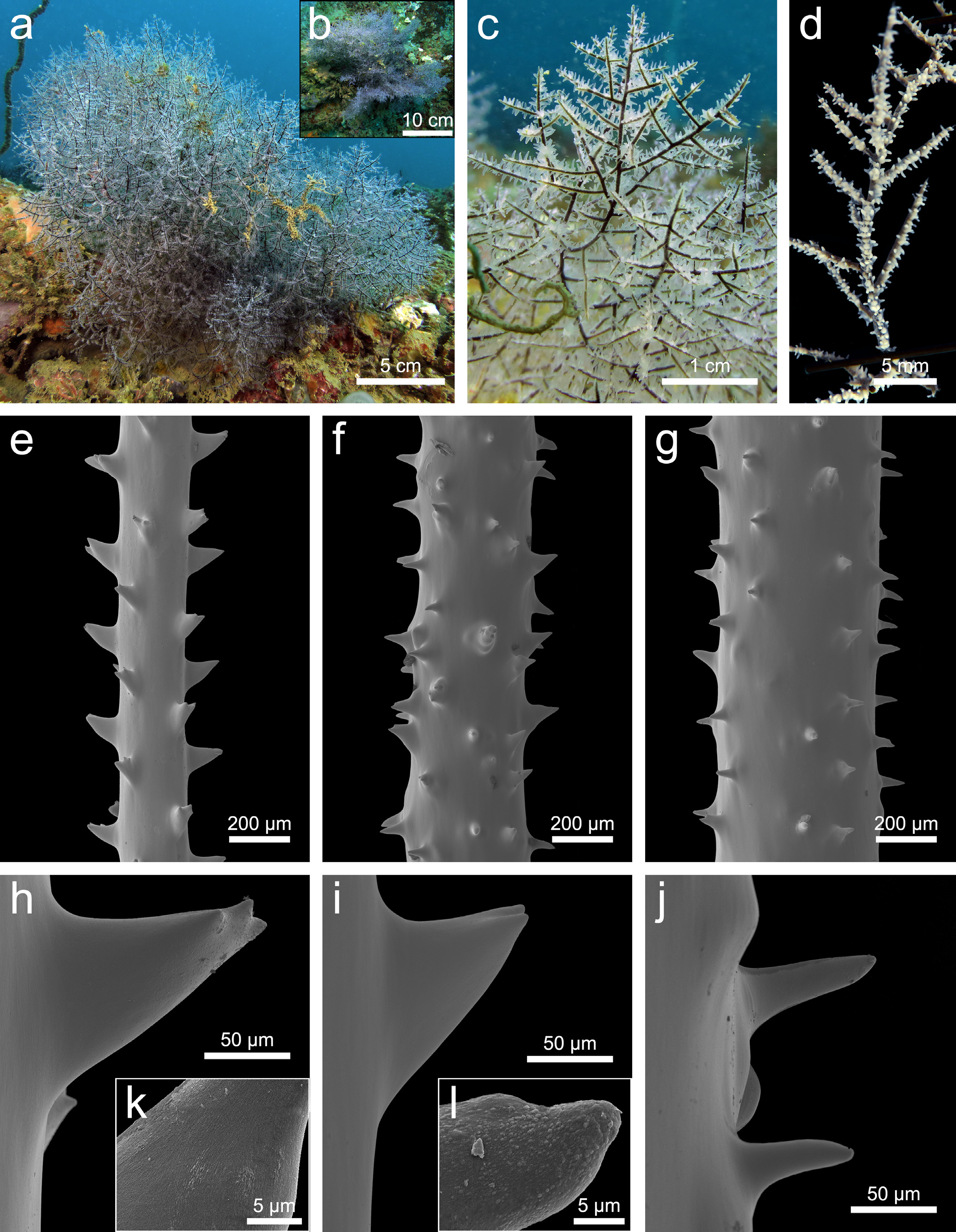
Description. A branched, bushy and highly anastomosed colony measuring 25 cm in width and 20 cm in height, with basal diameter of 4 mm ( Fig. 13 View FIGURE 13 , a), but larger colonies up to 40 cm in height can be found. The colony forms distinct thick masses of branches when seen from the top ( Fig. 13 View FIGURE 13 , b). When lower thick branches are close to the substrate, they can form new basal plates. The basal part of the colony shows highly anastomosed, curved thick branches that arise from the main stem. Adjacent branches and branchlets tend to fuse together in a flabellate shape, and then the flabellate parts are fused together by the branchlets sitting out of the plane. This branching pattern is less visible in large colonies where the fusions are numerous, but rather seen in young, small colonies. The diameter of the branches and branchlets appears to be similar, the latter only tapering at their end and giving a spiny appearance to the colony ( Fig. 13 View FIGURE 13 , c). The branchlets are growing in four directions and are inserted at an angle of 60–90°. Branchlets are either straight or slightly curved upwards ( Fig. 13 View FIGURE 13 , c). The longest measure up to 4 cm and are always curved upwards, 3 to 7 are found per cm counting those on every side (mostly 5–7). Branchlets are irregularly spaced (1–7 mm) and arranged, some being inserted opposite one another ( Fig. 13 View FIGURE 13 , c, d). This irregular arrangement makes it difficult to determine the length at which a branchlet becomes a branch, as these long branchlets can bear a second order of branchlets. These are usually uniserial, short and straight, but some of them are divided. The polyps are white, thick with rounded tentacles and measure up to 1.2 mm in transverse diameter. They occur in a single row on the branches and branchlets with 9–10 polyps per cm, but on thicker branches they are found all around the axis ( Fig. 13 View FIGURE 13 , c, d). Smaller polyps occur between larger ones, the mutual distance is irregular: either they sit close to each other, or they are spaced up to 0.8 mm apart ( Fig. 13 View FIGURE 13 , d).
The spines are varying in size and shape depending on the branch where they occur ( Fig. 13 View FIGURE 13 , e–j). On a branch measuring 0.23 mm in diameter, the spines are slightly inclined upwards and there is no difference between polypar and abpolypar spines. The spines appear smooth, triangular and some of them are forked at their apex ( Fig. 13 View FIGURE 13 , e, h), but regardless of the branch and the polypar/abopolypar side, extremely faint striations ( Fig. 13 View FIGURE 13 , k) or small papillae ( Fig. 13 View FIGURE 13 , l) are sometimes visible. Four longitudinal rows are seen from one aspect, they measure 0.10–0.12 mm and are spaced 0.21–0.34 mm apart. On larger branches, the spines are smooth, and their morphology is either triangular, conical, hooked or almost cylindrical ( Fig. 13 View FIGURE 13 , f, g, i, j). Some of them are forked at their apex ( Fig. 13 View FIGURE 13 , f). They are inclined in different directions ( Fig. 13 View FIGURE 13 , f, g) and they measure 0.07–0.11 mm in height. On such branches, the longitudinal arrangement is lost, and some bumps are seen on the axial surface, often at the base of the spines ( Fig. 13 View FIGURE 13 , g, j).
Taxonomic remarks. The species assigned to the genus Arachnopathes are differentiated based on the thickness of the branchlets, the extent of anastomosing and the size and morphology of the spines. Only the features of the spines are well-defined taxonomic traits, and the original descriptions of Ar. ericoides and Ar. clathrata made by Pallas (1766) lack valuable taxonomic information to clearly separate these two species. To date, only Brook’s descriptions of non-type material have enough information to define both, as well as Ar. aculeata ( Brook 1889) . Arachnopathes aculeata is characterized branchlets varying in length and density and by spines measuring up to 0.06 mm. Two to four delicate branchlets, measuring 0.5–1.5 cm, are found per cm, but they are longer, thicker and more numerous in the middle of the colony ( Brook 1889). Arachnopathes clathrata is differentiated from Ar. ericoides solely by having a marked contrast between the thickness of the branches and those of the branchlets defined as “needle-like” by Brook (1889), which is not the case for the present specimen. The latter closely matches the descriptions made by Brook (1889) and van Pesch (1914). The only noticeable differences are the spiral arrangement of the branchlets described by Brook (1889), which is not clearly seen in the material from Madagascar, as well as the lack of information regarding which branch he was analyzing when he described the spines as “laterally compressed, ending in a blunt point formed by the lower margin taking a sharp curve upwards to join the upper margin”. Information about the polyps was missing from Brook’s description and is given here for the first time.
Distribution. Indian Ocean (type locality, Pallas 1766), Indonesia ( van Pesch 1914); Singapore ( Pax & Muller 1955); Madagascar (present study).
Genus Cirrhipathes de Blainville, 1834
Cirrhipathes was established amongst antipatharians by de Blainville (1834). It is characterized by unbranched and unpinnulated corallums, with polyps irregularly found on all sides of the stem. The colonies can be straight, slightly to highly contorted, curved, coiled or forming spirals. The spines on the skeleton are not arranged in verticils ( Brook 1889; Bo et al. 2009; Wagner 2015a). They can be variously papillose and may be knobbed or lobed at the apex. This genus currently encompasses 16 nominal species, of which four type specimens are lost or missing, including the type of Cirrhipathes spiralis ( Linnaeus, 1758) . All species have been described from the Indian and Pacific Oceans, except Cirrhipathes secchini Echeverria, 2002 from the SW Atlantic. However, a redescription of the type material is needed for the latter as it might be related to the genus Stichopathes rather than Cirrhipathes due to the arrangement of the polyps and the occurrence of secondary spines.
No known copyright restrictions apply. See Agosti, D., Egloff, W., 2009. Taxonomic information exchange and copyright: the Plazi approach. BMC Research Notes 2009, 2:53 for further explanation.
|
Kingdom |
|
|
Phylum |
|
|
Class |
|
|
Order |
|
|
Family |
|
|
Genus |
Arachnopathes ericoides ( Pallas, 1766 )
| Terrana, Lucas, Bo, Marzia, Opresko, Dennis M. & Eeckhaut, Igor 2020 |
Euantipathes ericoides
| van Pesch 1914: 82 |
Arachnopathes ericoides
| Milne-Edwards & Haime 1857: 320 |
Antipathes ericoides
| Pallas 1766: 208 |