
Centromochlus orca, Sarmento-Soares & Lazzarotto & Py-Daniel & Leitão, 2016
|
publication ID |
https://doi.org/ 10.1590/1982-0224-20160030 |
|
publication LSID |
lsid:zoobank.org:pub:C13F0385-E7A2-443D-9821-F95C71B7EA4E |
|
persistent identifier |
https://treatment.plazi.org/id/2C7587E1-0C31-FFDB-6B96-E86F6E035434 |
|
treatment provided by |
Carolina |
|
scientific name |
Centromochlus orca |
| status |
sp. nov. |
Centromochlus orca , new species
urn:lsid:zoobank.org:act:3C7F0FEA-CEBF-4FDA-A489-62D8089E2DF9
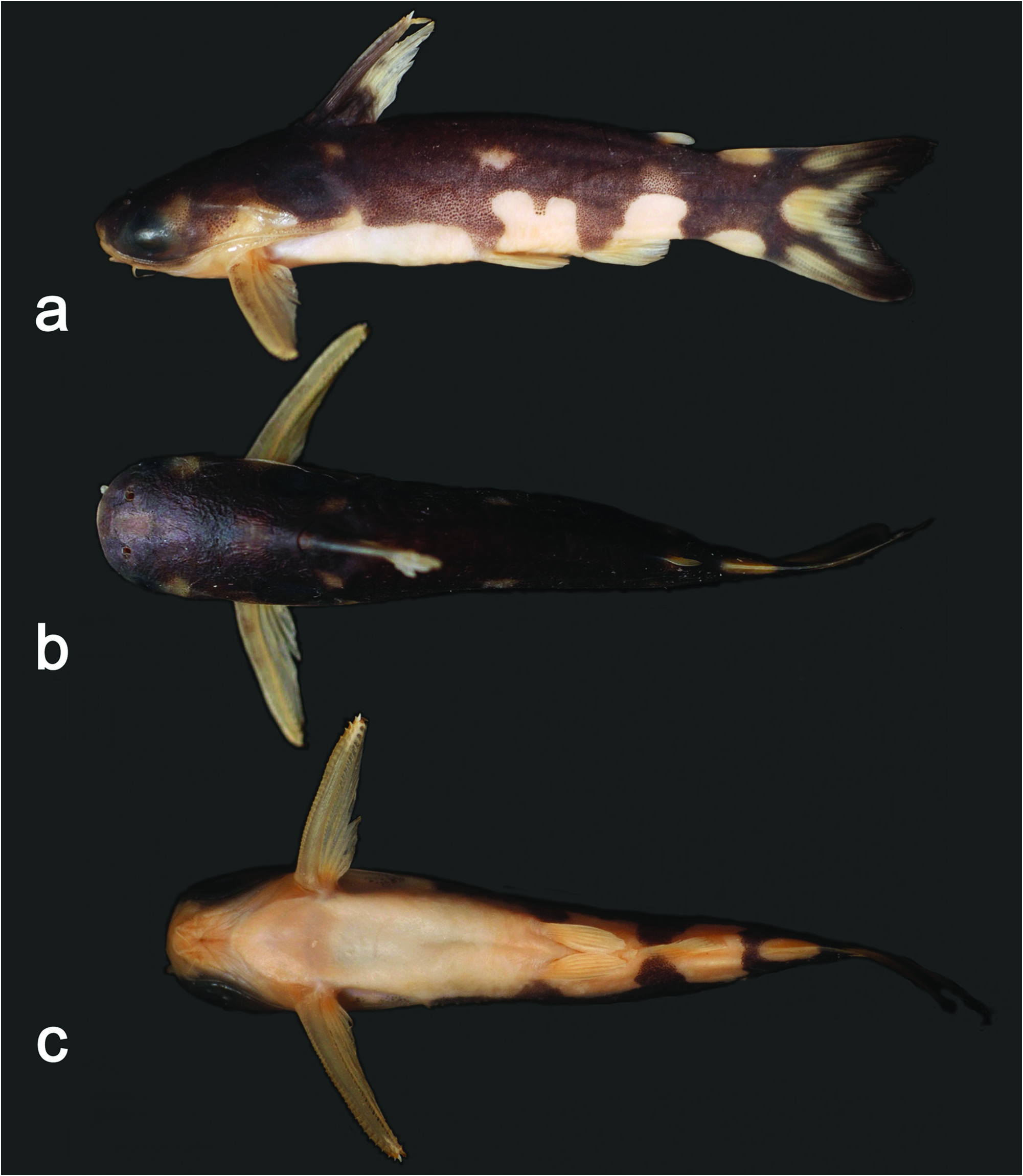
Fig. 1 View Fig
Holotype. INPA 50870 View Materials , 1 View Materials , 56.8 mm SL, Brazil, Pará , Terra Santa , mouth of Igarapé Jamari with lago de Terra Santa, rio Nhamundá basin, 02°00’04’’S 56°31’08’’W, 22 Sep. 2009, R. P. Leitão & H. Lazzarotto. GoogleMaps
Paratypes. All from Brazil. Pará : Terra Santa : INPA 35086 View Materials , 14 View Materials +1 CS, 40.5-56.8 mm SL, INPA 35087 View Materials , 4 View Materials , 21.0- 47.7 mm SL, MBML 11220 , 1 , 51.0 mm SL, MBML 11221 , 1 CS, 50.6 mm SL, MNRJ 45072 View Materials , 5 View Materials +1 CS, 38.2-52.3 mm SL, MPEG 33927 View Materials , 3 View Materials , 39.0- 50.8 mm SL, MZUEL 15706 , 1 , 42.8 mm SL, collected with the holotype. Amazonas : Nhamundá : INPA 43875 View Materials , 4 View Materials , 42.7-53.8 mm SL, lagoa Sete Ilhas upstream from Nhamundá, 01°50’28’’S 57°04’20’’W, 12 Nov. 2013, R. A. Collins. INPA 43883 View Materials , 3 View Materials , 37.0- 48.1 mm SL, Lagoa Sete Ilhas upstream from Nhamundá, on boundary between Amazonas and Pará, 01°43’04’’S 57°22’07’’W, 12 Nov. 2013, R. A. Collins GoogleMaps .
Non-type material. Brazil. Amazonas . Presidente Figueiredo: MZUEL 10467 , 11 , 25.7-46.2 mm SL, rio Uatumã , near mouth of rio Pitinga, at base WABA of ReBio Uatumã, in the reservoir of UHE, 01°31’19.7”S 69°49’17.9”W, 18 Sep. 2014, J. L. O. Birindelli, F. C. Jerep, L. Rapp Py-Daniel GoogleMaps & V. N. Machado.
Diagnosis. Centromochlus orca is distinguished from all congeners by its unique color pattern, black ground color sharply delimited from a white underside by conspicuous wavy border. Additional features for recognition of C. orca include the absence of anterior nuchal plate ( Fig. 2 View Fig ) and eye diameter large, between 24.4 to 29.9% in HL.
The boldly contrasted black and white color pattern is unique to C. orca , but a similar pattern is found in T. musaica , from rio Orinoco, T. carolae Vari & Ferraris 2013 , from rio Cuyuni, lower Essequibo, and T. melanoleuca , from rio Teles Pires, upper Tapajós. All those species lack the anterior nuchal plate. Centromochlus orca is distinguished from T. musaica by coloration, as in C. orca the pattern is contrasted with white areas extending dorsally onto lower sides of body (vs. a non-pigmented area in a mosaic pattern - compare Figs. 1 View Fig and 11 View Fig ). Notable differences among C. orca and T. musaica are observed in suspensorium bones (compare Figs. 3a and 3b View Fig ), with contact between metapterygoid and quadrate via suture and cartilage (vs. contact between metapterygoid and quadrate via cartilage only in T. musaica ), and hyomandibula broad, with a well developed medial lamina (vs. hyomandibula notched, lacking medial lamina in T. musaica ). The color pattern in C. orca is contrasting as black and white colors, differing from the faint coloration in T. carolae and T. melanoleuca (see Vari & Calegari, 2014, fig. 1; Vari & Ferraris, 2013, fig. 1). Centromochlus orca is further distinguished from both T. melanoleuca and T. carolae by shape of Müllerian ramus disc (bulbous in C. orca vs. spoon shaped in T. melanoleuca and T. carolae ) (compare Figs. 4a and 4b View Fig ). For further details, see discussion.
Description. Morphometric data presented in Table 1. Small size, adult specimens 47.7-56.8 mm SL. Body almost cylindrical from rear of head through abdomen and gradually compressed towards caudal fin. Dorsal profile slightly convex from snout tip to dorsal-fin origin, approximately straight from that point to vertical through pelvic-fin origin, slightly convex to adipose-fin terminus, then slightly concave on caudal peduncle. Greatest body width at pectoral-fin origin, 19.4-22.1% SL. Ventral profile of body gently concave between anal-fin base and caudal-fin origin. Greatest body depth at origin of dorsal fin, 17.3-20.8%SL. Head integument thick, obscuring bones of cranial roof; eyes large, laterodorsal on anterior portion of head, covered by thick layer of translucent skin. Snout margin broadly rounded in dorsal view; anterior nostril close to snout margin, with tubular skin flap anteriorly oriented; posterior nostril also with tubular skin flap, located at vertical through anterior border of orbit; transverse distance between anterior nostrils almost equal to distance between posterior ones. Three pairs of barbels: maxillary, inner and outer mental. Maxillary barbel short, surpassing membranous border of opercle, reaching approximately vertical through dorsal-fin origin; adpressed maxillary barbel fits in groove on lateral portion of head, immediately above rictal fold and below eye; mental barbels short, tips not reaching pectoral-fin base; barbels arranged in arc along ventral surface of jaw; inner mental barbel about half the length of outer one. When adpressed, inner and outer mental barbels received by groove in skin. Posterior process of cleithrum moderately large, almost reaching vertical through middle of dorsalfin base.
Rostral border of cranium with mesethmoid broader than long; premaxilla with synchondral articulation; cranial fontanel narrow and elliptical, enclosed by mesethmoid and frontals ( Fig. 2 View Fig ). Nasal ossified as short tubular bone situated between mesethmoid cornua and lateral ethmoid, not sutured to mesethmoid. Lateral ethmoid not participating in dorsal face of cephalic shield. Autopalatine rod-like, oriented almost parallel to longitudinal axis of body; maxilla slightly elongated, about two-thirds the size of autopalatine ( Fig. 3 View Fig , mx); vomer arrow-shaped with short rostral-lateral processes. Jaws of equal size; premaxilla and dentary slender, each with two or three rows of small conical teeth. Anterior nuchal plate absent; middle nuchal plate slightly concave along lateral margins; posterior nuchal plate short, projected laterally, with prominent tip. Epioccipital process small.
Hyomandibular broad, with anteriorly projected lamina, connected to quadrate only, through cartilage and deeply interdigitated suture ( Fig. 4a View Fig , hm). Quadrate large, trapezoidal, with broad base, sutured to preopercle, hyomandibular and metapterygoid. Metapterygoid somewhat rectangular, as short lamina, joined to quadrate via suture ( Fig. 4a View Fig , mt); entopterygoid in connective tissue linking metapterygoid to lateral process of vomer ( Fig. 4a View Fig , en). Long ventral margin of preopercle sutured to both quadrate and hyomandibula; suprapreopercle present as long canal bone; preopercular canal exiting on anterior portion of pterotic. Opercle laminate, ornamented and broadly subtriangular.
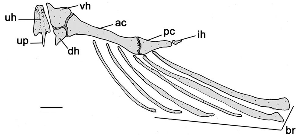
Hyoid arch with compact parurohyal with short ventral process; short dorsal hypohyal associated with comparatively large ventral hypohyal ( Fig. 6 View Fig , vh); anterior ceratohyal more developed than posterior ceratohyal. Six branchiostegal rays articulated with hyoid arch, four slender rays associated with anterior ceratohyal, two flattened rays with posterior ceratohyal ( Fig. 6 View Fig , br). Branchiostegal membrane broadly united to isthmus.
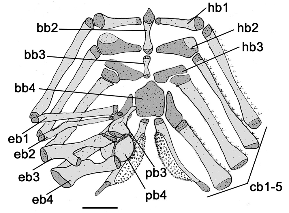
Branchial (gill) arches with urohyal close to basibranchial 2; basibranchial 2 mostly cartilaginous, broadest anteriorly, slightly separated from basibranchial 3 ( Fig. 7 View Fig , bb2); basibranchial 3 shorter, forming osseous rod; basibranchial 4 large, flattened and cartilaginous; basibranchial 2 bordered laterally by cartilaginous head of hypobranchial 1; basibranchial 3 between cartilaginous head of hypobranchial 2 and cartilaginous hypobranchial 3; basibranchial 4 bordered laterally by cartilaginous head of ceratobranchial 4 and caudally by cartilaginous head of ceratobranchial 5. Hypobranchials 1 and 2 subtriangular, mostly osseous, elongate and expanded laterally, with cartilaginous tips ( Fig. 7 View Fig , hb1, hb2); hypobranchial 3 mainly cartilaginous, trapezoidal; hypobranchial 4 absent. Five ceratobranchials, mostly ossified, with cartilage on both tips ( Fig. 7 View Fig , cb1-5). Ceratobranchials supporting single row of rakers; fifth ceratobranchial expanded postero-medially to support lower pharyngeal toothplate bearing short conical teeth. Four epibranchials, all largely ossified except for cartilaginous tips, each one supporting few rakers, close to articulation with ceratobranchials. Epibranchials 1 and 2 rod-like; epibranchial 3 with posterior uncinate process articulated to epibranchial 4; epibranchial 4 with laminar extension; reduced accessory cartilage located at angle between cartilaginous tips of epibranchial 4 and ceratobranchial 4. Pharyngobranchial 1 absent; pharyngobranchial 2 short, cartilaginous, somewhat ellipsoid, placed between anteromedial cartilaginous tips of epibranchials 1 and 2; pharyngobranchial 3 elongate, ossified, with expanded posterior border; pharyngobranchial 4 ossified. Upper pharyngeal tooth plate bearing conical teeth, supported by pharyngobranchial 3 and 4, and also epibranchials 3 and 4 ( Fig. 7 View Fig , fb3, fb4).
Müllerian ramus disc of parapophysis of fourth vertebrae with enlarged tip forming bulbous projection, directed posteriorly, extending into lumen of swim bladder ( Fig. 5a View Fig , pv4).
Laterosensory cephalic canal with infraorbital 1 bearing a ventrolateral process restricted to anterior border of eye, and followed by four canal-like bones, in incomplete infraorbital series. Dorsal rim of orbit formed mainly by frontal, with sphenotic contributing the posteriormost limit. Infraorbital canal exiting on middle portion of sphenotic. Lateral line weakly sinuous and with ossified canal bones on anteriormost portion of trunk, then approximately straight with singular terminus on base of caudal-fin rays.
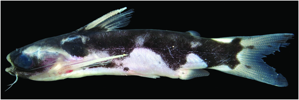
Coloration in alcohol. Centromochlus orca has unique color pattern, black ground color sharply delimited from white underside by conspicuous wavy border through lower half of body side. Dorsal cloak uniformly dark or sometimes with white rounded blotch in larger specimens, rounded extensions of dark coloration reach the base of both anal and pelvic fins. Dorsal surface of head also largely black, with tips of third nuchal plate whitish-brown. Latero-ventral sides and belly whitish-yellow, strongly contrasting dark cloak. Posterior process of cleithrum pale to mostly black. Paired and anal fins whitish-yellow with hyaline tips. Dorsal fin with dark base. Dorsal fin spine mostly dark, branched rays hyaline. Adipose fin with dark base and hyaline tips. Caudal peduncle with white half-circle on upper and lower margins. Caudal fin with white circle on fin base; each lobe with pale outer streak and broader dark inner stripe ( Fig. 8 View Fig ).
Dorsal fin II,5; dorsal-fin spine strong, slightly shorter than first branched ray, anterior face with about 20 minute dentations becoming progressively smaller towards fin base; posterior face mostly smooth except for 3-8 retrorse dentations distally; fin tip cartilaginous. Pectoral fin I,5. Pectoral-fin spine strong with 25-33 antrorse dentations on anterior face; 20-22 retrorse dentations on posterior face; dentations on both faces become larger towards tip. Pelvic fin i,5, lateral margin rounded. Adipose fin teardrop shaped, with free posterior margin. Anal fin iii,7; anal-fin pterygiophores with eight rod-like proximal radials and six cartilaginous distal radials. Caudal fin moderately forked, lobes with rounded tips, 8+9 principal rays, all rays branched except for outermost ray of upper and lower lobes; 20-22 upper and 17-19 lower procurrent rays.
Nine ribs attached to consecutive vertebrae 6-14, becoming progressively smaller posteriorly. Total vertebrae 30 (observed in three cleared and stained specimens).
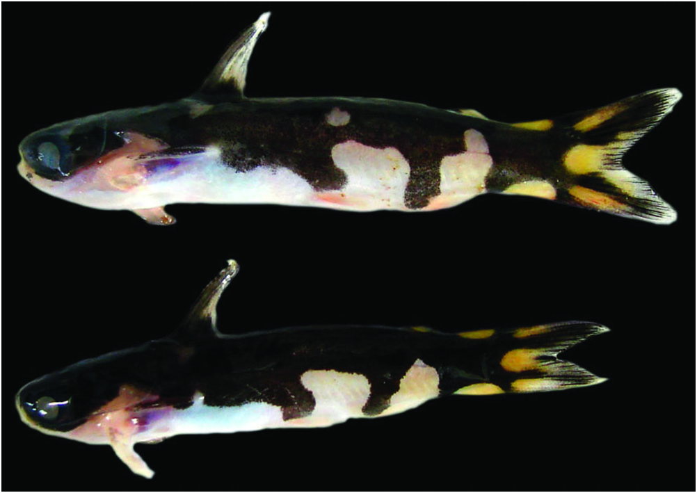
Live coloration. Bold pattern of black and white more highly contrasted than in preserved specimens. Dorsal and lateral surfaces of head with distinctive dark coloration; translucent areas on tip of snout, posterior nuchal plate region, and opercular area. Dark cloak with light rounded blotch present on each side of body between dorsal and adipose fins in large specimens. Belly, anal and paired fins white with traces of pink. Dorsal and pectoral fin spines dark. Adipose fin with dark base and yellow tip. Half circles on caudal peduncle, rounded blotch on caudal-fin base and outer streak on each lobe light-yellow ( Fig. 9 View Fig ).
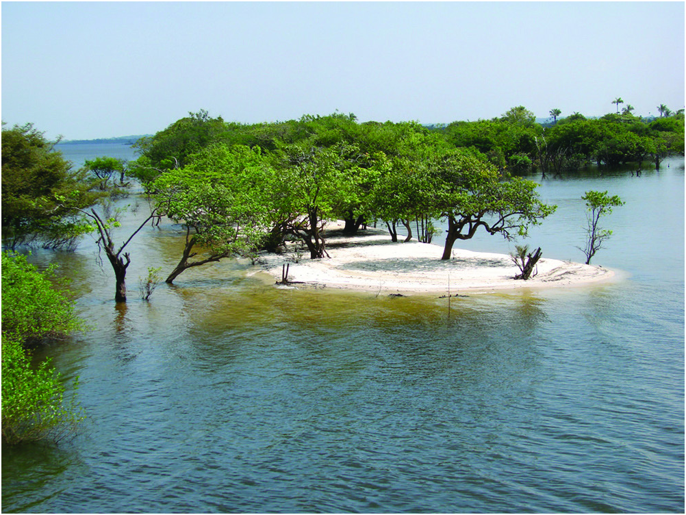
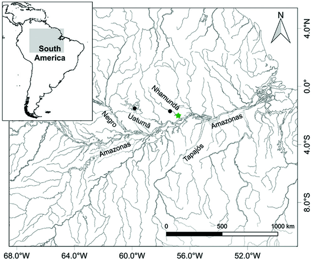
Distribution. Centromochlus orca is known from the mouth of igarapé Jamari, a tributary of the lower rio Nhamundá, in the western Pará, and from two close localities in lagoa Sete Ilhas upstream from Nhamundá, on boundary between Amazonas and Pará, in a region along the transition between the Amazonas floodplain and the Guiana Shield ( Fig. 10 View Fig ).
Ecological notes. In the rio Nhamundá basin, the specimens of C. orca were collected in the lower portions of igarapé Jamari, in the transition zone between lotic river habitats and lentic habitats of varzea lakes ( Fig. 11 View Fig ). In igarapé Jamari, the water current was virtually absent. Individuals of C. orca were observed actively swimming on the surface or in mid-column in places illuminated by light bulbs close to the shoreline or on boats. All specimens were collected during the night, either by seining (a sandy beach) or with dip nets while swimming (in illuminated areas). Fish assemblages at these localities are highly diverse, encompassing species mainly from the white-water floodplains, but including some taxa typical of clear water rivers that flow from the Guiana Shield, such as Hoplarchus psittacus , Hypancistrus sp. , Otocinclus mura and Peckoltia vittata . Among the other 151 species collected syntopically with C. orca , Tatia nigra was the only additional Centromochlinae . Local fishermen reported that C. orca is caught and sold for the aquarium trade, but is not a primary target.
Sexual dimorphism. Among all specimens examined, none have a fully modified anal fin for insemination. One maturing male exhibited partial modified anal fin ( Fig. 8 View Fig ).
Etymology. The specific epithet is an allusion to the coloration resembling that of the orca whales ( Orcinus orca ). A noun in apposition.
Conservation status. Centromochlus orca was recorded for lower portions of rio Nhamundá basin at western Pará, near border with Amazonas state. It corresponds to an species of small fish, with pelagic habits and apparently non migratory. Although reports of specimens caught for aquarium trade, such an activity do not represent a primary target, and possibily does not cause a severe impact in fish natural populations. The species is abundant in the places where it is found and no significative threatening were recognized for the species locally. Centromochlus orca was categorized as Least Concern (LC), according to the IUCN criteria for evaluation on threatening status ( IUCN, 2016) .


| R |
Departamento de Geologia, Universidad de Chile |
| CS |
Musee des Dinosaures d'Esperaza (Aude) |
No known copyright restrictions apply. See Agosti, D., Egloff, W., 2009. Taxonomic information exchange and copyright: the Plazi approach. BMC Research Notes 2009, 2:53 for further explanation.