
Chilotherium schlosseri (Weber, 1905)
|
publication ID |
https://doi.org/ 10.5252/g2016n2a6 |
|
publication LSID |
urn:lsid:zoobank.org:pub:256C1778-4D62-46B2-A292-95CB584FCC37 |
|
persistent identifier |
https://treatment.plazi.org/id/03F587FD-FFFE-9E0A-FF66-FFE3FD03F9F2 |
|
treatment provided by |
Felipe |
|
scientific name |
Chilotherium schlosseri (Weber, 1905) |
| status |
|
Chilotherium schlosseri (Weber, 1905)
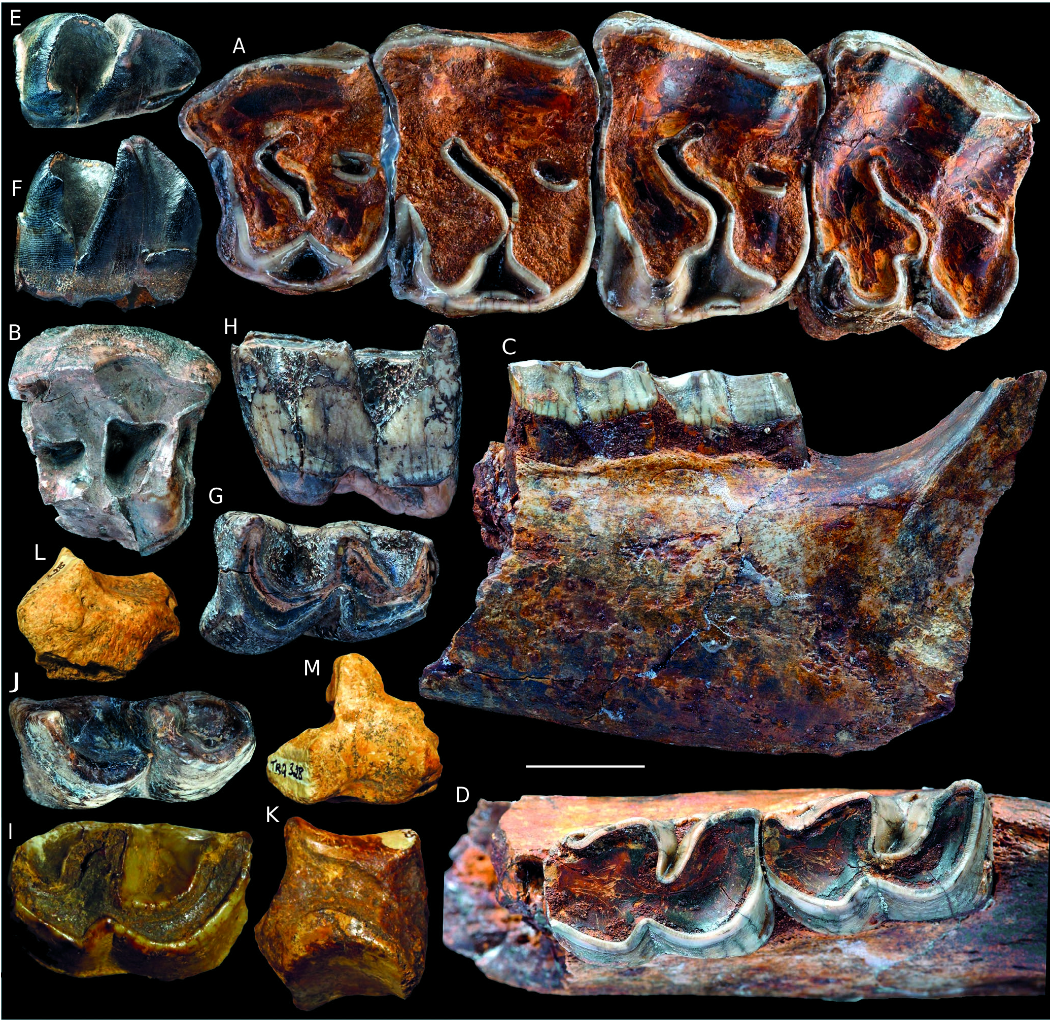
( Fig. 2 View FIG )
Chilotherium kowalevskii – Pavlow 1913: 48-67. pl. 4-5.
Aceratherium sp. – Malik & Nafiz 1933: 44-45, pl. VII, figs. 4-5.
Aceratherium cf. kowaleskii [partim] – Nicolas 1978: 456.
MATERIAL EXAMINED. — MNHN.F.TRQ339, right P2; TRQ341, left unworn P4; KÇ297, right M2;TRQ344, ectoloph of a left M1- 2; KÇ295, left D3; TRQ346, left D4 (without ectoloph); KÇ305, right D4 (broken); KÇ338, right i2; TRQ348, right i2 (anterior tip); TRQ349, right i2 (anterior tip); TRQ352, left mandible fragment with p3 (talonid)-m1; TRQ351, right mandible fragment with m2-3;TRQ353, right p4 (germ);TRQ357, left m2;TRQ354, right m2; TRQ358, left d3; TRQ330, right Mc3 (without distal end); TRQ329, right Mc4; TRQ364, left cuboid; TRQ335, left Mt3 (proximal fragment).
DESCRIPTION
Upper dentition
The upper teeth referable to this taxon are high-crowned ( Table 1), with thinly wrinkled enamel and scattered patches of cement on the ectoloph. The dental pattern is more complicated than in the other aceratheriine from Küçükçekmece ( Fig. 3 View FIG ), notably due to the presence of secondary folding and accessory enamel tubercles ( Fig. 2A, B View FIG , E-G). Apart from P2 (ectoloph convex, without any fold; Fig. 2A View FIG ), all other upper teeth have undulated ectolophs in occlusal view, with a parastyle and a long metastyle pointing labially, a shallow parastyle groove and a smooth paracone fold on P4 and M1-2 (deep/thick on D3), but also a smooth mesostyle on molars and D3 ( Fig. 2F View FIG ). On upper teeth (premolars, molars, and upper deciduous teeth), the lingual cingulum is low and reduced, restricted to the median part of the lingual wall, whereas the labial cingulum is restricted to enamel tubercles running along the anterior and posterior edges of the ectoloph (e.g., Fig. 2D View FIG ). The postfossette is narrow transversely and elongate sagittally, especially after early wear stages ( Fig. 2E View FIG ). The morphology of roots is essentially unknown. On upper premolars, the metaloph forms a dihedron, with a thick sagittal crochet located at the anterior angle, either right-angled (P2 MNHN.F.TRQ339) or open-angled (P4 TRQ341). The P2 TRQ339 is small and with a trapezoid occlusal outline (lingually compressed; Fig. 2A View FIG ). The parastyle is as thick as the metastyle. The prefossette is wide and deep, bordered lingually by a sharp groove, coinciding with the protocone constriction. The protoloph is complete. The protocone is partly broken. There is neither antecrochet nor crista. The metacone and the hypocone are located at the same level in occlusal view. The protocone and the hypocone, equally developed, are strongly connected by a thick lingual bridge. Accordingly, the median valley is closed and V-shaped in occlusal view. The P4 MNHN.F.TRQ341 is unworn ( Fig. 2 View FIG B-D), which allows for determining the hypsodonty index (H/L = 1.19). The ectoloph is very high, but convex and oblique, as is the lingual border, in sagittal view. As a consequence, the occlusal surface is much narrower at early wear stages than at late wear stages. At the same time, the ectoloph is shortening sagittally with wear, which leads to a drastic change in terms of W/L ratio during the lifetime of the individual. The crochet is simple, long and sharp, and sagittally oriented. The antecrochet is strong, sharp and developed at any wear stage. The protoloph is curved (convex anteriorly) and completely connected to the ectoloph. There is a shallow protocone constriction, restricted to the base of the crown. The protocone and the hypocone are distant and not connected (molariform pattern). There is neither crista nor medifossette. The metaloph is thin but lacking a constriction. It is very short with respect to the protoloph and pointing postero-lingually, in occlusal view. The posterior cingulum is very low and V-shaped in posterior view. KÇ297 is a right M2, damaged in its lingual and posterolabial parts ( Fig. 2E View FIG ). The antecrochet is strongly developed, sharp, and located labially with respect to the deep protocone constriction. The protoloph is sigmoid in occlusal view. The crochet is thick but short, closely appressed to the ectoloph and neighboring a small enamel fold (crista?). There is neither medifossette nor cristella. The median valley is sigmoid, narrow, and open lingually. The lingual cingulum is not preserved, but there is a small enamel tubercle at the bottom of the median valley. The metaloph is short with respect to the protoloph in occlusal view. The posterior cingulum is low but continuous, following the V-shaped pattern of the bottom of the postfossette. There is a deep hypocone constriction, restricted to the base of the crown. The antecrochet does not connect the metaloph, even at late wear stages.
The upper deciduous teeth are molariform, with patterns similar to those of permanent teeth (ectoloph undulated, with strong paracone and metacone folds and a smooth mesostyle; anterior cingulum strong and complete; protoloph curved; crochet long, sharp, and sagittal; protocone and hypocone constricted; antecrochet long and sharp, connecting the metaloph in late wear stages; lingual cingulum reduced; postfossette deep, narrow, and triangular in occlusal view; metastyle very long). Yet, they are more brachydont, with thinner enamel, and a lighter patina. The left D3 KÇ295 is complete, with a very long parastyle and a sharp crista, but no medifossette ( Fig. 2F View FIG ). The parastyle groove is much deeper than in permanent teeth (not preserved on D4s). Its median valley is open until late wear stages (no lingual wall), but much wider in occlusal view than in D4 (MNHN.F.TRQ346 and KÇ305). There is no crista on D4s. The lingual roots are connected but unfused on TRQ346 ( Fig. 2G View FIG ).
Mandible and lower dentition
The i2 KÇ338 is cracked (long emersion) and eroded (e.g., transported). Most enamel has disappeared with wear and the neck is not easily discernable. It has a straight and cylindrical root, and the tip of the crown is curved, in a similar way to MNHN.F.TRQ348 and TRQ349 (crowns preserved, root broken). The cross section of the crown is a right triangle. The wear facet is flat transversely and concave sagittally.
The mandibular fragments are badly broken and they yield no morphological feature.
Lower teeth are high-crowned (p4MNHN.F.TRQ353: HI = 1.17; m2 TRQ354: HI = 1.2). There is no lingual or labial cingulid, but a short anterolingual ridge borders the paralophid on TRQ351 (m2-m3). The ectolophid groove, deep and V-shaped in occlusal view, reaches the neck and points up- and frontward in labial view. There is no cement preserved. Lingual cusps are constricted on most fresh teeth: metaconid of p4 (TRQ353), m2 (TRQ357), and m3 (TRQ351); entoconid of m2 (TRQ351 and TRQ357), and m3 (TRQ351). The trigonid is smooth and right-angled, and U-shaped in occlusal view. The lingual valleys are narrow and V-shaped in lingual view. Lophids are oriented transversely on premolars ( Fig. 2H, I View FIG ). Premolars have wide hypolophids with respect to trigonids. On m2-m3, the hypolophid is very oblique, especially at early wear stages ( Fig. 2J View FIG ). There is no lingual groove on the entoconid. The p3TRQ352 lacks any external rugosity. The lingual border of the lingual cusps is flattened. The posterior cingulum is low and particularly smooth on m2-m3 (TRQ351, TRQ354, and TRQ357).
The left d3 MNHN.F.TRQ358 is elongate sagittally ( Fig. 2L View FIG ), and quite high-crowned for a deciduous tooth ( Table 1). The paralophid is forked, with a sharp sagittal spur. There is an incipient groove on the labial side of the trigonid. The labial cingulid is restricted to a short and low ridge closing the shallow ectolophid groove. There is no lingual cingulid ( Fig. 2L View FIG ). The lingual cusps are not constricted and there is no protoconid fold. The lingual valleys are V-shaped in lingual view.
Postcranial skeleton
All available metapodials have wide and sagittally-flattened diaphyses and long insertions for the m. interossei. Metacarpals have strong and thick insertions for the m. extensor carpalis, contrary to the Mt3 TRQ335 (flat surface) referred to as Persiatherium sp. ( Fig. 3L, M View FIG ). The Mc3 TRQ330 lacks its distal end ( Fig. 2M View FIG ). In proximal view, it is much wider (TD = 47) than deep (APD = 35), with a rounded aspect ( Fig. 2N View FIG ). There seems to be no scaphoid-facet. The semilunate-facet is quadrate in proximal view, concave transversely, and elongated and convex sagittally. The unciform-facet is flat transversely and strongly convex sagittally, separate from the former by a smooth ridge (c. 110°). The strong tubercle for the insertion of the m. extensor carpalis determines a deep groove proximal to it. In anterior view, the magnum-facet is visible. There is only one Mc2-facet, restricted to a low almond-shaped strip, elongate sagittally. The Mc4-facets are oval and distant one from another. The anterior one is located much more proximally. The medial border of the diaphysis is straight whereas the lateral border is strongly and regularly concave.
TRQ329 is a complete Mc4, with an eroded surface ( Fig. 2 View FIG O-Q). Its proximal and distal ends are not widened with respect to the diaphysis (L = 112; proxTD = 29; proxAPD = 29; diaDT = 25; diaAPD = 13.5; distTD = 28; distAPD = 30). The diaphysis is strongly curved in its proximal third (concave laterally; Fig. 2O View FIG ), which fits the shape of the Mc3 MNHN.F.TRQ330 ( Fig. 2M View FIG ). The proximal facet has a pentagonal outline, due to the Mc3-facets forming a 135° angle in proximal view. The Mc5-facet has no delimited outline: it coincides with the smooth and rounded lateral border of the proximal end. There is a thick tubercle at the antero-lateral angle of the bone, corresponding to the insertion for the m. extensor carpalis ( Fig. 2P View FIG ). In medial view, the Mc3-facets are disconnected and subvertical. The anterior facet is flat and elongate sagittally, whereas the posterior one is oval and elongate proximo-distally. In medial view, the diaphysis is straight, with a salient posterodistal tubercle on the median part of the diaphysis ( Fig. 3Q View FIG ), just proximal to the distal articulation. In distal view, the distal pulley has a quarter-circle outline, as wide as deep (TD = APD), and with a postero-medial right angle. The intermediate relief is sharp, separating two concave lips equally developed transversely.
The left cuboid MNHN.F.TRQ364 is robust and low anteriorly (H = 40; TD = 33.5; APD = 54.5; antH = 26.5), with salient muscular insertions ( Fig. 2R, S View FIG ). In proximal view, the proximal facet is heart-shaped (triangular, with an anterior inflection) and saddle-shaped. It is split into two equal parts by a shallow sagittal groove; the astragalus-facet is more developed sagittally than the calcaneus-facet. In anterior view, the anterior side has a convex proximal border, parallel medial and lateral sides, and a posterior border forming an open dihedron (c. 135°; Fig. 2R View FIG ). On the medial side, the antero-proximal facet is damaged. The postero-proximal facet (for the ectocuneiform) has a cochlear shape. It is much larger than the antero-distal facet, semi-oval and elongate proximodistally. The posterior tuberosity is well developed, with an oblique posterior border in lateral view ( Fig. 2S View FIG ). The distal tip of such tuberosity well overhangs the distal articulation. It is longer medially than laterally. The distal MtIV-facet is T-shaped in distal view, as deep as wide (APD = TD). It is slightly concave sagittally and strongly convex transversely.
MNHN.F.TRQ335 is a proximal fragment of a left Mt3 ( Fig. 2T, U View FIG ). In anterior view, the proximal border is sigmoid ( Fig. 2U View FIG ). The diaphysis was probably widened distally. In proximal view, the proximal end is almost as deep (APD = 36.5) as wide (TD = 38.5), due to posteriorly-projected posterior Mt3-facet ( Fig. 2T View FIG ); the ectocuneiform-facet is L-shaped, with an anterior border forming a widely open dihedron (c. 160°); there is no cuboid-facet. The medial surface is much eroded, which impedes any observation on Mt2-facet(s). In lateral view, the Mt4-facets are large and remote. The anterior one is vertical, with a semi-circular outline. It connects the proximal facet. The posterior facet is subvertical (visible in proximal view), and projected posteriorly. The diaphysis is flattened, but much more pinched laterally than medially.
DISCUSSION
The smallest rhinocerotid from Küçükçekmece is undoubtedly referable to Aceratheriina (sensu Antoine et al. 2003b, 2010) or Aceratheriini (sensu Lu 2013; Pandolfi 2016), due to the presence of wrinkled enamel on cheek teeth, of upturned i2s, of protocone and hypocone equally developed on P2, of a metaloph constriction on P2-4, of a weak paracone fold on M1-2, and of a constricted hypocone on M2. Morphological features, such as partial hypsodonty, the presence of a protocone constriction on P3-4 and a strong antecrochet on upper molars, of a deep ectolophid groove reaching the neck on lower cheek teeth, or the absence of labial cingulum on lower cheekteeth, allow for recognizing it as a “chilothere” sensu lato (i.e. an unformal cluster of aceratheriines including Chilotherium Ringström, 1924 , Acerorhinus Kretzoi, 1942 , Shansirhinus Kretzoi, 1942 , and Persiatherium Pandolfi, 2016 ). Among chilotheres, it can be identified as belonging to Chilotherium based on the presence of a reduced lingual cingulum on upper premolars, of protocone and hypocone separate at early wear stages and later connected by a lingual bridge on P2, of a strong antecrochet on P4, of a labial cingulum restricted to the anterior and posterior edges of the ectoloph on upper molars, of a strong protocone constriction on upper molars, and the absence of a metaconid constriction on lower milk teeth. The available remains share the closest affinities with Chilotherium schlosseri (Weber, 1905) , from the late Vallesian of Ukraine (upper MN9 and MN10; see Vangengeim &Tesakov 2013) and its coeval Chilotherium kowalevskii ( Pavlow, 1913) from Moldova and Ukraine (MN10; Vangengeim &Tesakov 2013). C. kowalevskii is considered here as a junior synonym of Chilotherium schlosseri . Both have strikingly similar craniodental features and concurring ranges (early late Miocene), north to the Black Sea in Ukraine and Moldova. Although not explicitly stated, this synonymy was already suggested by the results of the phylogenetic analysis of aceratheriines as proposed by Pandolfi (2016). Interestingly, Nicolas (1978) had assigned the chilothere remains from Küçükçekmece West to “ Aceratherium cf. kowalevskii ”.
The small chilothere from Küçükçekmece shares salient dental features with C. schlosseri , such as the presence of a lingually-projected antecrochet and of a continuous posterior cingulum on upper molars, of a deep and V-shaped ectolophid groove on lower cheekteeth, and of a strong mesostyle on D3. It further differs from other Chilotherium species in having no median incision of the lingual cingulum on upper premolars, no antecrochet on P2, protocone and hypocone equally developed on P2, lingual cusps separate on P3-P4, and a double paralophid on d3 (see comparison in Lu 2013 and Pandolfi 2016). The lingual border of the lingual cusps is flattened (contrary to what is observed on teeth from Küçükçekmece referred to as Persiatherium sp. ).

No known copyright restrictions apply. See Agosti, D., Egloff, W., 2009. Taxonomic information exchange and copyright: the Plazi approach. BMC Research Notes 2009, 2:53 for further explanation.
|
Kingdom |
|
|
Phylum |
|
|
Class |
|
|
Order |
|
|
Family |
|
|
Genus |
Chilotherium schlosseri (Weber, 1905)
| Antoine, Pierre-Olivier & Sen, Sevket 2016 |
Aceratherium
| NICOLAS J. 1978: 456 |
Aceratherium sp.
| MALIK A. & NAFIZ H. 1933: 44 |
Chilotherium kowalevskii
| PAVLOW M. A. 1913: 48 |